

In vitro protective effects of Paeonia mascula subsp. hellenica callus extract on human keratinocytes
- PMID: 33154501
- PMCID: PMC7645794
- DOI: 10.1038/s41598-020-76169-0
In vitro protective effects of Paeonia mascula subsp. hellenica callus extract on human keratinocytes
Erratum in
-
Author Correction: In vitro protective effects of Paeonia officinalis var. mascula callus extract on human keratinocytes.Sci Rep. 2023 May 19;13(1):8156. doi: 10.1038/s41598-023-34609-7. Sci Rep. 2023. PMID: 37208384 Free PMC article. No abstract available.
Abstract
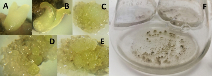
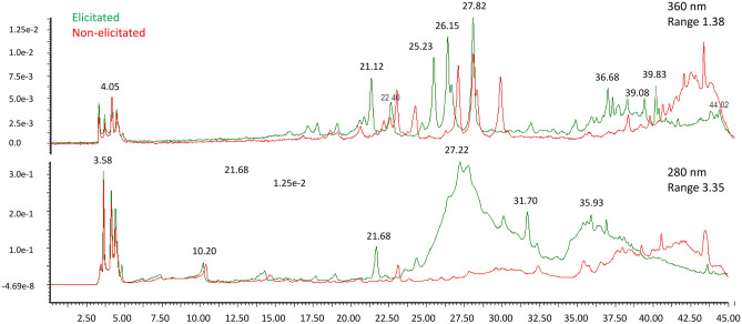
Natural ingredients have been used to improve the state of health in humans. The genus Paeonia has been studied only limited yet it's reported to have many activities such as antioxidant and anti-inflammatory. To this context, here we focused on an endemic Paeonia species in Attica. This study aims to present the development of the Paeonia mascula subsp. hellenica callus extract and its pleiotropic bioactivity on human primary keratinocytes exploring its potential application as an active agent in skin-related products. This extract showed a high scavenging activity with high phenolic content and an interesting metabolic profile. At a molecular level, the study on the transcript accumulation of genes revealed that this extract exhibits in vitro skin-related protection properties by mediating mitochondrial energy, cell proliferation, immune and inflammatory response and positively regulates genes involved in epidermal and in stratum corneum function. Besides, the extract is proven not skin irritant on reconstructed human skin model. These findings indicate that the specific P. mascula subsp. hellenica extract possesses significant in vitro protection activity on human epidermis and provides new insights into its beneficial role in skin confirming that the advent of biotechnology contribution the past few decades.
Conflict of interest statement
The authors declare no competing interests.
Figures


References
-
- Fowler JF, Woolery-Lloyd H, Waldorf H, Saini R. Innovations in natural ingredients and their use in skin care. J. Drugs Dermatol. 2010;9:S72–S81. - PubMed
Publication types
MeSH terms
Substances
LinkOut - more resources
Full Text Sources
Other Literature Sources
Miscellaneous
