. 2020 Nov 5;10(1):19201.
doi: 10.1038/s41598-020-76062-w.

Establishment of a heart-on-a-chip microdevice based on human iPS cells for the evaluation of human heart tissue function

Affiliations

Establishment of a heart-on-a-chip microdevice based on human iPS cells for the evaluation of human heart tissue function

Mosha Abulaiti et al. Sci Rep. .

Abstract

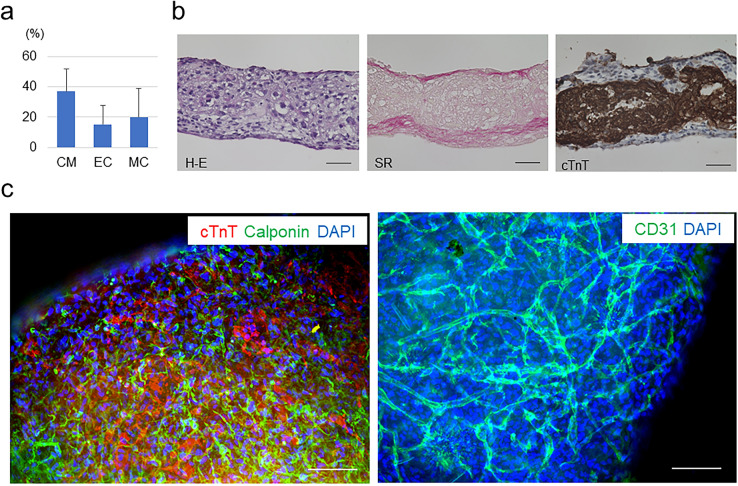
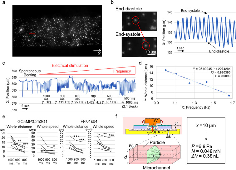
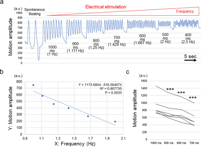
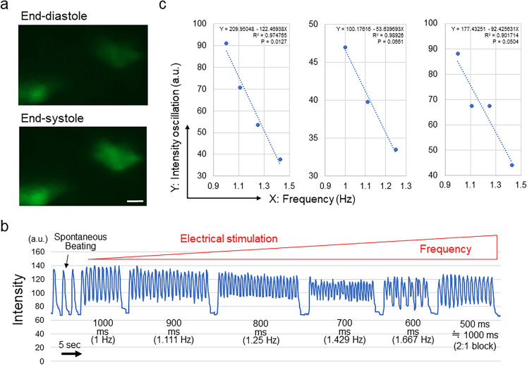
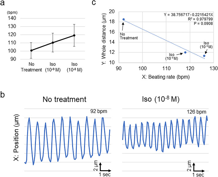
Human iPS cell (iPSC)-derived cardiomyocytes (CMs) hold promise for drug discovery for heart diseases and cardiac toxicity tests. To utilize human iPSC-derived CMs, the establishment of three-dimensional (3D) heart tissues from iPSC-derived CMs and other heart cells, and a sensitive bioassay system to depict physiological heart function are anticipated. We have developed a heart-on-a-chip microdevice (HMD) as a novel system consisting of dynamic culture-based 3D cardiac microtissues derived from human iPSCs and microelectromechanical system (MEMS)-based microfluidic chips. The HMDs could visualize the kinetics of cardiac microtissue pulsations by monitoring particle displacement, which enabled us to quantify the physiological parameters, including fluidic output, pressure, and force. The HMDs demonstrated a strong correlation between particle displacement and the frequency of external electrical stimulation. The transition patterns were validated by a previously reported versatile video-based system to evaluate contractile function. The patterns are also consistent with oscillations of intracellular calcium ion concentration of CMs, which is a fundamental biological component of CM contraction. The HMDs showed a pharmacological response to isoproterenol, a β-adrenoceptor agonist, that resulted in a strong correlation between beating rate and particle displacement. Thus, we have validated the basic performance of HMDs as a resource for human iPSC-based pharmacological investigations.


Conflict of interest statement

M.A., Y.Y., A.S., Y.T. and H.M. are inventors of the patent related to the HMD. The rest of the authors declare no competing interests.

Figures

Figure 1
Figure 1
Preparation of 3D cardiac microtissues. (a) Cellular components of prepared human iPSC-derived generated cardiac tissue sheets (n = 13). CM, cardiomyocytes; EC, vascular endothelial cells; MC, vascular mural cells. (b) Representative histological evaluations of 3D cardiac microtissues. Left: haematoxylin–eosin (H-E) staining; Middle: Sirius red (SR) staining; Right: cardiac isoform of troponin-T (cTnT) immunostaining. Scale bars = 50 µm. (c) Representative fluorescent immunostaining of 3D cardiac microtissues. Left: Double staining of cTnT (CMs) and calponin (MCs). Right: Staining of CD31 (ECs). DAPI, 4′,6-diamidino-2-phenylindole. Scale bars = 100 µm. In (b) and (c), Biorevo BZ-9000 (https://www.keyence.com/products/microscope/fluorescence-microscope/bz-9000/) (Keyence) was used.
Figure 2
Figure 2
Preparation of HMDs. (a) Representative macroscopic view of the microfluidic chip. (b) Representative macroscopic view of the HMD. The arrow indicates the human iPSC-derived 3D cardiac microtissues attached to the microfluidic chip. The dotted circle indicates the position of the push bar. Scale bar = 2 mm. (c) (d) Schematic of the structure and working machinery of the HMD. (c) Top view. (d) Side view.
Figure 3
Figure 3
Detection of the particle displacement of the HMD and the analysis of tissue function. (a) Representative observation of microchannels loaded with particles. X indicates the axis of the particle position. The red square indicates the selected location in (b). (b) Representative pattern of particle displacement. The red dotted circles indicate the same particle at the end-diastolic (top) and end-systolic (bottom) phases during the pulsation of 3D cardiac microtissues. (c) Representative changes in the particle position in accordance with the interval and frequency of the electrical stimulation. After 500-ms interval stimulation, the HMD could capture 1 time point in 2 stimulations (2:1 block), which is equivalent to 1000-ms interval stimulation. (d) Representative relationship between the frequency of electrical stimulation (X) and the particle displacement distance in the whole beating cycle (Y). The results of the regression analysis are shown. All data are shown in Supplementary Fig. 2a. (e) Particle displacement distance and speed of the whole beating cycle at each electrical stimulation interval in HMDs constructed from the GCaMP3-253G1 (n = 7) and FFI01 s04 (n = 6) human iPSC lines, respectively. *P < 0.05. **P < 0.01. ***P < 0.001. (f) Calculation of the physiological parameters. ΔV = stroke volume, z = vertical displacement of the diaphragm, P = applied pressure, F = applied force, r = radius of the chamber, w = width of the microchannel, d = depth of the microchannel, and x = particle displacement distance. In (a) and (b), cellSens Standard (version 1.18) (https://www.olympus-lifescience.com/en/software/cellsens/) (Olympus) was used.
Figure 4
Figure 4
MUSCLEMOTION analysis of 3D cardiac microtissues. (a) Representative changes in the motion amplitude in accordance with the interval and frequency of electrical stimulation. (b) Representative relationship between the frequency of electrical stimulation (X) and the motion amplitude in the whole beating cycle (Y). The results of the regression analysis are shown. All data are shown in Supplementary Fig. 3. (c) Motion amplitude of the whole beating cycle at each electrical stimulation interval in 3D cardiac microtissues (n = 6). ***P < 0.001.
Figure 5
Figure 5
Intracellular calcium oscillation analysis of 3D cardiac microtissues. (a) Representative visualization of human iPSC (GCaMP3-253G1)-derived CMs at end-diastole (top) and end-systole (bottom). Scale bar = 10 µm. (b) Representative changes in the signal intensity in accordance with the interval and frequency of electrical stimulation. Upon 500-ms interval stimulation, 3D cardiac microtissues could capture 1 time point in 2 stimulations (2:1 block), which is equivalent to 1000-ms interval stimulation. (c) Relationship between the frequency of electrical stimulation (X) and the oscillation of the signal intensity in the whole beating cycle (Y). The results of the regression analysis are shown. In (a), cellSens Standard (version 1.18) (https://www.olympus-lifescience.com/en/software/cellsens/) (Olympus) was used.
Figure 6
Figure 6
Pharmacological responses of HMDs to isoproterenol. (a) Dose-dependent change in the beating rate of HMDs. Bpm, beats per minute; Iso, isoproterenol. (b) Representative changes in the particle position in accordance with the administration of isoproterenol. Left: no treatment; Right, administration of isoproterenol (10–8 M). (c) Representative relationship between the beating rate of the HMD (X) and the particle displacement distance in the whole beating cycle (Y). The results of the regression analysis are shown. All data are shown in Supplementary Fig. 4.

References

    1. 1Causes of death, 2000–2016, Global Health Estimates (GHE), World Health Organiation (WHO). https://www.who.int/healthinfo/global_burden_disease/estimates/en/. Accessed on 3rd October, 2020.
    1. Packer M, et al. The effect of carvedilol on morbidity and mortality in patients with chronic heart failure. U. S. Carvedilol Heart Failure Study Group. N. Engl. J. Med. 1996;334:1349–1355. doi: 10.1056/NEJM199605233342101. - DOI - PubMed
    1. 3The Cardiac Insufficiency Bisoprolol Study II (CIBIS-II): a randomised trial. Lancet353, 9–13, (1999). - PubMed
    1. Kotecha D, et al. Efficacy of beta blockers in patients with heart failure plus atrial fibrillation: an individual-patient data meta-analysis. Lancet. 2014;384:2235–2243. doi: 10.1016/S0140-6736(14)61373-8. - DOI - PubMed
    1. Metra M, Teerlink JR. Heart failure. Lancet. 2017;390:1981–1995. doi: 10.1016/S0140-6736(17)31071-1. - DOI - PubMed

Publication types

MeSH terms