

Fast In Situ Image Reconstruction for Proton Radiography
- PMID: 33154789
- PMCID: PMC7641509
- DOI: 10.1007/s13566-019-00387-x
Fast In Situ Image Reconstruction for Proton Radiography
Abstract
Objective: Proton beam therapy is an emerging modality for cancer treatment that, compared to X-ray radiation therapy, promises to provide better dose delivery to clinical targets with lower doses to normal tissues. Crucial to accurate treatment planning and dose delivery is knowledge of the water equivalent path length (WEPL) of each ray, or pencil beam, from the skin to every point in the target. For protons, this length is estimated from relative stopping power based on X-ray Hounsfield units. Unfortunately, such estimates lead to 3 to 4% uncertainties in the proton range prediction. Therefore, protons in the Bragg peak may overshoot (or undershoot) the desired stopping depth in the target causing tissue damage beyond the target volume. Recent studies indicate that tomographic imaging using protons has the potential to provide directly more accurate measurement of RSPs with significantly lower radiation dose than X-rays. We are currently working on a proton radiography system that promises to provide accurate two-dimensional (2D) images of WEPL values for protons that pass through the body. These will be suitable for positioning and range verification in daily treatments. In this study, we demonstrate that this system is capable of rapidly achieving such accurate images in clinically meaningful times.
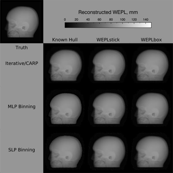
Methods: We have developed a software platform to characterize the potential performance of the prototype proton radiography system. We use Geant4 to simulate raw data detected by the device. An especially-written software - pRad - was written to process these data as they are received and uses iterative methods to generate radiographs. The software has been designed to generate a radiograph from a few million protons in under a minute after receiving the first proton from the device. We used a head phantom with known chemical compositions that could be modelled quite accurately in Geant4 simulations of proton radiographs. The radiographs are displayed as pixelated WEPL values displayed on a 2D gray scale image of WEPL values.
Results: Rapid radiograph reconstruction of 3D phantoms using simulated proton pencil beams have been achieved with our software platform. On a modest desktop computer with a single central processing unit (CPU) and a single graphics processing unit (GPU), it takes about 11 seconds to reconstruct images using iterative linear algorithms to reconstruct a radiograph from 7.6 million protons. For the radiographic reconstructions of the head phantom described here, the mean WEPL errors, in the proton radiograph using a large majority of the pixels in the complete image were less than 1 mm when compared to images obtained without proton scattering and without detector resolution included.
Conclusion: We have demonstrated, through computer simulations of proton irradiation of a pediatric head phantom using the newly built pRad detector and image reconstruction software, that high quality proton radiographs can be generated for patient alignment and verification of water equivalent thickness of the patient before each treatment.
Keywords: Patient Alignment; Proton Computed Tomography; Proton Radiography; Range Verification; Relative Stopping Power; Treatment Planning.
Conflict of interest statement
Conflict of Interest Statement The authors have intellectual property rights to the innovations described in this paper. James S. Welsh has served as a medical advisor to ProTom International. Don F. Dejongh is a co-owner of ProtonVDA Inc.
Figures


References
-
- Particle Therapy Co-operative Group, Particle Therapy Centers, Facilities in Operation. https://www.ptcog.ch. Accessed 16 April 2018.
-
- Seco J, Spadea MF (2015) Imaging in particle therapy: State of the art and future perspective. Acta Oncol 54:1254–1258. - PubMed
-
- Knopf AC, Lomax A (2013) In vivo proton range verification: a review. Phys Med Biol 58:R131–160. - PubMed
-
- Bär E, Lalonde A, Royle G, Lu HM, Bouchard H (2017) The potential of dual-energy CT to reduce proton beam range uncertainties. Med Phys 44:2332–2344. - PubMed
-
- Xie Y, Bentefour EH, Janssens G, Smeets J et al. (2017) Prompt Gamma Imaging for In Vivo Range Verification of Pencil Beam Scanning Proton Therapy. Int J Radiat Oncol Biol Phys 99:210–218. - PubMed
Grants and funding
LinkOut - more resources
Full Text Sources