. 2020 May 14;8(5):e2834.
doi: 10.1097/GOX.0000000000002834. eCollection 2020 May.

Establishing Photographic Standards for Facial Transplantation: A Systematic Review of the Literature

Affiliations

Establishing Photographic Standards for Facial Transplantation: A Systematic Review of the Literature

Erin M Wolfe et al. Plast Reconstr Surg Glob Open. .

Abstract

Photography provides a means for objective assessment and dissemination of clinical information. The American Society of Plastic Surgeons (ASPS) and Plastic Surgery Foundation (PSF) published photography guidelines in 2006 to optimize its clinical use. However, photographic documentation of outcomes in facial transplantation (FT) continues to lack standardization. We therefore appraised the current state of FT photography in the peer-reviewed literature.

Methods: A PubMed search was conducted from July 2005 to July 2019. Studies containing photographs of partial or full FT recipients were included. Non-English language articles, cadaveric and animal studies, technique papers, and case reports were excluded. Data were extracted from 91 articles. Adherence rates were calculated to determine whether published FT photographs followed ASPS/PSF guidelines. Proposed photographic standards particular to FT were then formulated to guide standardization of practice.

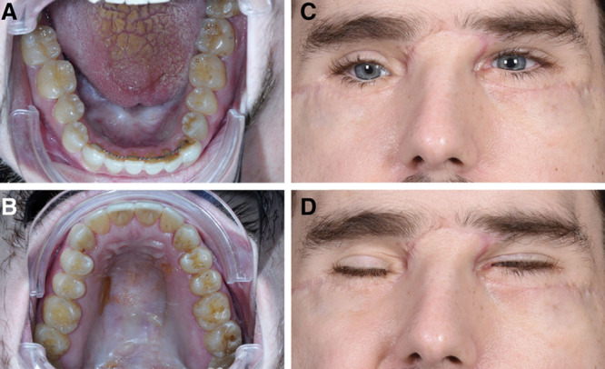
Results: Only 28.6% (26/91) of articles adhered to the photographic conventions of preparation, positioning, and views. Of 162 patient appearances in the 91 articles, 95% (154/162) met the criteria for preparation, 98.8% (160/162) met the criteria for positioning, but only 24.7% (40/162) met the criteria for views.

Conclusions: Photographic documentation of FT outcomes in the peer-reviewed literature is limited, with inconsistent adherence to ASPS/PSF guidelines. There is substantial deficiency in provision of alternative views, with the majority of publications only depicting the frontal view. FT photography standards should specifically incorporate alternative views, proper lighting and framing, and elimination of distractions, and accommodate for remote patient self-photography. This will promote a transparent and consistent longitudinal evaluation of the surgical results.


Conflict of interest statement

Disclosure: The authors have no financial interest to declare in relation to the content of this article.

Figures

Fig. 1.
Fig. 1.
Preferred reporting items for systematic reviews and meta-analyses (PRISMA) diagram.
Fig. 2.
Fig. 2.
Adherence to photographic standards, by guideline. The percentage of patient appearances with correct photographic documentation per guideline. Most published photographs met the criteria for patient preparation and relevant sub-categories such as lighting and background. The guideline least frequently met was that for cast shadows (64%).
Fig. 3.
Fig. 3.
Adherence to photographic standards, by view. The percentage of patients with photographic documentation by view. While patient preparation and positioning were adequate, sufficient alternative views were lacking. While most patients had a frontal view (96.3%), few had accompanying profile (16.0%), oblique (14.8%), anatomical (20%), or functional views (8.6%).
Fig. 4.
Fig. 4.
Proposed photographic standards for facial transplantation.
Fig. 5.
Fig. 5.
Representative FT recipient image series. Images in this series were taken directly from the patient’s medical record, with copyrights retained by the senior author as stated. Images are representative of patient appearance 1-year status post partial facial transplantation after injuries sustained from high-energy ballistic trauma. This 25-year-old patient had undergone multiple reconstructive procedures before presentation, including maxillary, mandibular, zygomatic, and right orbital floor open reduction and internal fixation with bilateral anterolateral thigh and supraclavicular flaps to the midface and lower face. In preparation for partial facial transplantation, he required removal of exposed hardware, debridement of facial fractures, bilateral naso-orbito-ethmoid osteotomies, medial canthal tendon repositioning, and bilateral orbital floor reconstruction with alloplastic titanium implants. Partial face, double jaw, and teeth transplantation were performed in January of 2018. A, Frontal view, neutral expression (published with permission from and copyrights retained by Dr. Rodriguez). B, Frontal view, smiling (published with permission from and copyrights retained by Dr. Rodriguez). C, Profile view, right (published with permission from and copyrights retained by Dr. Rodriguez). D, Profile view, left (published with permission from and copyrights retained by Dr. Rodriguez). E, Oblique view, right (published with permission from and copyrights retained by Dr. Rodriguez). F, Oblique view, left (published with permission from and copyrights retained by Dr. Rodriguez). G, Vertex view (published with permission from and copyrights retained by Dr. Rodriguez). H, Basal view (published with permission from and copyrights retained by Dr. Rodriguez).
Fig. 6.
Fig. 6.
Representative FT recipient image series. Images in this series were taken directly from the patient’s medical record, with copyrights retained by the senior author as stated. Images are representative of patient appearance 1-year status post partial facial transplantation after injuries sustained from high-energy ballistic trauma. This 25-year-old patient had undergone multiple reconstructive procedures before presentation, including maxillary, mandibular, zygomatic, and right orbital floor open reduction and internal fixation with bilateral anterolateral thigh and supraclavicular flaps to the midface and lower face. In preparation for partial facial transplantation, he required removal of exposed hardware, debridement of facial fractures, bilateral naso-orbito-ethmoid osteotomies, medial canthal tendon repositioning, and bilateral orbital floor reconstruction with alloplastic titanium implants. Partial face, double jaw, and teeth transplantation were performed in January of 2018. A, Intraoral: floor of mouth (published with permission from and copyrights retained by Dr. Rodriguez). B, Intraoral: palate (published with permission from and copyrights retained by Dr. Rodriguez). C, Subunit: eyes, open (published with permission from and copyrights retained by Dr. Rodriguez). D, Subunit: eyes, functional: voluntary blink (published with permission from and copyrights retained by Dr. Rodriguez).

References

    1. Zarem HA.Standards of photography. Plast Reconstr Surg. 1984;74:137–146. - PubMed
    1. Williams R, Photography L, So M.Medical Photography Study Guide. 1984Boston, Mass: MTP Press;
    1. Sanniec KJ, Velazco CS, Macias LH, et al. Adherence to photographic standards: a review of ASPS and ASAPS member Surgeons’ websites. J Aesthet Reconstr Surg. 2016;2:11.
    1. American Society of Plastic Surgeons and the Plastic Surgery Foundation. Photographic Standards in Plastic Surgery. https://drsunol.com/pdf/fotografia-cirugia-estetica-plastica-joaquim-sun.... 2006. Accessed July 1, 2019.
    1. Ettorre G, Weber M, Schaaf H, et al. Standards for digital photography in cranio-maxillo-facial surgery—Part I: basic views and guidelines. J Craniomaxillofac Surg. 2006;34:65–73. - PubMed