Review
. 2021 Jan;47(1):23-36.
doi: 10.3892/ijmm.2020.4781. Epub 2020 Nov 4.

BCL2‑regulated apoptotic process in myocardial ischemia‑reperfusion injury (Review)

Affiliations
Review

BCL2‑regulated apoptotic process in myocardial ischemia‑reperfusion injury (Review)

Anna Yu Korshunova et al. Int J Mol Med. 2021 Jan.

Abstract

The leading cause of death in developed countries is cardiovascular disease, where coronary heart disease is the main cause of death. Myocardial reperfusion is the most significant method to prevent cell death after ischemia. However, restoration of blood flow may paradoxically lead to myocardial ischemia‑reperfusion injury (MI/RI) accompanied by metabolic disturbances and cardiomyocyte death. As the myocardium has an extremely limited ability to regenerate, the mechanisms of regulated cell death, including apoptosis, are the most significant for contemporary research due to their reversibility. BCL2 is a key anti‑apoptotic protein. There are several signaling pathways and compounds regulating BCL2, including PI3K/AKT and MEK1/ERK1/2, JAK2/STAT3, endothelial nitric oxide synthase, PTEN, cardiac ankyrin repeat protein and microRNA, which can serve as targets for modern methods of cardioprotective therapy inhibiting intrinsic apoptosis and saving viable cardiomyocytes after MI/RI. The present review considers the mechanisms of Bcl2‑regulated apoptosis in the development and treatment of MI/RI.

Keywords: myocardial ischemia-reperfusion injury; BCL2; apoptosis; regulated cell death; cardioprotective therapy.


Figures

Figure 1
Figure 1
BCL2 proteins and homology domains. The BCL-2 family of proteins is divided into three groups based on their functional role in the regulation of apoptosis and the number of BH domains they bear. Pro-apoptotic BCL2 proteins include: Multidomain proteins BAX, BAK, BOX and BH3-only proteins BID, BIM, BAD, BIK, BMF, HRK, BNIP3, NIX, NOXA and PUMA; anti-apoptotic multidomain BCL2 proteins BCL2, BCL-XL, BCL-W, MCL1 and A1/BFL-1. BH, BCL2 homology; BAK, BCL2 antagonist/killer; BOX, BCL2-related ovarian killer; BID, BH3-interacting domain death agonist; BIM, BCL2-interacting mediator of cell death; BAD, BCL2-associated agonist of cell death; BIK, BCL2-interacting killer; BMF, BCL2-modifying factor; HRK, activator of apoptosis hara-kiri; BNIP3, BCL2-interacting protein 3; NIX, BNIP3-like; NOXA, phorbol-12-my-ristate-13-acetate-induced protein 1; PUMA, p53-upregulated modulator of apoptosis; BCL-XL; BCL2 X-linked protein; BCL-w, BCL2-like protein 2; MCL1, myeloid cell leukemia 1; A1/BFL-1, BCL2-related protein A1.
Figure 2
Figure 2
Main pathways of BCL2 regulation in MI/RI. The figure shows the simplified scheme of BCL2 signal transduction regulation in MI/RI. The RISK pathway is in red and the SAFE pathway is in blue. MI/RI, myocardial ischemia-reperfusion injury; RISK, reperfusion injury salvage kinase; SAFE, survivor activating factor enhancement; PIP3, phosphatidylinositol-triphosphate 3; PIP2, phosphatidylinositoltriphosphate 2; eNOS, endothelial nitric oxide synthase; PKG, protein kinase G; miR, microRNA; MOMP, mitochondrial outer membrane permeabilization.
Figure 3
Figure 3
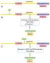
Effects of IPC and IPostC on BCL2-regulated apoptosis in MI/RI. (A) The progress of MI/RI without any treatment. (B) The effects of IPC on BCL2-regulated apoptosis in MI/RI. (C) The effects of IPostC on BCL2-regulated apoptosis in MI/RI. IPC, ischemic preconditioning; IPostC, ischemic postconditioning; MI/RI, myocardial ischemia-reperfusion injury; RISK, reperfusion injury salvage kinase; SAFE, survivor activating factor enhancement.

References

    1. Kerr JF, Wyllie AH, Currie AR. Apoptosis: A basic biological phenomenon with wide-ranging implications in tissue kinetics. Br J Cancer. 1972;26:239–257. doi: 10.1038/bjc.1972.33. - DOI - PMC - PubMed
    1. Rogalińska M. Alterations in cell nuclei during apoptosis. Cell Mol Biol Lett. 2002;7:995–1018. - PubMed
    1. Galluzzi L, Vitale I, Aaronson SA, Abrams JM, Adam D, Agostinis P, Alnemri ES, Altucci L, Amelio I, Andrews DW, et al. Molecular mechanisms of cell death: Recommendations of the Nomenclature Committee on Cell Death 2018. Cell Death Differ. 2018;25:486–541. doi: 10.1038/s41418-017-0012-4. - DOI - PMC - PubMed
    1. Tait SW, Green DR. Mitochondria and cell death: Outer membrane permeabilization and beyond. Nat Rev Mol Cell Biol. 2010;11:621–632. doi: 10.1038/nrm2952. - DOI - PubMed
    1. Kalkavan H, Green DR. MOMP, cell suicide as a BCL-2 family business. Cell Death Differ. 2018;25:46–55. doi: 10.1038/cdd.2017.179. - DOI - PMC - PubMed

MeSH terms