

Evidence of Duodenal Epithelial Barrier Impairment and Increased Pyroptosis in Patients With Functional Dyspepsia on Confocal Laser Endomicroscopy and "Ex Vivo" Mucosa Analysis
- PMID: 33156108
- PMCID: PMC8409129
- DOI: 10.14309/ajg.0000000000000827
Evidence of Duodenal Epithelial Barrier Impairment and Increased Pyroptosis in Patients With Functional Dyspepsia on Confocal Laser Endomicroscopy and "Ex Vivo" Mucosa Analysis
Abstract
Introduction: Duodenal epithelial barrier impairment and immune activation may play a role in the pathogenesis of functional dyspepsia (FD). This study was aimed to evaluate the duodenal epithelium of patients with FD and healthy individuals for detectable microscopic structural abnormalities.
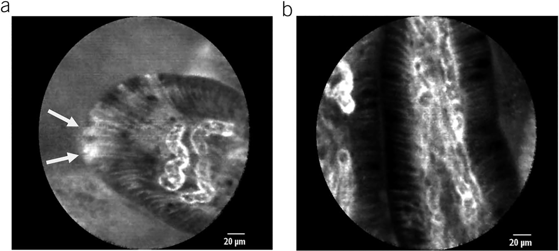
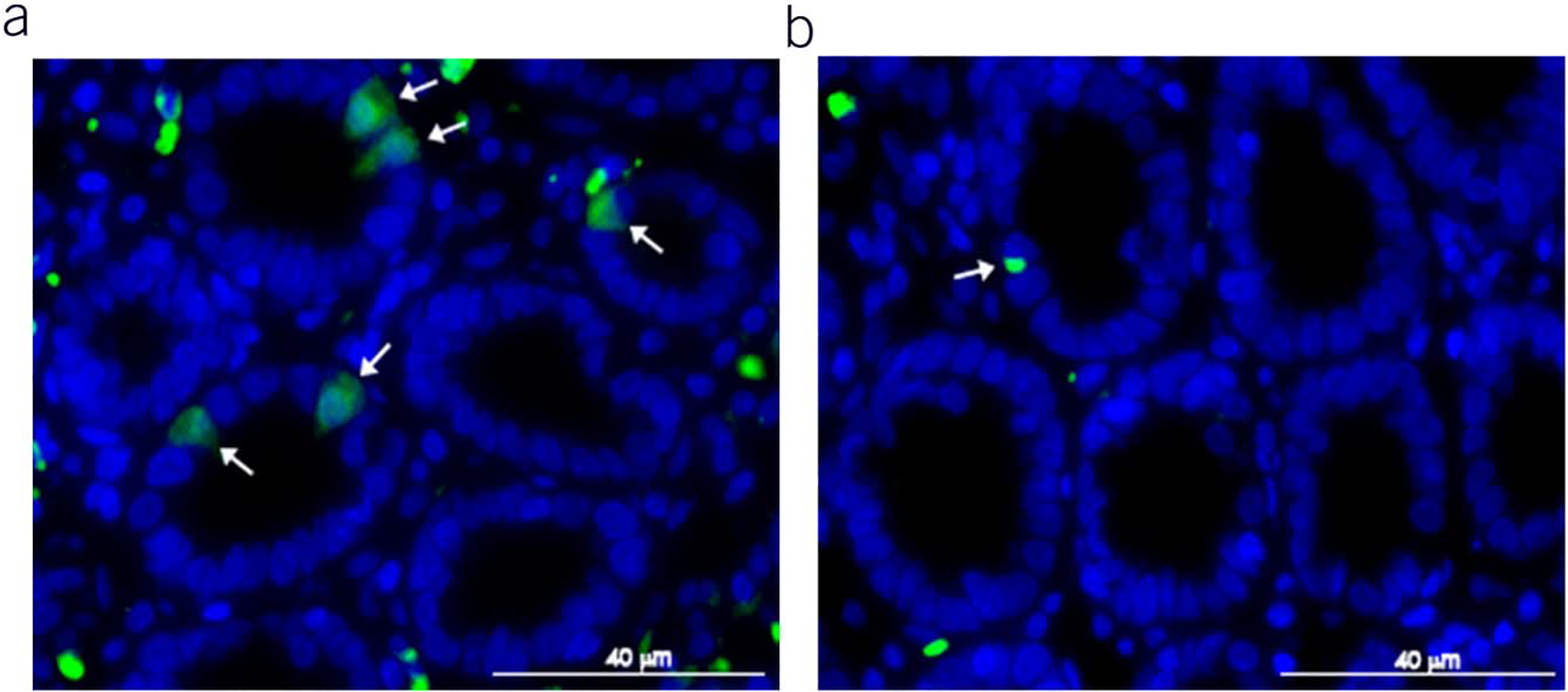
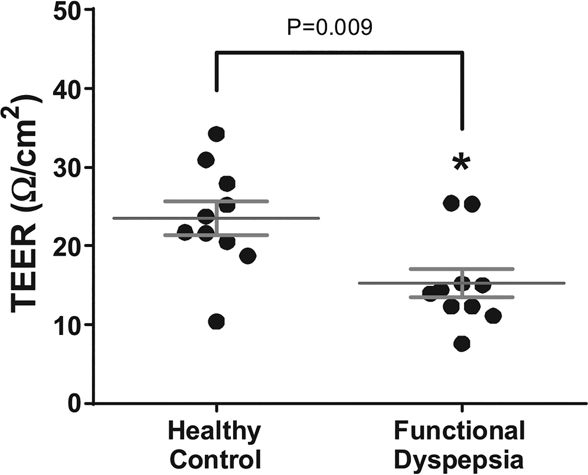
Methods: This is a prospective study using esophagogastroduodenoscopy enhanced with duodenal confocal laser endomicroscopy (CLE) and mucosal biopsies in patients with FD (n = 16) and healthy controls (n = 18). Blinded CLE images analysis evaluated the density of epithelial gaps (cell extrusion zones), a validated endoscopic measure of the intestinal barrier status. Analyses of the biopsied duodenal mucosa included standard histology, quantification of mucosal immune cells/cytokines, and immunohistochemistry for inflammatory epithelial cell death called pyroptosis. Transepithelial electrical resistance (TEER) was measured using Ussing chambers. Epithelial cell-to-cell adhesion proteins expression was assessed by real-time polymerase chain reaction.
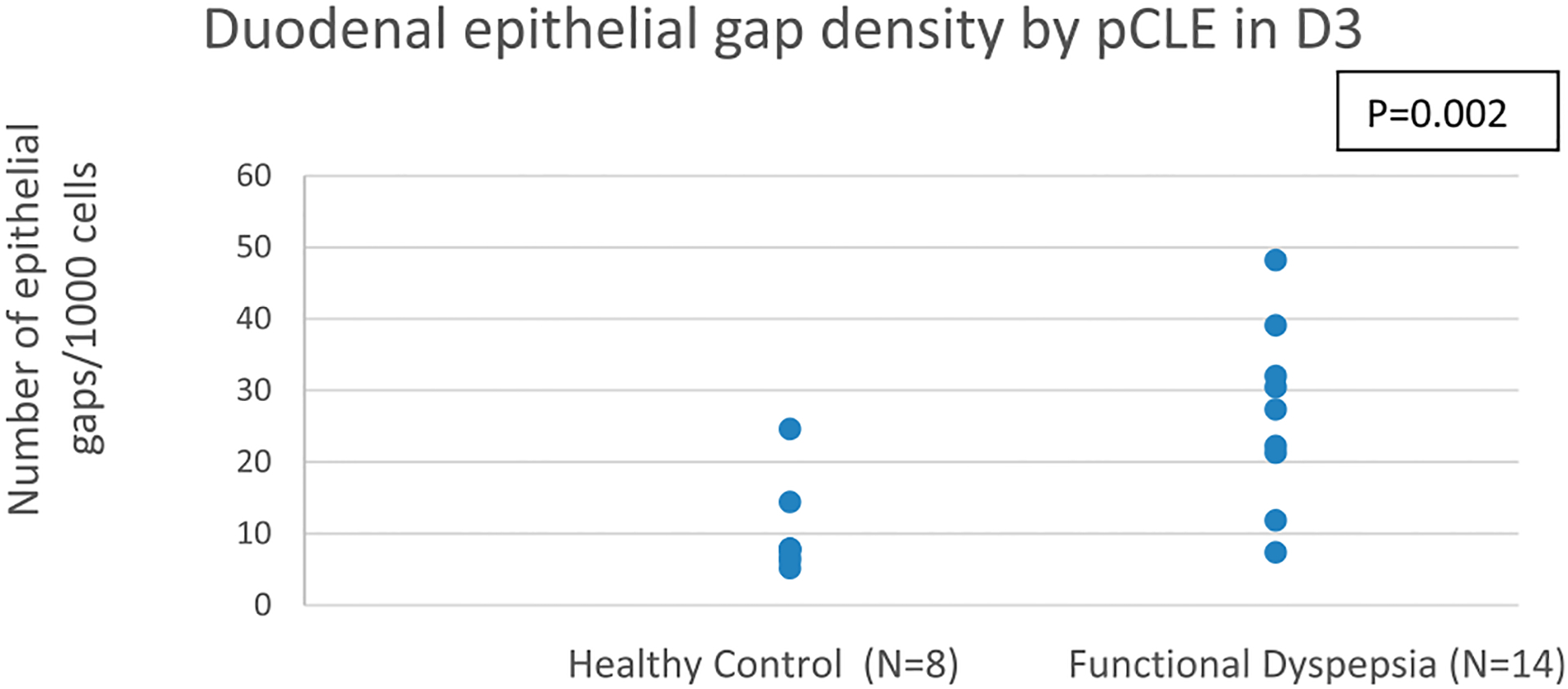
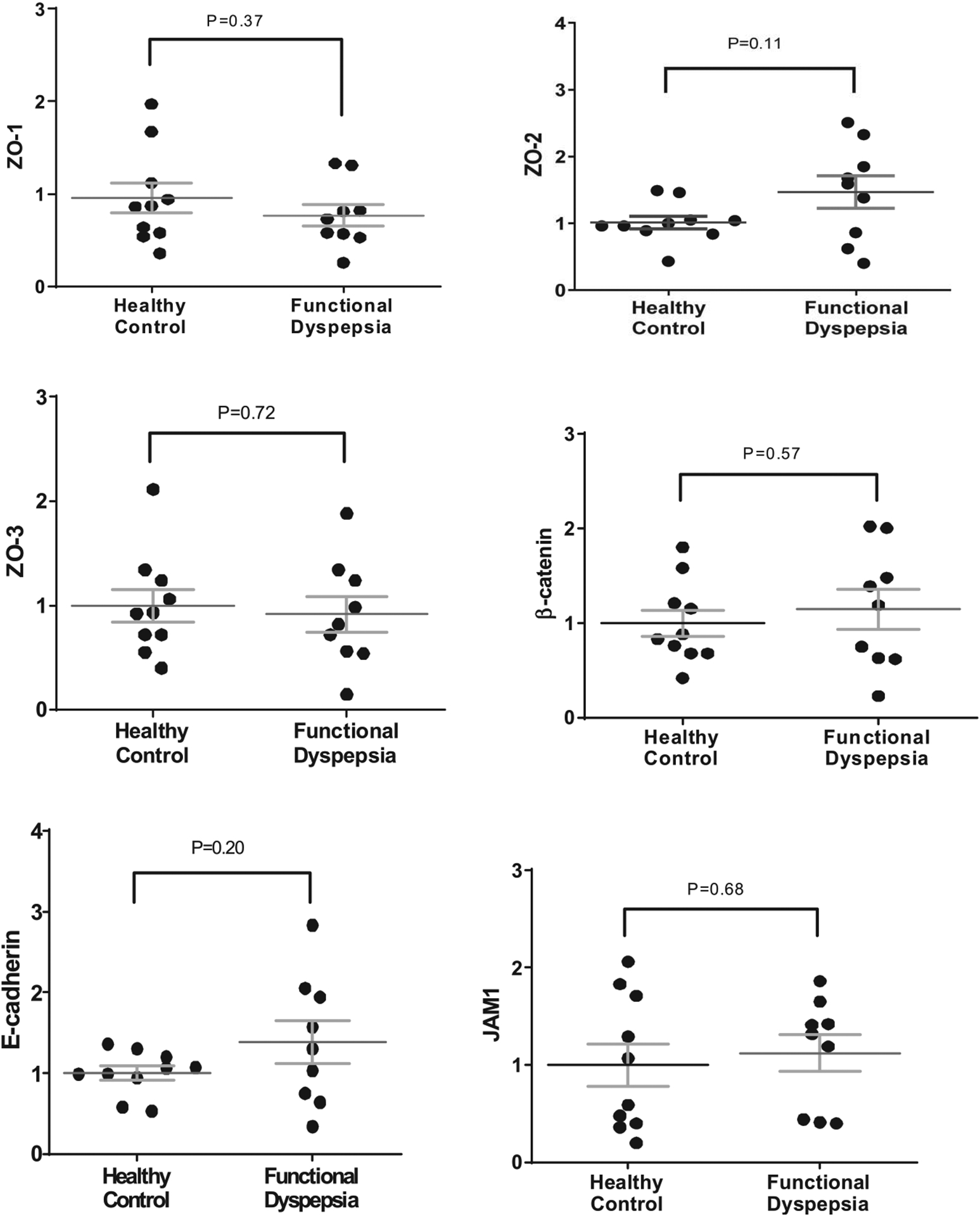
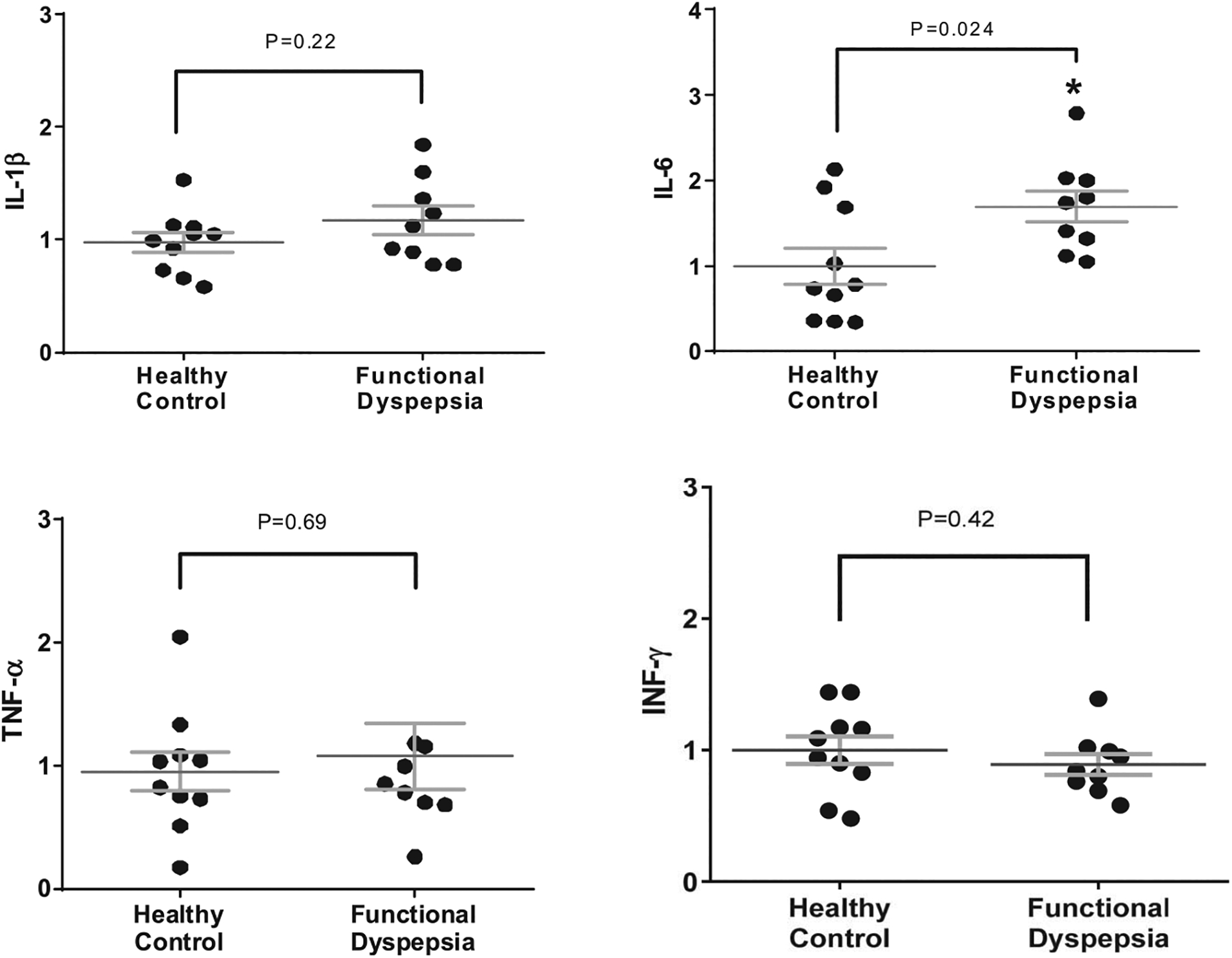
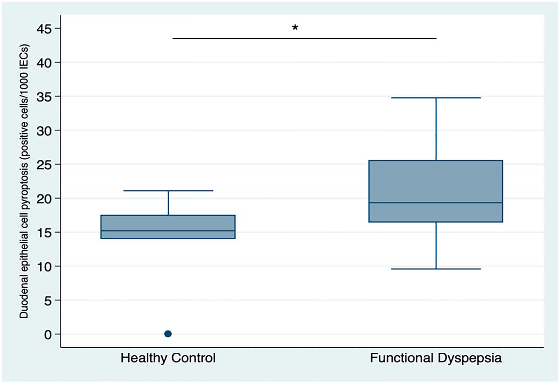
Results: Patients with FD had significantly higher epithelial gap density on CLE in the distal duodenum than that of controls (P = 0.002). These mucosal abnormalities corresponded to significant changes in the duodenal biopsy samples of patients with FD, compared with controls, including impaired mucosal integrity by TEER (P = 0.009) and increased number of epithelial cells undergoing pyroptosis (P = 0.04). Reduced TEER inversely correlated with the severity of certain dyspeptic symptoms. Furthermore, patients with FD demonstrated altered duodenal expression of claudin-1 and interleukin-6. No differences in standard histology were found between the groups.
Discussion: This is the first report of duodenal CLE abnormalities in patients with FD, corroborated by biopsy findings of epithelial barrier impairment and increased cell death, implicating that duodenal barrier disruption is a pathogenesis factor in FD and introducing CLE a potential diagnostic biomarker in FD.
Conflict of interest statement
Figures
Comment in
-
Paradigm Shift: Functional Dyspepsia-A "Leaky Gut" Disorder?Am J Gastroenterol. 2021 Feb 1;116(2):274-275. doi: 10.14309/ajg.0000000000001077. Am J Gastroenterol. 2021. PMID: 33298703
-
Response to Keszthelyi.Am J Gastroenterol. 2021 Jul 1;116(7):1557. doi: 10.14309/ajg.0000000000001251. Am J Gastroenterol. 2021. PMID: 33767099 No abstract available.
-
Irritable Bowel Syndrome and Functional Dyspepsia: Different Ends of the Same Spectrum of Intestinal Barrier Dysfunction?Am J Gastroenterol. 2021 Jul 1;116(7):1556. doi: 10.14309/ajg.0000000000001174. Am J Gastroenterol. 2021. PMID: 34183583 No abstract available.
References
-
- Stanghellini V, Chan FD, Hasler WL et al. Gastroduodenal disorders. Gastroenterology 2016;150(6):1380–92. - PubMed
-
- Ford AC, Marwaha A, Sood R, et al. Global prevalence of, and risk factors for, uninvestigated dyspepsia: A meta-analysis. Gut 2015;64:1049–57. - PubMed
-
- Ford AC, Marwaha A, Lim A, et al. What is the prevalence of clinically significant endoscopic findings in subjects with dyspepsia? Systematic review and meta-analysis. Clin Gastroenterol Hepatol 2010;8:830–7. - PubMed
-
- Sander GB, Mazzoleni LE, Francesconi CF, et al. Influence of organic and functional dyspepsia on work productivity: The HEROES-DIP study. Value Health 2011;14(Suppl 1):S126–9. - PubMed
-
- Lacy BE, Weiser KT, Kennedy AT et al. Functional dyspepsia: The economic impact to patients. Aliment Pharmacol Ther 2013;38:170. - PubMed
Publication types
MeSH terms
Substances
Grants and funding
LinkOut - more resources
Full Text Sources
Medical
Miscellaneous
