. 2020 Nov 6;15(11):e0241837.
doi: 10.1371/journal.pone.0241837. eCollection 2020.

Relative expression of microRNAs, apoptosis, and ultrastructure anomalies induced by gold nanoparticles in Trachyderma hispida (Coleoptera: Tenebrionidae)

Affiliations

Relative expression of microRNAs, apoptosis, and ultrastructure anomalies induced by gold nanoparticles in Trachyderma hispida (Coleoptera: Tenebrionidae)

Saeed El-Ashram et al. PLoS One. .

Abstract

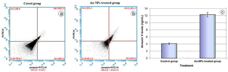
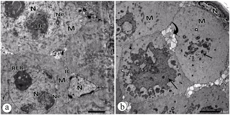
The extensive use of nanomaterials generates toxic effects on non-target species and the ecosystem. Although gold nanoparticles (Au-NPs) are generally expected to be safe, the recent study contains conflicting data regarding their cytotoxicity in the darkling beetles Trachyderma hispida. The study postulated cellular perturbation in the ovarian tissue of the beetles induced by a sublethal dose of Au-NPs (0.01 mg/g). When compared with the controls, a significant inhibition in the activities of the antioxidant enzymes selenium-dependent (GPOX) and selenium-independent (GSTP) glutathione peroxidases (GPx) was observed in the treated beetles. The study proposed microRNAs (miRNA-282 and miRNA-989) as genotoxic markers for the first time, reporting a significant suppression in their transcriptional levels in the treated beetles. Furthermore, TUNEL (Terminal deoxynucleotidyl transferase-mediated dUTP nick end labeling) and flow cytometry assays (annexin V-Fitc) indicated a significant increase in ovarian cell apoptosis in the treated beetles. Additionally, an ultrastructure examination revealed pathological changes in the ovarian cells of the treated beetles. The resulting anomalies in the present study may interrupt the fecundity of the beetles and lead to the future suppression of beetle populations.


Conflict of interest statement

The authors have declared that no competing interests exist.

Figures

Fig 1
Fig 1. Transmission electron micrograph of Au-NPs.
Fig 2
Fig 2. Cumulative mortality percentage in the control group and the Au-NPs treated group till 30 days.
Data are expressed as mean ± SE.
Fig 3
Fig 3. Energy-dispersive X-ray spectra revealing the qualitative elemental composition as measured in counts per second in ovarian tissues of a: Control group and b: Au-NPs treated group.
Horizontal scale, X-ray energy; vertical scale, X-ray counts. Nd: not detected.
Fig 4
Fig 4. a: Activities of antioxidant enzymes (GPOX and GSTP) (nmol NADPH/min/mg. protein) in the ovarian tissues of the control group and the Au-NPs treated group, b: Relative gene expression of miRNA-282, and c: Relative gene expression of miRNA-989 in the ovarian tissues of the control group and the Au-NPs treated group (expression was measured relative to U6 small nuclear RNA (RNU6B) using qPCR).
Data are expressed as mean ± SE.
Fig 5
Fig 5
a: TUNEL assay for detection apoptosis in the ovarian tissues of beetles. Under fluorescent microscopy light green: normal DNA (non-apoptotic cell) and bright green: damaged DNA (apoptotic cell) (× 100). b: Apoptotic rate in the ovarian tissues of the control group and the Au-NPs treated group. Data are expressed as mean ± SE.
Fig 6
Fig 6
Flow cytometry analysis of annexin-V-FITC and propidium iodide staining of ovarian cells in the control group (a) and Au-NPs treated group (b). The upper left (UL) quadrant (PI+/Annexin V−) represents necrotic cells, the left lower (LL) quadrant (PI−/Annexin V−) represents healthy cells, the upper right (UR) quadrant (PI+/Annexin V+) represents early apoptotic cells and the lower right (LR) quadrant (PI−/Annexin V+) represents late apoptotic cells. Apoptosis was calculated as summation of UR + LR. Values represent average percentage (± SE) of at least three samples. c: Annexin V levels in the ovarian tissues of the control group and the Au-NPs treated group. Data are expressed as the mean ± SE.
Fig 7
Fig 7
Electron micrographs a: Trophocytes in the ovary of the control group showing heterochromatic (HC) Nucleus (N), nuclear envelope (Ne), mitochondria (M), rough endoplasmic reticulum (RER), and free ribosomes (r). Interstitial cells (IC). b: Trophocytes in the ovary of Au-NPs treated group showing pyknotic nuclei (arrow) and disintegrated mitochondria (M). Note: degenerated Interstitial cells (IC) (curved arrow).
Fig 8
Fig 8. Electron micrographs in the oocyte of the control group and Au-Nps treated group.
a, b: Follicular epithelial cells (FEC) and oocyte in the control group. c, d, & e: Follicular epithelial cells (FEC) and oocyte in the ovary of the Au-NPs treated group. c: intended nuclear envelope (Ne), vacuolated cytoplasm (V), distorted microvilli (MV), and vacuolated ooplasm (V). d: abnormal nuclei (N) of the follicular epithelial cells (FEC) with undefined nuclear envelopes (Ne), disintegrated mitochondria (M), dilated smooth endoplasmic reticulum (SER), and vacuolated cytoplasm (V). Note: distorted microvilli (MV) and degenerated yolk granules (DYG). Arrow pointed at electron dense particles. e: follicular epithelial cell (FEC) with intended nuclear envelopes (Ne), enlarged mitochondria with distorted cristae (M), dilated rough (RER) and smooth (SER) endoplasmic reticulum, and vacuolated cytoplasm (V). Arrow pointed at electron dense particles. HC: Heterochromatin, N: nucleus, Ne: nuclear envelope, M: mitochondria, SER: smooth endoplasmic reticulum, RER: rough endoplasmic reticulum, MV: Microvilli, OC: Oocyte, OP: ooplasm, YG: yolk granules, FD: fat droplets, LD: lipid droplets, Pi: pinosomes, r: free ribosomes.

Similar articles

Cited by

References

    1. Savolainen K, Alenius H, Norppa H, Pylkkä L, Tuomi T, Kasper G. Risk assessment of engineered nanomaterials and nanotechnologies—A review. TOX Toxicology. 2010;269(2):92–104. 10.1016/j.tox.2010.01.013 - DOI - PubMed
    1. Chifiriuc MC, Ratiu AC, Popa M, Ecovoiu AA. Drosophotoxicology: An Emerging Research Area for Assessing Nanoparticles Interaction with Living Organisms. Int J Mol Sci. 2016;17(2):36 Epub 2016/02/26. 10.3390/ijms17020036 - DOI - PMC - PubMed
    1. Pan Y, Neuss S, Leifert A, Fischler M, Wen F, Simon U, et al. Size-dependent cytotoxicity of gold nanoparticles. Small (Weinheim an der Bergstrasse, Germany). 2007;3(11):1941–9. Epub 2007/10/30. 10.1002/smll.200700378 . - DOI - PubMed
    1. Nel A, Xia T, Mädler L, Li N. Toxic potential of materials at the nanolevel. Science (New York, NY). 2006;311(5761):622–7. Epub 2006/02/04. 10.1126/science.1114397 . - DOI - PubMed
    1. Yadav T, Mungray AA, Mungray AK. Fabricated nanoparticles: current status and potential phytotoxic threats. Reviews of environmental contamination and toxicology. 2014;230:83–110. Epub 2014/03/13. 10.1007/978-3-319-04411-8_4 . - DOI - PubMed

Publication types