. 2020 Nov 5;107(5):963-976.
doi: 10.1016/j.ajhg.2020.10.002.

NCKAP1 Disruptive Variants Lead to a Neurodevelopmental Disorder with Core Features of Autism

Affiliations

NCKAP1 Disruptive Variants Lead to a Neurodevelopmental Disorder with Core Features of Autism

Hui Guo et al. Am J Hum Genet. .

Abstract

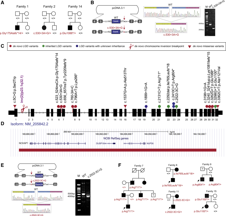
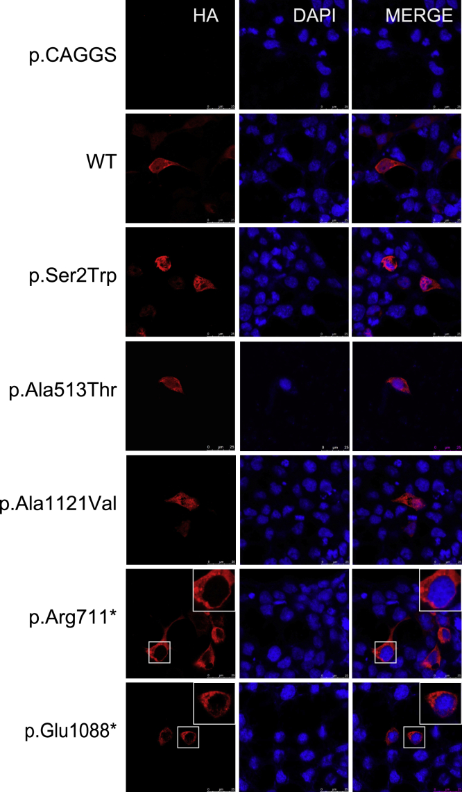
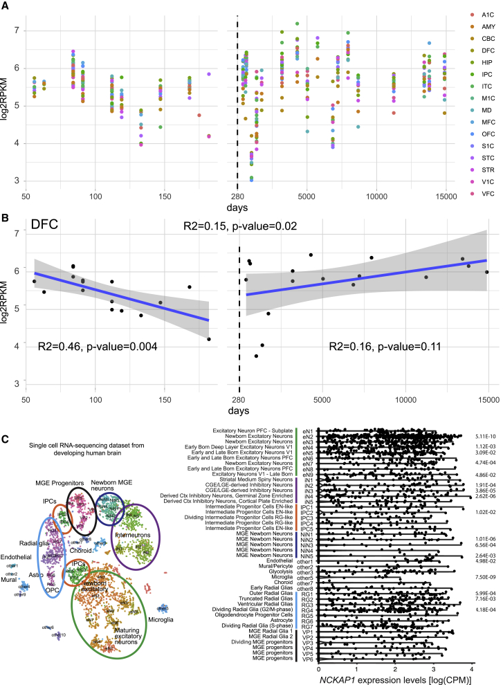
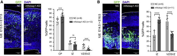
NCKAP1/NAP1 regulates neuronal cytoskeletal dynamics and is essential for neuronal differentiation in the developing brain. Deleterious variants in NCKAP1 have been identified in individuals with autism spectrum disorder (ASD) and intellectual disability; however, its clinical significance remains unclear. To determine its significance, we assemble genotype and phenotype data for 21 affected individuals from 20 unrelated families with predicted deleterious variants in NCKAP1. This includes 16 individuals with de novo (n = 8), transmitted (n = 6), or inheritance unknown (n = 2) truncating variants, two individuals with structural variants, and three with potentially disruptive de novo missense variants. We report a de novo and ultra-rare deleterious variant burden of NCKAP1 in individuals with neurodevelopmental disorders which needs further replication. ASD or autistic features, language and motor delay, and variable expression of intellectual or learning disability are common clinical features. Among inherited cases, there is evidence of deleterious variants segregating with neuropsychiatric disorders. Based on available human brain transcriptomic data, we show that NCKAP1 is broadly and highly expressed in both prenatal and postnatal periods and demostrate enriched expression in excitatory neurons and radial glias but depleted expression in inhibitory neurons. Mouse in utero electroporation experiments reveal that Nckap1 loss of function promotes neuronal migration during early cortical development. Combined, these data support a role for disruptive NCKAP1 variants in neurodevelopmental delay/autism, possibly by interfering with neuronal migration early in cortical development.

Keywords: NCKAP1; autism spectrum disorder; de novo variants; disruptive variant; genotype-phenotype correlation; neurodevelopmental disorder.


Conflict of interest statement

E.E.E. is on the scientific advisory board (SAB) of DNAnexus, Inc. M.J.G.S., A. Begtrup, R.E.S., S.P., I.M.W., and L.R. are employees of GeneDx, Inc. All of the remaining authors declare no conflict of interest.

Figures

Figure 1
Figure 1
NCKAP1 Disruptive or De Novo Missense Variants and Phenotype Spectrum (A) Three families with de novo disruptive (families 1 and 14) or intronic (family 2) variants in SSC cohort. (B) Minigene assay shows that de novo intronic variant c.530+3A>G impairs normal splicing. Sequence highlighted by green above the Sanger trace is from exon 5. Sequence highlighted by pink is from exon 7. (C) Distribution of NCKAP1 disruptive and de novo missense variants within the gene. (D) A de novo deletion (red bar) removed three genes, including NCKAP1, and was identified in family 16. (E) Minigene assay shows that the intronic variant c.2522−3C>G impairs normal splicing. Sequence highlighted by green above the Sanger trace is from exon 23. Sequence highlighted by pink is from exon 25. (F) Pedigree plots of families with dominantly transmitted NCKAP1 disruptive variants and neuropsychiatric disorders.
Figure 2
Figure 2
Subcellular Localization of NCKAP1 Wild-Type and Variants Proteins in HEK293 Cells Two LGD variants (c.2131C>T [p.Arg711] and c.3262G>T [p.Glu1088]) and three missense variants (c.5C>G [p.Ser2Trp], c.1537G>A [p.Ala513Thr], c.3362C>T [p.Ala1121Val]) were analyzed. HA-tagged NCKAP1 is shown in red and nuclei were stained by 4,6-diamidino-2-phenylindole in blue. The merged images show the subcellular localization of NCKAP1 in HEK293. Scale bar = 25 μm.
Figure 3
Figure 3
Expression Pattern of NCKAP1 in the Developing Human Brain (A) Expression of NCKAP1 across development periods by brain regions. The x axis is the age of samples in days and y axis is the log2-transformed reads per kilobase million (RPKM) of NCKAP1. The dashed line shows the day of birth. A1C, primary auditory cortex; AMY, amygdaloid complex; CBC, cerebellar cortex; DFC, dorsolateral prefrontal cortex; HIP, hippocampus; IPC, inferior parietal cortex; ITC, inferolateral temporal cortex; M1C, primary motor cortex; MD, mediodorsal nucleus of thalamus; MFC, medial prefrontal cortex; OFC, orbital frontal cortex; S1C, primary somatosensory cortex; STC, superior temporal cortex; STR, striatum; V1C, primary visual cortex; VFC, ventrolateral prefrontal cortex. (B) Expression of NCKAP1 in the DFC region. Univariate regression analysis was conducted in prenatal brains (right), postnatal brains (left), and all brains (top) separately. Blue line indicates the regression line and the gray region indicates 95% confidence interval. The text shows the R2 and p value of regression. (C) Cell-type-specific expression of NCKAP1 in developing human brain. Left: The t-SNE plot shows the the major cell types in the developing human cerebral cortex. Cluster numbers and biological interpretations are derived from the source study. Right: Violin plot showing NCKAP1 expression across the cell types. Bonferroni adjusted p values calculated by Wilcoxon rank sum test are shown.
Figure 4
Figure 4
Disruption of Nckap1 Promotes Neuronal Migration in Mouse Embryonic Cortical Development (A) Wild-type mouse (B6) brains were electroporated at E13.5 with Nckap1-shRNA and collected at E17.5. More GFP-positive neurons appear in the cortical plate (CP), while fewer fluorescent cells are located at the intermediate zone (IZ) and ventricular/subventricular zones (VZ/SVZ) in Nckap1-shRNA transferred brains compared to WT controls. (B) Reducing the interval time for harvesting (E14.5–E16.5), we find more positive cells at IZ in Nckap1-shRNA transferred brains compared to WT controls. The statistical results for the proportion of GFP-positive neurons in CP, IZ, and VZ/SVZ are shown. The bar represents standard deviation. p < 0.05, ∗∗p < 0.01, ∗∗∗p < 0.001, ∗∗∗∗p < 0.0001. Scale bar = 100 μm.

References

    1. De Rubeis S., He X., Goldberg A.P., Poultney C.S., Samocha K., Cicek A.E., Kou Y., Liu L., Fromer M., Walker S., DDD Study. Homozygosity Mapping Collaborative for Autism. UK10K Consortium Synaptic, transcriptional and chromatin genes disrupted in autism. Nature. 2014;515:209–215. - PMC - PubMed
    1. Iossifov I., O’Roak B.J., Sanders S.J., Ronemus M., Krumm N., Levy D., Stessman H.A., Witherspoon K.T., Vives L., Patterson K.E. The contribution of de novo coding mutations to autism spectrum disorder. Nature. 2014;515:216–221. - PMC - PubMed
    1. Satterstrom F.K., Kosmicki J.A., Wang J., Breen M.S., De Rubeis S., An J.-Y., Peng M., Collins R., Grove J., Klei L., Autism Sequencing Consortium. iPSYCH-Broad Consortium Large-Scale Exome Sequencing Study Implicates Both Developmental and Functional Changes in the Neurobiology of Autism. Cell. 2020;180:568–584.e23. - PMC - PubMed
    1. Lai M.C., Lombardo M.V., Baron-Cohen S. Autism. Lancet. 2014;383:896–910. - PubMed
    1. Barnard R.A., Pomaville M.B., O’Roak B.J. Mutations and Modeling of the Chromatin Remodeler CHD8 Define an Emerging Autism Etiology. Front. Neurosci. 2015;9:477. - PMC - PubMed

Publication types

MeSH terms

Substances