. 2021 Jan 11;31(1):138-149.e5.
doi: 10.1016/j.cub.2020.10.013. Epub 2020 Nov 5.

Phosphatase of Regenerating Liver-1 Selectively Times Circadian Behavior in Darkness via Function in PDF Neurons and Dephosphorylation of TIMELESS

Affiliations

Phosphatase of Regenerating Liver-1 Selectively Times Circadian Behavior in Darkness via Function in PDF Neurons and Dephosphorylation of TIMELESS

Elżbieta Kula-Eversole et al. Curr Biol. .

Abstract

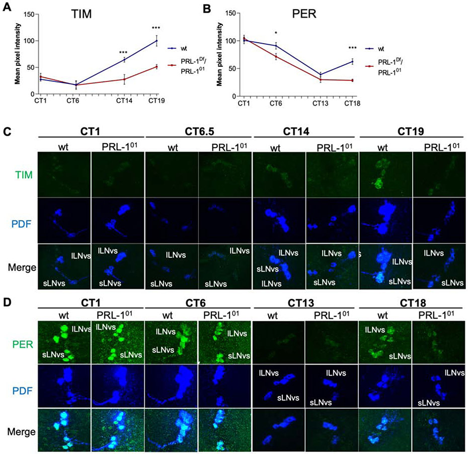
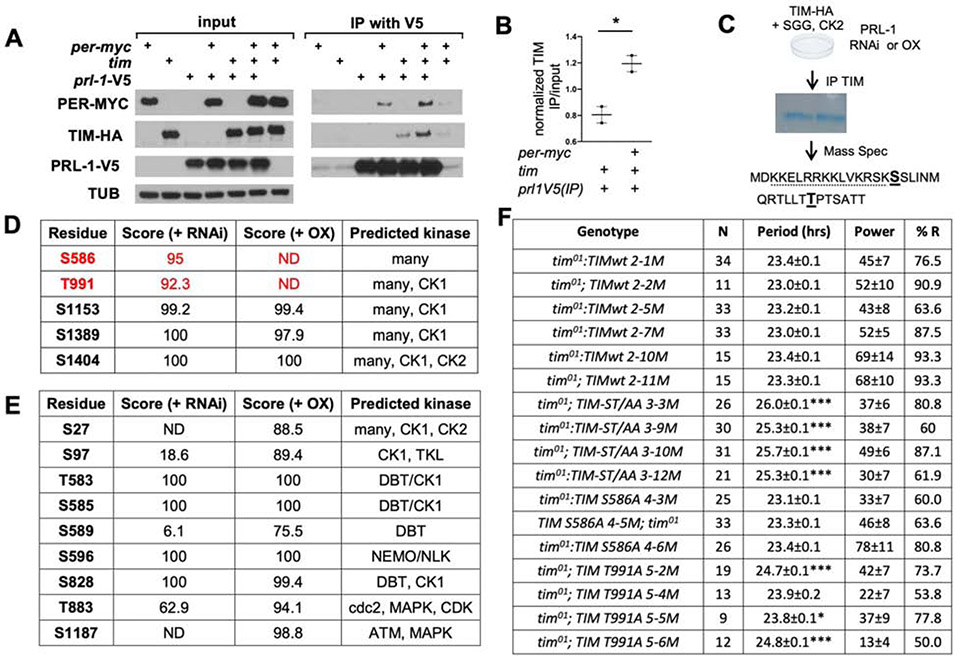
The timing of behavior under natural light-dark conditions is a function of circadian clocks and photic input pathways, but a mechanistic understanding of how these pathways collaborate in animals is lacking. Here we demonstrate in Drosophila that the Phosphatase of Regenerating Liver-1 (PRL-1) sets period length and behavioral phase gated by photic signals. PRL-1 knockdown in PDF clock neurons dramatically lengthens circadian period. PRL-1 mutants exhibit allele-specific interactions with the light- and clock-regulated gene timeless (tim). Moreover, we show that PRL-1 promotes TIM accumulation and dephosphorylation. Interestingly, the PRL-1 mutant period lengthening is suppressed in constant light, and PRL-1 mutants display a delayed phase under short, but not long, photoperiod conditions. Thus, our studies reveal that PRL-1-dependent dephosphorylation of TIM is a core mechanism of the clock that sets period length and phase in darkness, enabling the behavioral adjustment to change day-night cycles.

Keywords: Drosophila; circadian; phosphorylation; photoperiod; seasonality.


Conflict of interest statement

Declaration of Interest The authors declare no competing interests.

Figures

Figure 1.
Figure 1.. PRL-1 mRNA cycles in PDF cells but not in DN1 cells or whole heads.
(A-C) qPCR analysis of PRL-1 mRNA expression in adult Drosophila tissues. ZT indicates Zeitgeber time. PRL-1 expression rhythms are detected in (A) FACS-isolated PDF cells (pdfGAL4 UAS-mGFP; p < 0.05), but not in (B) isolated DN1p clock cells (clk4.1GAL4 UAS-mGFP, p = 0.57), or (C) whole head tissue (p = 0.65), as determined using one way ANOVA and Tukey’s post-hoc test, n=2. PRL-1 expression in DN1p neurons (B) is ~60% of levels observed in PDF cells (A). (D) PRL-1 mRNA expression in PDF cells in wild-type (wt), per01, and ClkJrk mutants at ZT12, n=2, assessed by Student’s t-test (*** p < .001, * p < .05). All data reported as mean expression levels with error bars indicating SEM. See also Figure S1 and Table S8.
Figure 2.
Figure 2.. PRL-1 mutant flies exhibit a long period phenotype.
Representative double-plotted locomotor activity profiles of individual males over 4 days LD followed by 7 days constant darkness (DD). Period and rhythmic power determined from DD data using Chi-squared periodogram analysis. (A) w1118: n = 38, (B) PRL-101/+: n= 27, (C) PRL-101/PRL-101: n= 23, (D) PRL-1Df/+: n= 51, (E) PRL-1Df/PRL-101: n= 38. Period measurements displayed above panels ± SEM, and comparisons made using one-way ANOVA and Tukey’s post-hoc test, *** p < 0.001. See also Figures S1, S2 and Tables S1,S2,S3, and S4.
Figure 3.
Figure 3.. PRL-1 more strongly affects TIM accumulation relative to PER in the sLNv.
Quantification (A,B) and representative images (C,D) of clock protein expression in wild-type (wt) vs. PRL-1 mutants (PRL-101/PRL-1Df) in adult Drosophila brains. (A-B) Quantification of TIM (A) and PER (B) levels in sLNv neurons under DD1 conditions. CT indicates circadian time. Plots report average intensity and error bars indicate SEM. Statistical comparisons made using Student’s t test (*** p < .001, * p < .05). Number of cells analyzed: (A) CT1: 49-56, CT6.5: 24-25, CT14: 29-47, CT19: 37-5; (B) CT1: 57-85, CT6: 50-59, CT13: 60-49, CT18: 63-62. (C-D) Representative images showing (C) TIM or (D) PER (green) with PDF (blue), and merged images in co-labeled sLNv and lLNv neurons in wild-type and PRL-1 mutants. See also Figures S3 and S4 and Table S5.
Figure 4.
Figure 4.. PRL-1 interacts with PER and TIM in S2R+ cells and selectively dephosphorylates TIM.
(A) Western blot analyses of Drosophila S2R+ cell extracts transfected with the constructs indicated. Extracts assayed directly (left panels) or immunoprecipitated (IP) with anti-V5 (right panels) followed by blotting with anti-MYC, anti-TIM, anti-V5 or anti-TUBULIN (TUB). (B) Quantification of TIM IP/ input levels, comparing co-transfection of tim-HA with Prl-1-V5 alone vs. Prl-1-V5 and per-MYC, n= 2. (C) Schematic of IP and mass spectrometry (MS) analysis performed on S2 cells transfected with TIM-HA, GSK3β, and CK2, in the presence of PRL-1 knockdown (RNAi) or overexpression (OX). Two putative PRL-1 dephosphorylation sites identified on TIM using this analysis indicated in bold. Dotted line represents TIM nuclear localization signal (NLS) sequence. (D-E) Probability scores (max 100) for phosphorylation of specific residues in TIM (D) or PER (E), as determined from MS data using PhosphoRS [95]. (C) List of differentially phosphorylated sites in TIM protein in PRL1 OX vs. RNAi. n=2, data shown for one experiment. Peptide coverage 42.9 (RNAi) and 40.8 (OX) for this experiment. (D) No PER sites identified as dephosphorylated by PRL-1 using a similar approach, peptide coverage 34.1 (RNAi) and 44.0 (OX), n=1. (F) DD locomotor rhythmicity analysis in tim01 mutants containing wild-type (wt) or phosphosite mutant TIM rescue constructs. ST/AA strains contain both the S586A and T991A mutation. ‘N’ indicates number of flies analyzed; ‘%R’ refers to percentage of rhythmic flies. Period and power shown as average ± SEM. Asterisks indicate significant difference between wt and phosphosite mutant rescue (* p< 0.05,***p < 0.001). See also Figures S3 and S5.
Fig 5.
Fig 5.. PRL-1 effects on period are absent under LL.
Representative double-plotted activity profiles of PRL-1; cry double mutants vs. single mutant controls under 12H light:12H dark (LD) followed by either 7 days DD or 7 days LL as indicated. A) cryb/cry01, B) PRL-1Df/PRL-101, C) PRL-101/+; cryb/cry01, D) PRL-1Df/PRL-101; cryb/cry01. Number of flies examined DD/LL: A) 51/48, B) 35/22, C) 24/19, D) 58/32. See also Figure S6 and Table S6.
Figure 6.
Figure 6.. PRL-1 mutants display delayed phases of morning and evening activity under winter, but not summer photoperiods.
Normalized group average activity profiles for (A) wild-type, (B) PRL-1Df/+, and (C) PRL-101/ PRL-1Df mutant flies in either 6L:18D (winter), 12:12 (intermediate), or 18L:6D (summer) photoperiods as indicated. Red arrows indicate morning onset of activity for conditions/genotypes in which morning anticipatory behavior detected (see Table S7). Blue arrows indicate time of evening activity offset (for 6L:18D) or onset (for 12L:12D and 18L:6D). Error bars indicate SEM. n= 36 to 153 per genotype. See also Figure S6 and Table S7.
Figure 7.
Figure 7.. Knockdown of human PRL-1 orthologs in U2OS cells lengthens period of per2∷luc rhythms.
A) Traces of luciferase reporter activity in mammalian U2OS cells expressing Per2∷luc, assayed for 6 days after synchronization. Cells transfected with siRNAs as indicated. B) Quantification of period length in transfected U2OS per2∷luc cells, as determined using LumiCycle analysis software (Actimetrics) from Day 1.5-4.5; n=4 for all samples except siCry2 (n=3) ( *** one-way ANOVA, Dunnett’s post-hoc test, p < 0.001). See also Figure S7 and Table S8.

References

    1. Mohawk JA, Green CB, and Takahashi JS (2012). Central and peripheral circadian clocks in mammals. 35, 445–462. - PMC - PubMed
    1. Allada R, and Chung BY (2010). Circadian organization of behavior and physiology in Drosophila. Annu Rev Physiol 72, 605–624. - PMC - PubMed
    1. Dubowy C, and Sehgal A (2017). Circadian Rhythms and Sleep in Drosophila melanogaster. Genetics 205, 1373–1397. - PMC - PubMed
    1. So WV, and Rosbash M (1997). Post-transcriptional regulation contributes to Drosophila clock gene mRNA cycling. Embo J 16, 7146–7155. - PMC - PubMed
    1. Curtin KD, Huang ZJ, and Rosbash M (1995). Temporally regulated nuclear entry of the Drosophila period protein contributes to the circadian clock. Neuron 14, 365–372. - PubMed

Publication types

LinkOut - more resources