. 2021 Jan 25;31(2):283-296.e7.
doi: 10.1016/j.cub.2020.10.025. Epub 2020 Nov 5.

The Proteomic Landscape of Centromeric Chromatin Reveals an Essential Role for the Ctf19CCAN Complex in Meiotic Kinetochore Assembly

Affiliations

The Proteomic Landscape of Centromeric Chromatin Reveals an Essential Role for the Ctf19CCAN Complex in Meiotic Kinetochore Assembly

Weronika E Borek et al. Curr Biol. .

Abstract

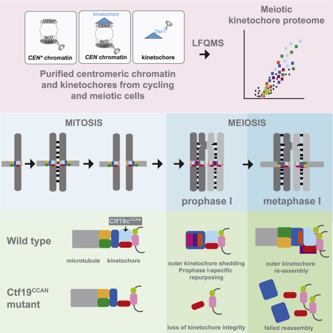
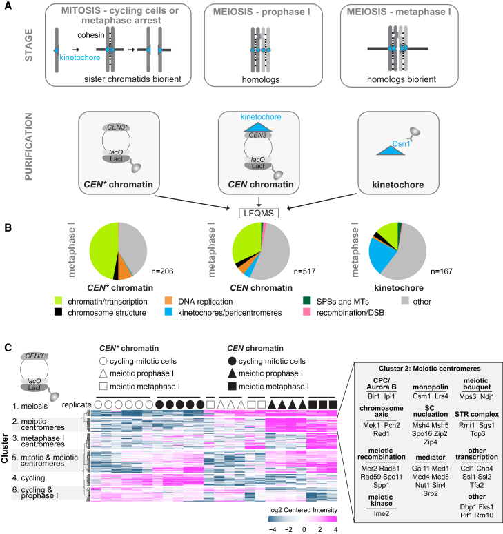
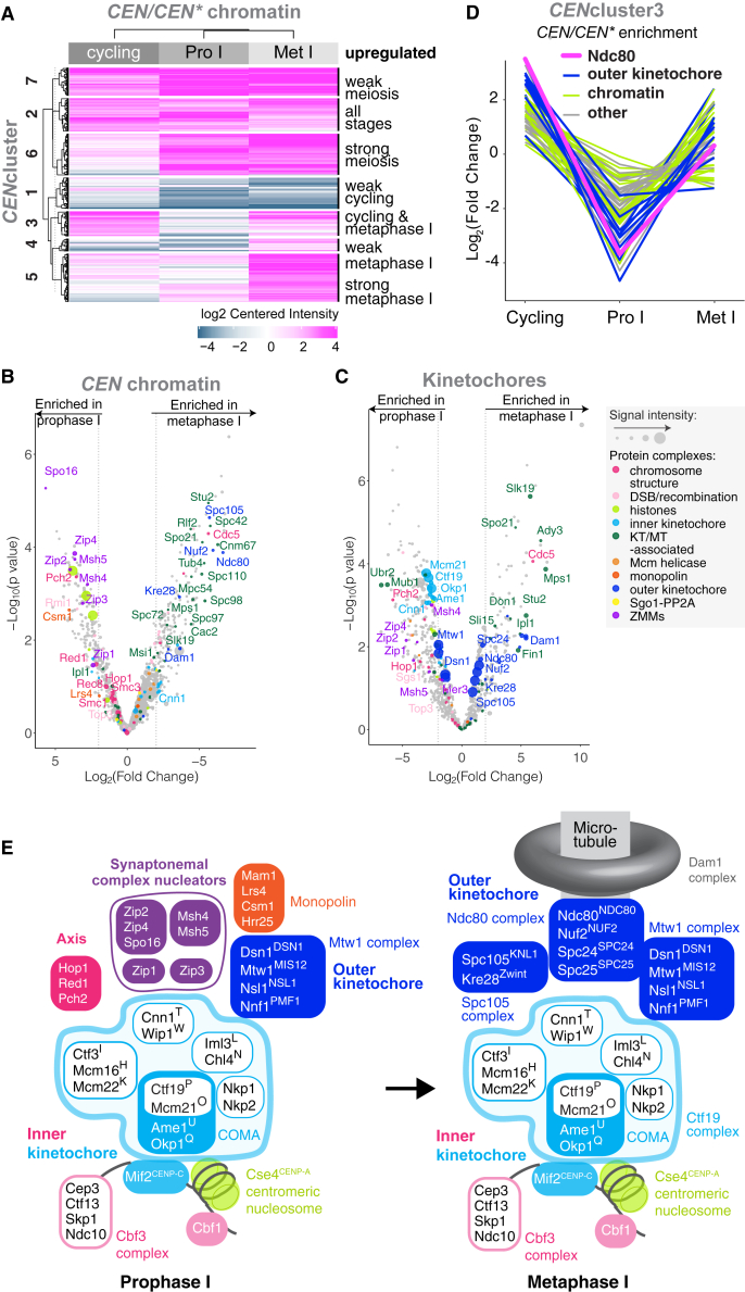
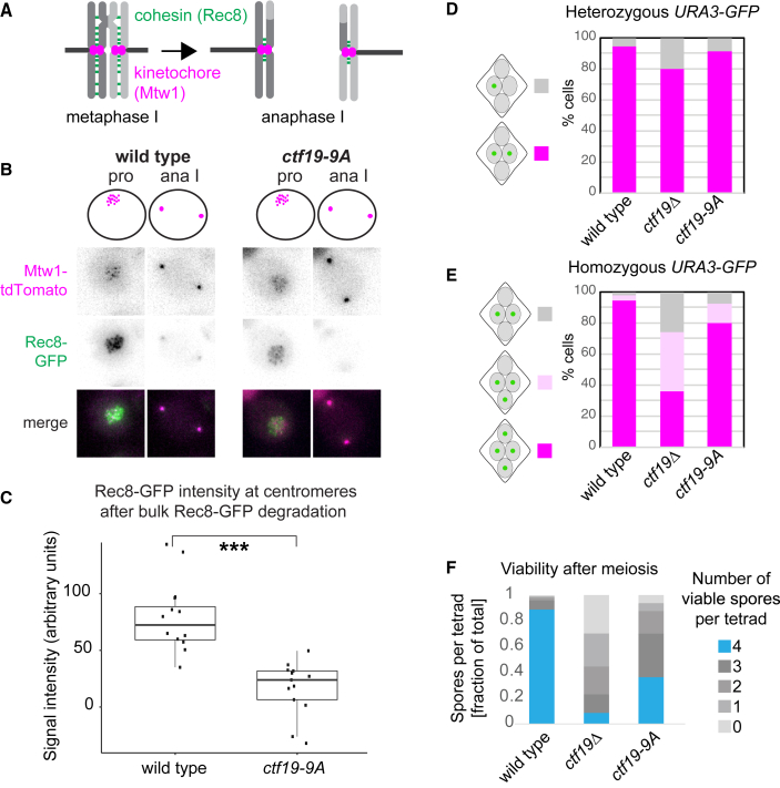
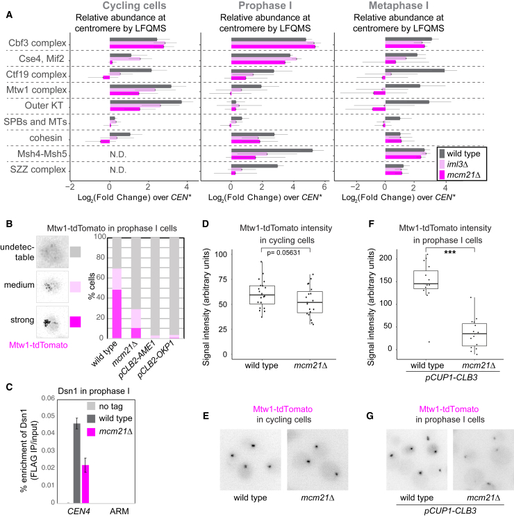
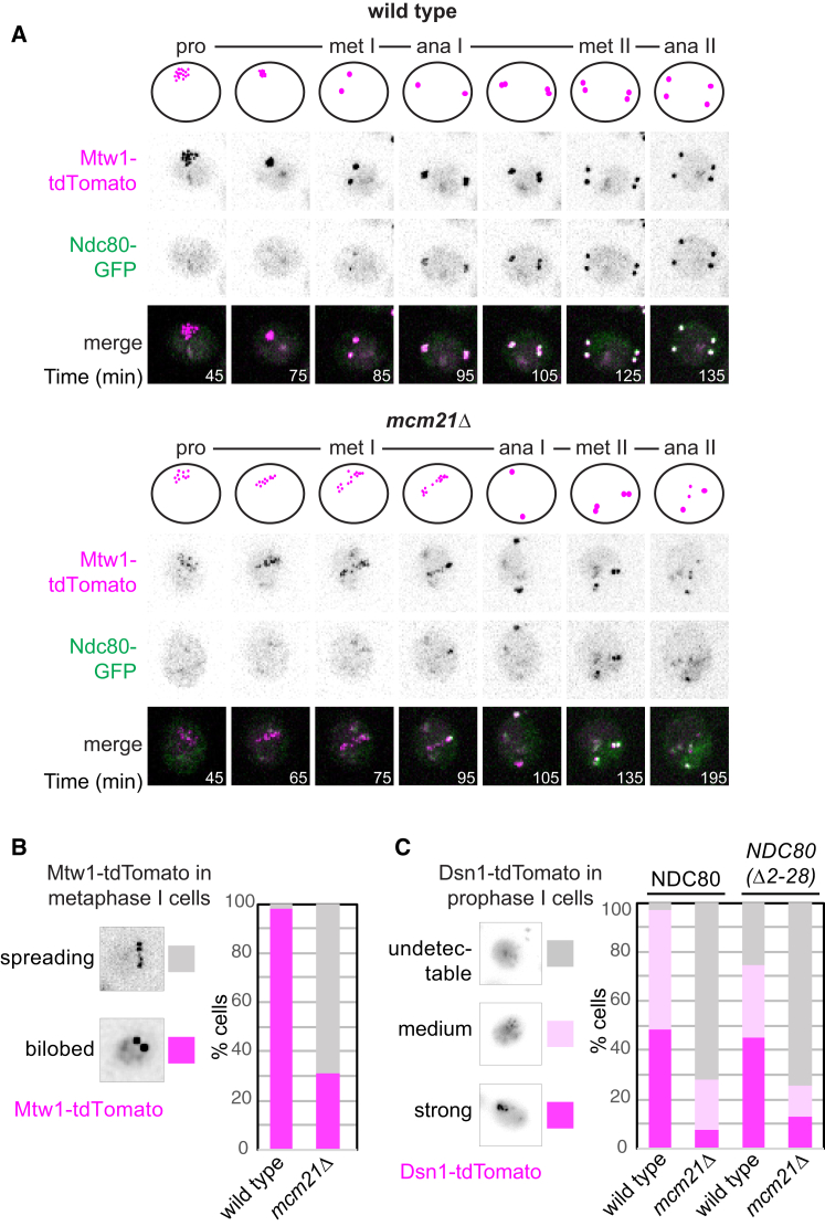
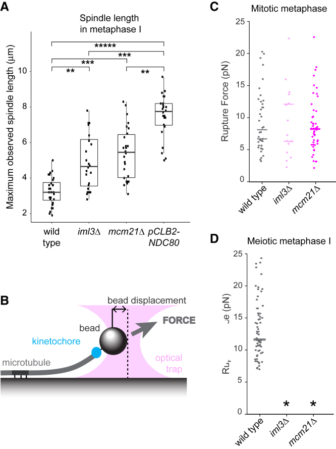
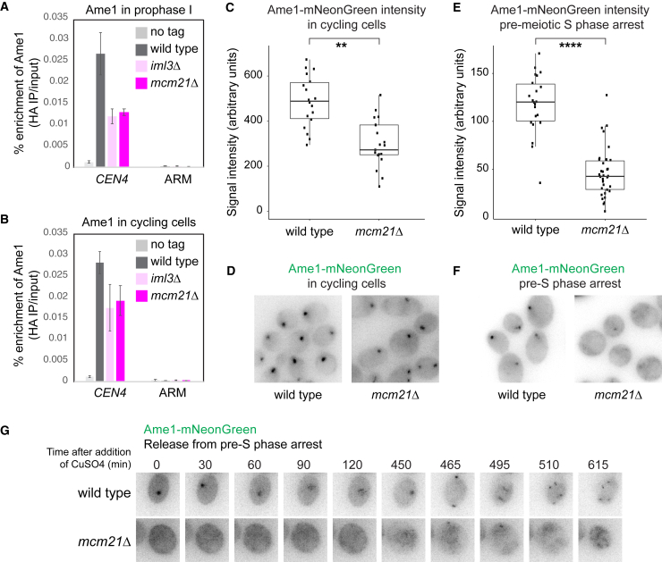
Kinetochores direct chromosome segregation in mitosis and meiosis. Faithful gamete formation through meiosis requires that kinetochores take on new functions that impact homolog pairing, recombination, and the orientation of kinetochore attachment to microtubules in meiosis I. Using an unbiased proteomics pipeline, we determined the composition of centromeric chromatin and kinetochores at distinct cell-cycle stages, revealing extensive reorganization of kinetochores during meiosis. The data uncover a network of meiotic chromosome axis and recombination proteins that bind to centromeres in the absence of the microtubule-binding outer kinetochore sub-complexes during meiotic prophase. We show that the Ctf19cCCAN inner kinetochore complex is essential for kinetochore organization in meiosis. Our functional analyses identify a Ctf19cCCAN-dependent kinetochore assembly pathway that is dispensable for mitotic growth but becomes critical upon meiotic entry. Therefore, changes in kinetochore composition and a distinct assembly pathway specialize meiotic kinetochores for successful gametogenesis.

Keywords: CCAN; Ctf19; budding yeast; centromere; kinetochore; meiosis; metaphase I; prophase I; proteomics.


Conflict of interest statement

Declaration of Interests The authors declare no competing interests.

Figures

None
Graphical abstract
Figure 1
Figure 1
Quantitative Label-Free Mass Spectrometry (LFQMS) Reveals the Complexity of the Centromere and Kinetochore-Associated Proteomes (A) Schematic representation of determined proteomes. CEN chromatin, CEN chromatin, and kinetochores were isolated from cycling, prophase I-arrested, and metaphase I-arrested cells and subjected to LFQMS. (B) CEN chromatin, CEN chromatin, and kinetochores show respective increases and decreases in the fraction of enriched proteins that are associated with chromatin or kinetochores. Following immunoprecipitation of LacI-3FLAG (CEN chromatin and CEN chromatin) and Dsn1-6His-3FLAG (kinetochores), proteins were quantified using LFQMS, and those enriched over respective negative controls with a cut-off of Log2(fold change) > 4 and p < 0.01 were categorized in the indicated groups. (C) Stage-specific functional groups of proteins associating with CEN chromatin and CEN chromatin. k-means clustering with a cut-off of Log2(fold change) > 2 and p < 0.05 was used. Cluster 2 proteins are listed in the inset. See also Table S1.
Figure 2
Figure 2
Changes in the Centromeric and Kinetochore Proteomes between Meiotic Prophase I and Metaphase I (A) CEN chromatin exhibits distinct composition signatures at different stages. The CEN chromatin/CEN chromatin enrichment values for each of cycling, prophase I-arrested, and metaphase I-arrested conditions were clustered (k-means) to identify groups of proteins with similar behavior. A cut-off of Log2(fold change) > 2 and p < 0.05 was used. (B and C) Composition of CEN chromatin (B) or kinetochore particles (C) isolated from prophase I and metaphase I is strikingly different. Volcano plot presenting the LFQMS-identified proteins co-purifying with CEN plasmids (B) or Dsn1-6His-3FLAG (C) immunopurified from cells arrested in prophase I (ndt80Δ, B; inducible-NDT80, C) and metaphase I (pCLB2-CDC20). Log2(fold change) between conditions is shown with corresponding p values (STAR Methods). Dashed line indicates |Log2(fold change)| = 2. (D) Several proteins exhibit Ndc80NDC80-like depletion from centromeres specifically during meiotic prophase. Mean-centered Log2(fold change) from CENcluster3 is plotted to show abundance of individual proteins in the indicated stages. (E) Schematic illustrating changes in the kinetochore association of some key complexes between meiotic prophase I and metaphase I observable by proteomics. See also Figures S1 and S2 and Tables S2 and S3.
Figure 3
Figure 3
Pericentromeric Cohesion Is Absent in ctf19-9A Anaphase I Cells (A–C) Pericentromeric cohesin is reduced in ctf19-9A anaphase I cells. Wild-type and ctf19-9A cells expressing Rec8-GFP and Mtw1-tdTomato were imaged throughout meiosis. (A) Schematic showing Rec8REC8 loss from chromosome arms, but not pericentromeres in anaphase I. (B) Representative images are shown. (C) Quantification of Rec8-GFP signal in the vicinity of Mtw1-tdTomato foci immediately following bulk Rec8-GFP degradation. Whiskers represent 1.5 IQR (interquartile range), the middle line is median, and the box encompasses two middle quartiles of the data. ∗∗∗p < 10−5; Mann-Whitney test. n > 11 cells. (D and E) ctf19-9A cells show less severe meiosis II chromosome segregation defects than ctf19Δ cells. The percentage of tetra-nucleate cells with the indicated patterns of GFP dot segregation was determined in wild-type and ctf19-9A cells with either one copy (heterozygous, D) or both copies (homozygous, E) of chromosome V marked with GFP at URA3 locus. n = 2 biological replicates, 100 tetrads each; mean values are shown. (F) Spore viability of ctf19-9A cells is impaired, but less severe than ctf19Δ cells. The number of viable progeny was scored following tetrad dissection. n = 3 biological replicates, >70 tetrads each; mean values are shown. See also Figure S3.
Figure 4
Figure 4
Proteomics Identifies a Critical Role of Ctf19cCCAN in Meiotic Kinetochore Assembly (A) Global CEN/CEN proteomics reveals that kinetochore composition is altered in mcm21Δ and iml3Δ meiotic prophase I and metaphase I cells. The sum of LFQMS abundance of protein complexes on CEN chromatin in wild-type, iml3Δ, and mcm21Δ cells is shown as enrichment over CEN chromatin isolated from wild-type cells. The abundance of Iml3 CENP-L and Mcm21CENP-O proteins was not included in the total Ctf19cCCAN count, as these proteins are missing in iml3Δ and mcm21Δ cells, respectively (STAR Methods). Error bars represent SD. KT, kinetochore; MT, microtubule; SPB, spindle pole body; SZZ, Spo16SPO16, Zip2SHOC1, Zip4TEX11. (B–G) A functional Ctf19cCCAN is critical for Mtw1cMIS12c association with centromeres in meiotic prophase I, but not cycling cells. (B) Wild-type, mcm21Δ, pCLB2-AME1, and pCLB2-OKP1 cells were imaged immediately after release from prophase I arrest. Representative images and scoring of cells with Mtw1-tdTomato signal are shown. n > 58 cells. (C) Prophase I-arrested wild-type and mcm21Δ cells carrying ndt80Δ and DSN1-6His-3FLAG, together with untagged control, were subjected to anti-FLAG ChIP-qPCR. Error bars represent SE (n = 4 biological replicates). p < 0.05, paired t test. (D–G) Mtw1-tdTomato signal intensity in cycling (D and E) and prophase I-arrested (F and G) wild-type and mcm21Δ cells. In (F) and (G), cells were engineered to ectopically produce Clb3 to maintain kinetochore clustering and allow signal quantification. In (D) and (F), whiskers represent 1.5 IQR, the middle line is median, and the box encompasses the two middle quartiles of the data. ∗∗∗p < 10−5; Mann-Whitney test. n > 19 (D) or n = 15 cells (F). See also Figure S4.
Figure 5
Figure 5
An Intact Ctf19cCCAN Is Required for Functional Outer Kinetochore Assembly in Meiosis I (A and B) Abnormal kinetochore behavior in the absence of MCM21. (A) Representative images of wild-type and mcm21Δ cells carrying Mtw1-tdTomato and Ndc80-GFP after release from prophase I arrest and imaged throughout meiosis. Time after release from prophase I is indicated. (B) Scoring of Mtw1-tdTomato signal in (A). Cells showing kinetochore spreading in at least one time point during the time-lapse were included in the “spreading” category. n > 49 cells. (C) Non-degradable ndc80(Δ2-28) does not rescue kinetochore function upon the loss of MCM21. Dsn1-tdTomato signal was scored in prophase I wild-type and mcm21Δ cells expressing either Ndc80-GFP or Ndc80(Δ2-28)-GFP. n > 56 cells. See also Figure S5.
Figure 6
Figure 6
Essential Role for Ctf19cCCAN in Establishment of Kinetochore Microtubule Attachments in Meiosis I (A) Metaphase I spindles are elongated in iml3Δ and mcm21Δ cells. Wild-type, iml3Δ, mcm21Δ, and pCLB2-NDC80 cells carrying pCLB2-CDC20 and expressing Mtw1-tdTomato and GFP-Tub1 were imaged undergoing meiosis and the maximum observed spindle length was measured. Whiskers represent 1.5 IQR, the middle line is median, and the box encompasses the two middle quartiles of the data. ∗∗∗∗∗p < 10−15, ∗∗∗p < 10−7, ∗∗p < 10−4; t test. n > 23. (B–D) Purified kinetochore particles (Dsn1-6His-3FLAG immunoprecipitation) from iml3Δ and mcm21Δ cells fail to attach to microtubules in a single-molecule assay. (B) Schematic of assay showing the optical trap pulling on a bead attached to a coverslip-immobilized microtubule. The bead-microtubule interaction is facilitated by purified kinetochores. (C and D) Kinetochore particles isolated from metaphase I-arrested cells lacking IML3 and MCM21 are not able to form kinetochore-microtubule attachments in vitro. Rupture force measurements of kinetochore particles isolated from mitotically arrested (by the addition of benomyl, C) or meiosis metaphase I-arrested (due to the presence of pCLB2-CDC20, D) wild-type, iml3Δ, and mcm21Δ cells are shown. Total particles analyzed: n = 65 (wild type, meiosis), n = 41 (wild type, mitosis), n = 15 (iml3Δ, mitosis), and n = 49 (mcm21Δ, mitosis) from 2 biological replicates; bars represent medians for each replicate. Asterisks indicate conditions for which no initial kinetochore-microtubule attachment was formed and thus rupture force could not be measured. See also Figure S6 and Table S7.
Figure 7
Figure 7
Ctf19cCCAN Subunits that Are Dispensable for Mitosis Become Essential for Inner Kinetochore Retention upon Entry into Gametogenesis (A and B) Loss of essential inner kinetochore component Ame1CENP-U in cycling and prophase I cells lacking IML3 and MCM21. Prophase I-arrested (A) or cycling (B) wild-type, iml3Δ, and mcm21Δ cells carrying AME1-6HA, together with untagged control, were subjected to anti-HA ChIP-qPCR. Error bars represent SE (n = 3 or 4 biological replicates, cycling and prophase I-arrested cells, respectively). (C–F) Ame1-mNeonGreen imaging in cycling (C and D) and pre-S phase-arrested (E and F) wild-type and mcm21Δ cells. Signal quantification (C and E) and representative images (D and F) are shown. In (C) and (E), whiskers represent 1.5 IQR, the middle line is median, and the box encompasses the two middle quartiles of the data. ∗∗∗∗p < 10−10, ∗∗p < 10−4; t test. n > 13 (C) or n > 17 cells (E). (G) Wild-type and mcm21Δ cells expressing Ame1-mNeonGreen were allowed to synchronously enter pre-meiotic S-phase through induction of IME1 and IME4, followed by live-cell imaging. See also Figure S7.

Similar articles

Cited by

References

    1. Hinshaw S.M., Harrison S.C. Kinetochore function from the bottom up. Trends Cell Biol. 2018;28:22–33. - PubMed
    1. Furuyama S., Biggins S. Centromere identity is specified by a single centromeric nucleosome in budding yeast. Proc. Natl. Acad. Sci. USA. 2007;104:14706–14711. - PMC - PubMed
    1. Carroll C.W., Silva M.C.C., Godek K.M., Jansen L.E.T., Straight A.F. Centromere assembly requires the direct recognition of CENP-A nucleosomes by CENP-N. Nat. Cell Biol. 2009;11:896–902. - PMC - PubMed
    1. Xiao H., Wang F., Wisniewski J., Shaytan A.K., Ghirlando R., FitzGerald P.C., Huang Y., Wei D., Li S., Landsman D. Molecular basis of CENP-C association with the CENP-A nucleosome at yeast centromeres. Genes Dev. 2017;31:1958–1972. - PMC - PubMed
    1. Hornung P., Troc P., Malvezzi F., Maier M., Demianova Z., Zimniak T., Litos G., Lampert F., Schleiffer A., Brunner M. A cooperative mechanism drives budding yeast kinetochore assembly downstream of CENP-A. J. Cell Biol. 2014;206:509–524. - PMC - PubMed

Publication types

MeSH terms

LinkOut - more resources