. 2020 Dec 15:409:115317.
doi: 10.1016/j.taap.2020.115317. Epub 2020 Nov 4.

4-methylpyrazole protects against acetaminophen-induced acute kidney injury

Affiliations

4-methylpyrazole protects against acetaminophen-induced acute kidney injury

Jephte Y Akakpo et al. Toxicol Appl Pharmacol. .

Abstract

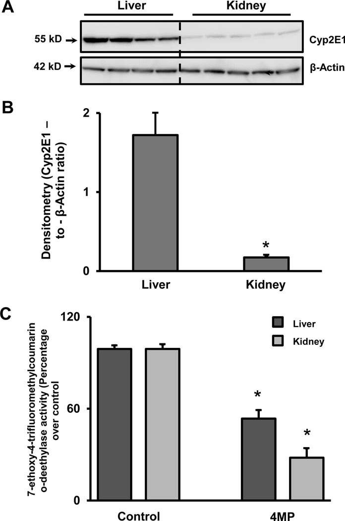
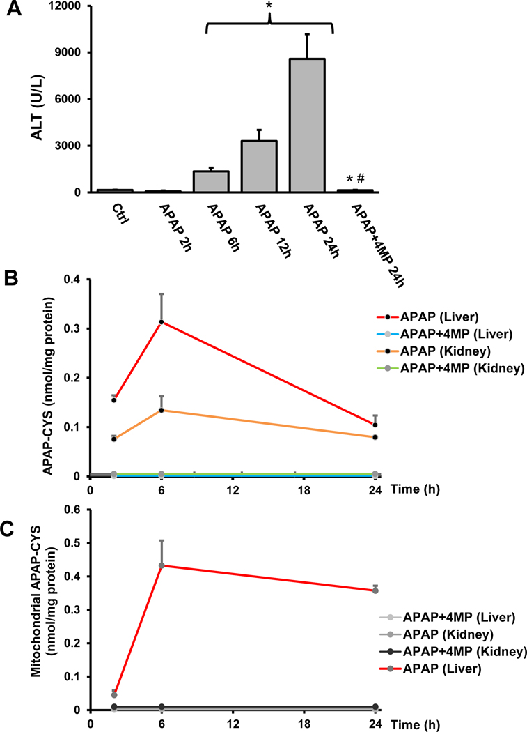
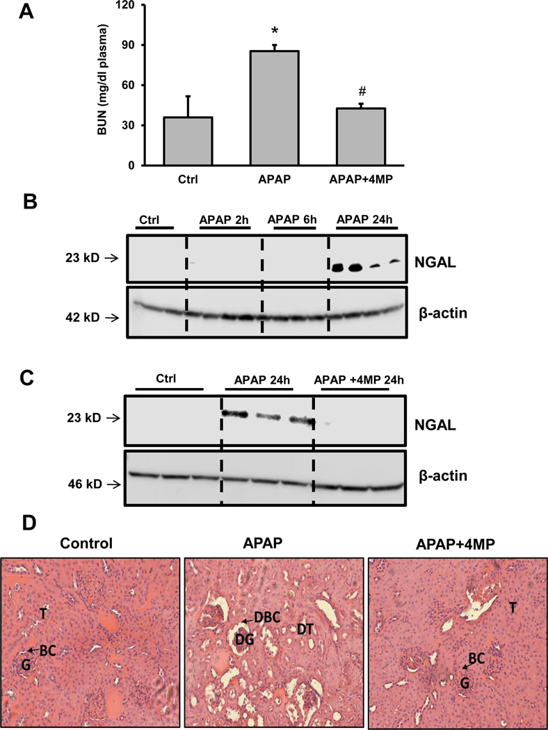
Acetaminophen (APAP) hepatotoxicity is the most common cause of acute liver failure in the United States, and while a significant percentage of APAP overdose patients develop kidney injury, molecular mechanisms involved in APAP-induced nephrotoxicity are relatively unknown. We have shown that 4-methylpyrazole (4MP, Fomepizole) protects against APAP-induced liver injury by inhibiting reactive metabolite formation through Cyp2E1, and analysis of data from APAP overdose patients indicated that kidney dysfunction strongly correlated with severe liver injury. Since Cyp2E1 is also expressed in the kidney, this study explored protection by 4MP against APAP-induced nephrotoxicity. Male C57BL/6 J mice were treated with either 300 or 600 mg/kg APAP with or without 4MP for 2, 6 or 24 h, followed by measurement of APAP metabolism and tissue injury. Interestingly, levels of APAP and its non-oxidative metabolites were significantly higher in kidneys when compared to the liver. APAP-protein adducts were present in both tissues within 2 h, but were absent in kidney mitochondria, unlike in the liver. While GSH depletion was seen in both tissues, activation of c-jun N-terminal kinase and its translocation to the mitochondria, which is a critical feature of APAP-induced liver injury, was not detected in the kidney. Treatment with 4MP attenuated APAP oxidative metabolite generation, GSH depletion as well as kidney injury indicating its potential use in protection against APAP-induced nephrotoxicity. In conclusion, since reactive metabolite formation seems to be common in both liver and kidney, 4MP mediated inhibition of Cyp2E1 protects against APAP-induced nephrotoxicity. However, downstream mechanisms of APAP-induced nephrotoxicity seem distinct from the liver.

Keywords: Acetaminophen; Fomepizole; Hepatotoxicity; N-Acetylcysteine; Nephrotoxicity; Protein Adducts.


Figures

Figure 1:
Figure 1:. Renal and hepatic Cyp2E1 protein expression levels and Cyp enzyme activities.
Liver and kidney homogenates from untreated C57BL/6J mice were subjected to western blot analysis to assess baseline expression of Cyp2E1 (A). Densitometric evaluation of Cyp2E1 and β-actin. The data are expressed as the mean ± SEM of the ratio for liver and kidney of 4–5 animals per group. *P<0.05 (compared to liver) (B). Cyp enzyme activities were assayed in liver and kidney homogenate using the fluorogenic substrate, 7-ethoxy-4-trifluoromethyl coumarin (7-EFC), which detects Cyp2E1 and Cyp1A2 activities, in the presence or absence of 50 μM 4MP. The enzyme activities were 260 and 500 RFU/min/mg protein for the control liver and the kidney respectively, which were normalized to 100%. (C). Data represent means ± SEM of 3 separate measurements. *P< 0.05 (compared to the respective control).
Figure 2:
Figure 2:. Formation of APAP metabolites in the kidney: effect of 4MP.
C57BL/6J mice were treated with 300 mg/kg APAP and either 50 mg/kg 4MP or 10 ml/kg saline. Renal concentrations of the parent compound APAP (A), the non-oxidative metabolites APAP-Glucuronide (B) and APAP-Sulfate (C) as well as its oxidative metabolite APAP-Cysteine (D) were measured by LC/MS/MS within the first 2 h after APAP administration. In a second experiments, renal concentrations of APAP (E), APAP-Glucuronide (F), APAP-Sulfate (G) and APAP-Cysteine (H) were measured 2 h after treatment with APAP and either 4MP or its solvent. Concentrations of all metabolites are expressed in nmol/mg protein. Data represent means ± SEM of n= 6 animals per group. *P< 0.05 (compared with APAP alone)
Figure 3:
Figure 3:. Formation of non-oxidative APAP metabolites in the liver: effect of 4MP.
C57BL/6J mice were treated with 300 mg/kg APAP and either 50 mg/kg 4MP or 10 ml/kg saline. Hepatic concentrations of the parent compound APAP (A) and its non-oxidative metabolites APAP-Glucuronide (B) and APAP-Sulfate (C) were measured by LC/MS/MS within the first 2 h after APAP administration. In a second experiments, hepatic concentrations of APAP (D), APAP-Glucuronide (E) and APAP-Sulfate (F) were measured 2 h after treatment with APAP and either 4MP or its solvent. Concentrations of all metabolites are expressed in nmol/mg protein. Data represent means ± SEM of n= 6 animals per group.
Figure 4:
Figure 4:. Formation of oxidative APAP metabolites in the liver: effect of 4MP.
C57BL/6J mice were treated with 300 mg/kg APAP and either 50 mg/kg 4MP or 10 ml/kg saline. Hepatic concentrations of the oxidative metabolites APAP-Cysteine (A), APAP-N-Acetylcysteine (B) and APAP-GSH (C) were measured by LC/MS/MS within the first 2 h after APAP administration. In a second experiments, hepatic concentrations of APAP-Cysteine (D), APAP-N-Acetylcysteine (E) and APAP-GSH (F) were measured 2 h after treatment with APAP and either 4MP or its solvent. Concentrations of all metabolites are expressed in nmol/mg protein. Data represent means ± SEM of n= 6 animals per group. *P< 0.05 (compared to APAP).
Figure 5:
Figure 5:. Renal glutathione and APAP protein adducts levels: effect of 4MP.
C57BL/6J mice were treated with 300 mg/kg APAP and either 50 mg/kg 4MP or 10 ml/kg saline. Total GSH was measured in renal tissue homogenate at 2 h post-APAP (A). Concentration of protein-derived APAP-cysteine adducts (APAP-CYS) in tissue homogenate (B) as well as the mitochondrial fractions (C) were measured by HPLC-ECD at 2 h post APAP treatment. APAP-CYS concentrations are expressed in nmol/mg protein. Data represent means ± SEM of n= 4 animals per group. *P< 0.05 (compared with control). #P< 0.05 (compared to APAP)
Figure 6:
Figure 6:. JNK activation and JNK translocation to the mitochondria in liver and kidney.
C57BL/6J mice were treated with 300 mg/kg APAP and either 50 mg/kg 4MP or 10 ml/kg saline. JNK and P-JNK were assessed in the cytosolic fraction and isolated mitochondria by western blotting. The two bands reflect JNK1 (46kD) and JNK2 (54 kD). Liver (A) and kidney (B) samples were obtained from controls (Cont) and 2 h after APAP or APAP+4MP.
Figure 7:
Figure 7:. Liver and kidney injury after a moderate overdose of APAP: effect of 4MP.
C57BL/6J mice were treated with 300 mg/kg APAP and sacrificed at 2, 6, 12, or 24 h. In addition, a group was co-treated with APAP and 50 mg/kg 4MP for 24 h. Plasma ALT activities (A). Kidney homogenates were subjected to western blotting for NGAL and β-actin (B). Representative H&E-stained kidney sections of a control and 24 h post APAP (×50 magnification) (C). Data represent means ± SEM of n= 4 animals per group. *P< 0.05 (compared to control). #P< 0.05 (compared to APAP 24 h). Abbreviations: T=tubules, BC=Bowman capsule, G=Glomerulus.
Figure 8:
Figure 8:. Liver injury and APAP protein adducts in liver and kidney after a severe overdose.
C57BL/6J mice were co-treated with 600 mg/kg APAP and 50 mg/kg 4MP or 10 ml/kg saline for 2, 6, 12, or 24 h. Time course of plasma ALT activities (A). Time course of protein-derived APAP-cysteine adducts measured in liver and kidney homogenate (B) as well as their mitochondrial fractions (C). APAP-CYS concentrations are expressed in nmol/mg protein. Data represent means ± SEM of n= 4 animals per group. *P< 0.05 (compared to untreated controls). #P< 0.05 (compared to APAP 24 h).
Figure 9:
Figure 9:. JNK activation and JNK translocation to the mitochondria in liver and kidney after a severe APAP overdose.
C57BL/6J mice were treated with 600 mg/kg APAP and either 50 mg/kg 4MP or 10 ml/kg saline. JNK and P-JNK were assessed in the cytosolic fraction and isolated mitochondria by western blotting. The two bands reflect JNK1 (46kD) and JNK2 (54 kD). Liver and kidney samples were obtained from controls (Cont) and 2, 6 or 24 h after APAP or APAP+4MP.
Figure 10:
Figure 10:. Kidney injury after a severe overdose of APAP: effect of 4MP.
C57BL/6J mice were treated with 600 mg/kg APAP and sacrificed at 2, 6, or 24 h. In addition, a group was co-treated with APAP and 50 mg/kg 4MP for 24 h. Blood Urea Nitrogen (BUN) levels at 24 h (A). Kidney homogenates were subjected to western blotting for NGAL and β-actin after APAP alone (B) or in combination with 4MP (C). Representative H&E-stained kidney sections of a control, APAP and APAP+4MP at 24 h post APAP (D) (×50 magnification). Data represent means ± SEM of n= 4 animals per group. *P< 0.05 (compared to control). #P< 0.05 (compared to APAP). Abbreviations: T=tubules, BC=Bowman capsule, G=Glomerulus, DT=Damaged tubules, DBC=Damaged Bowman capsule, DG=Damaged glomerulus.

References

    1. Akakpo JY, Ramachandran A, Duan L, Schaich MA, Jaeschke MW, Freudenthal BD, Ding WX, Rumack BH, Jaeschke H, 2019. Delayed Treatment With 4-Methylpyrazole Protects Against Acetaminophen Hepatotoxicity in Mice by Inhibition of c-Jun N-Terminal Kinase. Toxicol. Sci 170, 57–68. - PMC - PubMed
    1. Akakpo JY, Ramachandran A, Kandel SE, Ni HM, Kumer SC, Rumack BH, Jaeschke H, 2018. 4-Methylpyrazole protects against acetaminophen hepatotoxicity in mice and in primary human hepatocytes. Hum. Exp. Toxicol 37, 1310–1322. - PMC - PubMed
    1. Arzuk E, Turna B, Sözbilen M, Orhan H, 2018. Inter-individual and inter-organ variability in the bioactivation of paracetamol by human liver and kidney tissues. Environ. Toxicol. Pharmacol 61, 8–17. - PubMed
    1. Bhushan B, Walesky C, Manley M, Gallagher T, Borude P, Edwards G, Monga SP, Apte U, 2014. Pro-regenerative signaling after acetaminophen-induced acute liver injury in mice identified using a novel incremental dose model. Am. J. Pathol 184, 3013–325. - PMC - PubMed
    1. Björck S, Svalander CT, Aurell M, 1988. Acute renal failure after analgesic drugs including paracetamol (acetaminophen). Nephron 49, 45–53. - PubMed

Publication types

MeSH terms