

The Biological Function of Extracellular Vesicles during Fertilization, Early Embryo-Maternal Crosstalk and Their Involvement in Reproduction: Review and Overview
- PMID: 33158009
- PMCID: PMC7693816
- DOI: 10.3390/biom10111510
The Biological Function of Extracellular Vesicles during Fertilization, Early Embryo-Maternal Crosstalk and Their Involvement in Reproduction: Review and Overview
Abstract
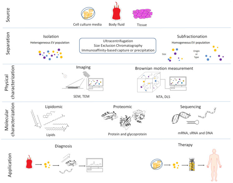
Secretory extracellular vesicles (EVs) are membrane-enclosed microparticles that mediate cell to cell communication in proximity to, or distant from, the cell of origin. Cells release a heterogeneous spectrum of EVs depending on their physiologic and metabolic state. Extracellular vesicles are generally classified as either exosomes or microvesicles depending on their size and biogenesis. Extracellular vesicles mediate temporal and spatial interaction during many events in sexual reproduction and supporting embryo-maternal dialogue. Although many omic technologies provide detailed understanding of the molecular cargo of EVs, the difficulty in obtaining populations of homogeneous EVs makes difficult to interpret the molecular profile of the molecules derived from a miscellaneous EV population. Notwithstanding, molecular characterization of EVs isolated in physiological and pathological conditions may increase our understanding of reproductive and obstetric diseases and assist the search for potential non-invasive biomarkers. Moreover, a more precise vision of the cocktail of biomolecules inside the EVs mediating communication between the embryo and mother could provide new insights to optimize the therapeutic action and safety of EV use.
Keywords: biomarker; diagnosis; embryo; exosomes; extracellular vesicles; miRNA; microvesicles; protein; reproduction; therapy.
Conflict of interest statement
The authors declare no conflict of interest.
Figures
Similar articles
-
Extracellular vesicles: roles in gamete maturation, fertilization and embryo implantation.Hum Reprod Update. 2016 Mar-Apr;22(2):182-93. doi: 10.1093/humupd/dmv055. Epub 2015 Dec 9. Hum Reprod Update. 2016. PMID: 26663221 Free PMC article. Review.
-
The evolving roles of extracellular vesicles in embryo-maternal communication.Commun Biol. 2024 Jun 21;7(1):754. doi: 10.1038/s42003-024-06442-9. Commun Biol. 2024. PMID: 38906986 Free PMC article. Review.
-
Extracellular vesicle research in reproductive science: Paving the way for clinical achievements†.Biol Reprod. 2022 Mar 19;106(3):408-424. doi: 10.1093/biolre/ioab245. Biol Reprod. 2022. PMID: 34982163 Review.
-
Extracellular vesicles: Mediators of embryo-maternal crosstalk during pregnancy and a new weapon to fight against infertility.Eur J Cell Biol. 2020 Nov;99(8):151125. doi: 10.1016/j.ejcb.2020.151125. Epub 2020 Oct 2. Eur J Cell Biol. 2020. PMID: 33059931 Review.
-
Global transcriptomic changes occur in uterine fluid-derived extracellular vesicles during the endometrial window for embryo implantation.Hum Reprod. 2021 Jul 19;36(8):2249-2274. doi: 10.1093/humrep/deab123. Hum Reprod. 2021. PMID: 34190319 Free PMC article.
Cited by
-
Extracellular vesicles from human Fallopian tubal fluid benefit embryo development in vitro.Hum Reprod Open. 2023 Feb 21;2023(2):hoad006. doi: 10.1093/hropen/hoad006. eCollection 2023. Hum Reprod Open. 2023. PMID: 36895886 Free PMC article.
-
The role of extracellular vesicles in animal reproduction and diseases.J Anim Sci Biotechnol. 2022 Jun 10;13(1):62. doi: 10.1186/s40104-022-00715-1. J Anim Sci Biotechnol. 2022. PMID: 35681164 Free PMC article. Review.
-
Extracellular vesicles from maternal uterine cells exposed to risk factors cause fetal inflammatory response.Cell Commun Signal. 2021 Oct 7;19(1):100. doi: 10.1186/s12964-021-00782-3. Cell Commun Signal. 2021. PMID: 34620169 Free PMC article.
-
Effect of cryopreservation and semen extender on extracellular vesicles isolated from bull semen.Front Vet Sci. 2024 Jul 30;11:1437410. doi: 10.3389/fvets.2024.1437410. eCollection 2024. Front Vet Sci. 2024. PMID: 39139604 Free PMC article.
-
Exploring the potential of in vitro extracellular vesicle generation in reproductive biology.J Extracell Biol. 2024 Sep 5;3(9):e70007. doi: 10.1002/jex2.70007. eCollection 2024 Sep. J Extracell Biol. 2024. PMID: 39238549 Free PMC article. Review.
