Review
. 2020 Nov 4;13(21):4946.
doi: 10.3390/ma13214946.

Silk Particles as Carriers of Therapeutic Molecules for Cancer Treatment

Affiliations
Review

Silk Particles as Carriers of Therapeutic Molecules for Cancer Treatment

Anna Florczak et al. Materials (Basel). .

Abstract

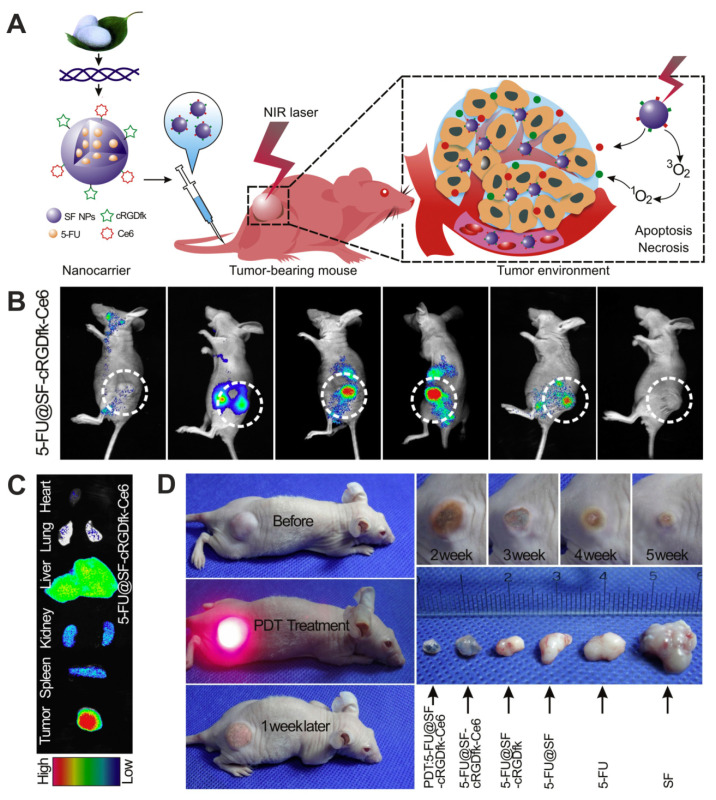
Although progress is observed in cancer treatment, this disease continues to be the second leading cause of death worldwide. The current understanding of cancer indicates that treating cancer should not be limited to killing cancer cells alone, but that the target is the complex tumor microenvironment (TME). The application of nanoparticle-based drug delivery systems (DDS) can not only target cancer cells and TME, but also simultaneously resolve the severe side effects of various cancer treatment approaches, leading to more effective, precise, and less invasive therapy. Nanoparticles based on proteins derived from silkworms' cocoons (like silk fibroin and sericins) and silk proteins from spiders (spidroins) are intensively explored not only in the oncology field. This natural-derived material offer biocompatibility, biodegradability, and simplicity of preparation methods. The protein-based material can be tailored for size, stability, drug loading/release kinetics, and functionalized with targeting ligands. This review summarizes the current status of drug delivery systems' development based on proteins derived from silk fibroin, sericins, and spidroins, which application is focused on systemic cancer treatment. The nanoparticles that deliver chemotherapeutics, nucleic acid-based therapeutics, natural-derived agents, therapeutic proteins or peptides, inorganic compounds, as well as photosensitive molecules, are introduced.

Keywords: bioengineering; cancer; drug delivery; particles; sericin; silk; silk fibroin; spidroin.


Conflict of interest statement

The authors declare no conflict of interest.

Figures

Figure 1
Figure 1
The schematic representation of the basic concept of the active and passive drug delivery systems in the tumor niche.
Figure 2
Figure 2
The proteins derived from natural silkworm cocoons, spider silks, and biotechnologically produced silk-derived proteins are used in the production of DDS for the systemic delivery of a variety of molecules used in cancer treatment.
Figure 3
Figure 3
Hydrophilic silk sericin-based nanoparticles (SSC@NPs) in the treatment of cancer. (A) Schematic illustration of SSC@NPs. (B) The fluorescence imaging of mice bearing MCF-7 tumors at 24 h after intravenous administration of free dye indocyanine green ICG or ICG-SSC@NPs. The tumors are pointed with white circles. (C) Top: ex vivo images of major organs and tumors resected from HepG2-bearing mice after 24 h of treatment with free Dox and Dox-loaded DOX-SSC@NPs (NPs contained 2.5 mg/kg of Dox); Bottom: the graph demonstrating Dox distribution in major organs and tumors analyzed at 24 h post-injection. (D) In vivo therapeutic efficacy Dox-loaded DOX-SSC@NPs in reference to control Phosphate buffered saline PBS and free Dox in HepG2 tumor-bearing animals. Top: The time dependent tumor volumes after treatment. Bottom: Image of resected tumors. NS, not significant; * p < 0.05. Reproduced with permission [66]. Copyright, 2018, Elsevier.
Figure 4
Figure 4
Functionalized spider silk nanospheres as drug carriers for targeted therapy of Her2-overexpressing tumors. (A) Schematic representation of DDS based on bioengineered spider silk that targets specifically Her2-overexpressing cancer cells. (B) Fluorescently labeled spheres that were functionalized with Her2-binding peptide H2.1 and H2.2 (H2.1MS1 and H2.2MS1) were effectively internalized into the cytoplasm of Her2(+) cells (SKOV-3 and SKBR-3) in contrast to non-functionalized spheres (MS1) and control Her2(−) (MSU1.1) cells. Cell membrane stained with ConA-FITC (green) and particles conjugated with ATTO 647N (red). Scale bar: 10 μm. (C) Dox was released from the H2.1MS1 spheres inside SKBR3 cells, and the colocalization of signals derived from Dox and the nucleus was observed. The nuclei stained with DAPI (blue) and autofluorescence of Dox (red). Scale bar: 10 μm. Reproduced with permission [71]. Copyright, 2014, American Chemical Society.
Figure 5
Figure 5
Doxorubicin-loaded magnetic silk fibroin nanoparticles (DMSs) in the targeted cancer treatment. (A) A scheme presenting the generation of DMSs: Dox is adsorbed in part on the surface of IONPs and then incorporated in the SF during the DMSs formation by using salting-out method. (B) Top panel: in vivo whole-body fluorescent imaging of MCF-7 breast cancer-bearing mice (tumors located on both sides) at 2 h after intravenous administration with DMSs or Dox-loaded SFNs; tumors location with and without magnet attachment are indicated with red and green circles, respectively. Bottom panel: tumors and major organs imaged ex vivo 12 h after intravenous administration of particles. (C) The biodistribution of the DMSs and Dox-loaded SFNs in the organs and tumors after particle administration and application of magnetic field at the tumor side; +MF and –MF: in the presence and absence of MF, respectively. (D) Schematic illustration of in vivo magnetic tumor targeting. (E) MCF-7/ADR tumors-bearing mice after injection of DMSs. DMSs contained 5 mg/kg of DOX. Reproduced with permission [140]. Copyright, 2014, Wiley.
Figure 6
Figure 6
Functionalized silk fibroin nanoparticles for multimodal targeted chemotherapy and photodynamic therapy. (A) Schematic illustration of the SF-based nanocarriers functionalized with cyclic cRGDfk peptide, chlorin e6 and 5-FU for in vivo MGC-803 gastric cancer treatment. (B) In vivo dynamic fluorescence of 5-FU@SF-cRGDfk-Ce6 NPs injected to MGC-803 tumor-bearing mice (5-FU dose = 5 mg/kg). (C) Ex vivo fluorescent images of organs and tumor tissue 24 h post-injection of 5-FU@SF-cRGDfk-Ce6 NPs. (D) In vivo therapeutic efficacy of SF-based NPs and PDT. The tumor volumes after the treatment with PDT and 5-FU@SG-cRGDfk-Ce6 NPs in reference to the application of control SF-based NPs derivates and free 5-FU. Reproduced with permission [89]. Copyright, 2018, Elsevier.

References

    1. Pérez-Herrero E., Fernández-Medarde A. Advanced targeted therapies in cancer: Drug nanocarriers, the future of chemotherapy. Eur. J. Pharm. Biopharm. 2015;93:52–79. doi: 10.1016/j.ejpb.2015.03.018. - DOI - PubMed
    1. Pucci C., Martinelli C., Ciofani G. Innovative approaches for cancer treatment: Current perspectives and new challenges. Ecancermedicalscience. 2019;13:961. doi: 10.3332/ecancer.2019.961. - DOI - PMC - PubMed
    1. Majumder J., Taratula O., Minko T. Nanocarrier-based systems for targeted and site specific therapeutic delivery. Adv. Drug Deliv. Rev. 2019;144:57–77. doi: 10.1016/j.addr.2019.07.010. - DOI - PMC - PubMed
    1. Murciano-Goroff Y.R., Taylor B.S., Hyman D.M., Schram A.M. Toward a More Precise Future for Oncology. Cancer Cell. 2020;37:431–442. doi: 10.1016/j.ccell.2020.03.014. - DOI - PMC - PubMed
    1. Murciano-Goroff Y.R., Warner A.B., Wolchok J.D. The future of cancer immunotherapy: Microenvironment-targeting combinations. Cell Res. 2020;30:507–519. doi: 10.1038/s41422-020-0337-2. - DOI - PMC - PubMed

LinkOut - more resources