Review
. 2020 Nov 4;8(4):46.
doi: 10.3390/medsci8040046.

The Evolving Role of Caveolin-1: A Critical Regulator of Extracellular Vesicles

Affiliations
Review

The Evolving Role of Caveolin-1: A Critical Regulator of Extracellular Vesicles

Kareemah Ni et al. Med Sci (Basel). .

Abstract

Emerging evidence suggests that extracellular vesicles (EVs) play an essential role in mediating intercellular communication and inter-organ crosstalk both at normal physiological conditions and in the pathogenesis of human diseases. EV cargos are made up of a broad spectrum of molecules including lipids, proteins, and nucleic acids such as DNA, RNA, and microRNAs. The complex EV cargo composition is cell type-specific. A dynamic change in EV cargos occurs along with extracellular stimuli and a change in the pathophysiological status of the host. Currently, the underlying mechanisms by which EVs are formed and EV cargos are selected in the absence and presence of noxious stimuli and pathogens remain incompletely explored. The term EVs refers to a heterogeneous group of vesicles generated via different mechanisms. Some EVs are formed via direct membrane budding, while the others are produced through multivesicular bodies (MVBs) or during apoptosis. Despite the complexity of EV formation and EV cargo selection, recent studies suggest that caveolin-1, a well-known structural protein of caveolae, regulates the formation and cargo selection of some EVs, such as microvesicles (MVs). In this article, we will review the current understanding of this emerging and novel role of cav-1.

Keywords: EV cargo; caveolin-1; extracellular vesicle (EVs).


Conflict of interest statement

The authors declare that they have no conflict of interest.

Figures

Figure 1
Figure 1
Schematic diagram of extracellular vesicle size and biogenesis. Extracellular vesicles (EVs) refer to a group of heterogeneous vesicles ranging from 20 nm to 5 μm. Small vesicles ranging from 20 to 200 nm used to be named exosomes, while small to medium-size vesicles ranging from 200 to 500 nm used to be called microvesicles (MVs). The largest EVs, which used to be called apoptotic bodies (ABs), are often generated from dying cells. The biogenesis of these EVs has distinct pathways. As illustrated here, small EVs are often generated via a long journey including endosomes, ER/Golgi, and multivesicular bodies (MVBs). On the other hand, the other larger EVs can be produced via direct budding from the plasma membrane. It is commonly facilitated by lipid raft proteins as discussed in this review. The largest-size EVs, or previously referred to as ABs, are broken down from apoptotic cells.
Figure 2
Figure 2
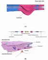
(A) Schematic illustration of caveolae and planar lipid rafts on the cell membranes. (B) Schematic diagram of caveolin-1 primary structure and predicted topology. Schematic showing that cav-1 is an integral membrane protein. The primary structure and predicted topology is illustrated here. Cav-1 often self-associates to a homo-oligomer which is called a caveolar assembly unit. Each caveolar assembly unit is composed of 14–16 monomers. Here, we show a dimer of cav-1 formed by two cav-1 monomers via the oligomerization domain (yellow color). The amino- and carboxyl-ends face towards the cytosol side. A hairpin loop (transmembrane domain, blue color) inserts into the membrane bilayer. The amino-terminal membrane-attachment domain is the caveolin scaffolding domain (CSD) (violet color).
Figure 3
Figure 3
Schematic diagram of cav-1-mediated selection of EV miRNAs via hnRNPA2B1. In the presence of noxious stimuli, such as oxidative stress, cav-1 Y14 is phosphorylated and results in conformational change of cav-1. This pY14-induced change exposes the CSD to bind with the hnRNPA2B1 RGG domain. The interaction of cav-1 and hnRNPA2B1 leads to the encapsulation of both hnRNPA2B1 and its bound miRNAs into the EVs along with cav-1.

References

    1. Chargaff E., West R. The biological significance of the thromboplastic protein of blood. J. Biol. Chem. 1946;166:189–197. - PubMed
    1. Harding C., Heuser J., Stahl P. Receptor-mediated endocytosis of transferrin and recycling of the transferrin receptor in rat reticulocytes. J. Cell Biol. 1983;97:329–339. doi: 10.1083/jcb.97.2.329. - DOI - PMC - PubMed
    1. Pan B.-T., Johnstone R.M. Fate of the transferrin receptor during maturation of sheep reticulocytes in vitro: Selective externalization of the receptor. Cell. 1983;33:967–978. doi: 10.1016/0092-8674(83)90040-5. - DOI - PubMed
    1. Pisitkun T., Shen R.-F., Knepper M.A. Identification and proteomic profiling of exosomes in human urine. Proc. Natl. Acad. Sci. USA. 2004;101:13368–13373. doi: 10.1073/pnas.0403453101. - DOI - PMC - PubMed
    1. Lässer C., Eldh M., Lötvall J. Isolation and Characterization of RNA-Containing Exosomes. J. Vis. Exp. 2012:e3037. doi: 10.3791/3037. - DOI - PMC - PubMed

LinkOut - more resources