Review
. 2020 Nov 4;21(21):8264.
doi: 10.3390/ijms21218264.

The Central Role of Cadherins in Gonad Development, Reproduction, and Fertility

Affiliations
Review

The Central Role of Cadherins in Gonad Development, Reproduction, and Fertility

Rafał P Piprek et al. Int J Mol Sci. .

Abstract

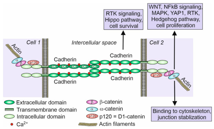
Cadherins are a group of membrane proteins responsible for cell adhesion. They are crucial for cell sorting and recognition during the morphogenesis, but they also play many other roles such as assuring tissue integrity and resistance to stretching, mechanotransduction, cell signaling, regulation of cell proliferation, apoptosis, survival, carcinogenesis, etc. Within the cadherin superfamily, E- and N-cadherin have been especially well studied. They are involved in many aspects of sexual development and reproduction, such as germline development and gametogenesis, gonad development and functioning, and fertilization. E-cadherin is expressed in the primordial germ cells (PGCs) and also participates in PGC migration to the developing gonads where they become enclosed by the N-cadherin-expressing somatic cells. The differential expression of cadherins is also responsible for the establishment of the testis or ovary structure. In the adult testes, N-cadherin is responsible for the integrity of the seminiferous epithelium, regulation of sperm production, and the establishment of the blood-testis barrier. Sex hormones regulate the expression and turnover of N-cadherin influencing the course of spermatogenesis. In the adult ovaries, E- and N-cadherin assure the integrity of ovarian follicles and the formation of corpora lutea. Cadherins are expressed in the mature gametes and facilitate the capacitation of sperm in the female reproductive tract and gamete contact during fertilization. The germ cells and accompanying somatic cells express a series of different cadherins; however, their role in gonads and reproduction is still unknown. In this review, we show what is known and unknown about the role of cadherins in the germline and gonad development, and we suggest topics for future research.

Keywords: cadherin; cell adhesion; fertilization; folliculogenesis; gamete; germ cells; gonads; ovary; primordial germ cells; spermatogenesis; testis.


Conflict of interest statement

The authors declare no conflict of interest.

Figures

Figure 1
Figure 1
Scheme of classical cadherin binding between two cells. Classical cadherins, such as E- and N-cadherin, possess five extracellular domains (EC1–EC5), which bind Ca2+ ions, a transmembrane domain, and two intracellular domains. Intracellular domains bind p120 protein (D1-catenin) and β-catenin that binds α-catenin, which in turn binds actin filaments. Although there are many examples of cadherin and associated catenin signaling participating in various pathways (indicated in the figure within the boxes) such as cell proliferation and cell survival (inhibition of apoptosis) in different cell types, very little is known about the signaling and pathways regulated by cadherins in the developing and mature gonads [24,27,28,29,30]. Inspired by and modified from [25].
Figure 2
Figure 2
Scheme of the mouse primordial germ cells (PGCs) fate. (A). In the mouse gastrula at E7.25, E-cadherin positive (+) PGCs are present at the site of their origin, the E-cadherin positive embryonic mesoderm (EM). At this stage, mesodermal cells segregate into a group of E-cadherin positive somatic cells, E-cadherin positive PGCs, and E-cadherin negative (−) cells that form the allantois (Aln). (B). In the mouse neurula at E8.0, PGCs translocate from the mesoderm to the E-cadherin positive endoderm, En (a region of hindgut formation). At this stage, the expression of E-cadherin in PGCs decreases. Subsequently, PGCs migrate individually in the anterior direction. (C) (lateral view) and (D) (cross section). In the mouse embryo at E10.5, PGCs leave the hindgut, increase E-cadherin expression and interconnect by filopodia. PGCs migrate dorsally through dorsal mesentery (DM) to the genital ridges (GR). Epi—epiblast; ExE—extraembryonic ectoderm; Mes—mesonephros; NT—neural tube; VE—visceral endoderm.
Figure 3
Figure 3
Scheme of contacts between germ and somatic cells in the gonads. (A). Adjacent germ cells (G) in the nest are connected by E-cadherin (red) and enclosed by the somatic cells (S). The N-cadherin (green) is present between somatic cells, and both E- and N-cadherin are present between somatic and germ cells. (B). Scheme of the mouse seminiferous epithelium. The spermatogonia (Sg) are located in the basal region of the seminiferous epithelium and adhere to the basement membrane (Bm) and Sertoli cells (Se). The accumulation of the adherence junctions forms the blood–testis barrier (BTB) above the spermatogonia. BTB divides the epithelium into two compartments: basal (Ba) and adluminal (Ad). The transitional disassembly of BTB allows pachytene spermatocyte (Sc) passage from the basal to the adluminal compartment. While spermatogenesis progresses, the round and elongated spermatids (Sd) become oriented toward the lumen of the seminiferous tubules. All the germ cells form contacts with Sertoli cells. Spermatids develop flagellum while still attached to the Sertoli cells. In the process of spermiation, the germ cells become released (detached) from the Sertoli cells, and from that moment, they are called the spermatozoa (Sp). The spermatogenesis, including BTB assembly and disassembly, the passage of germ cells toward the lumen, and spermatozoa release, are regulated by changes in cell adhesion.
Figure 4
Figure 4
Scheme of organization of the stem cell niche in Drosophila ovary and testis. (A) In the ovary germarium, the female stem cell niche at the apical tip of the ovariole is maintained by the signaling from the somatic cap cells (CC), which are located below the terminal filament (TF), and bind, through E-cadherin, the germline stem cells (GSC). The divisions of GSCs give rise to the cystoblasts (CB) that develop into germline cysts that are surrounded by the escort cells (EC) originating from the escort stem cells (ESCs). (B) The apical tip of testis contains the male stem cell niche in which the somatic hub cells (HC) bind germline stem cells (GSC) through E-cadherin. GSCs are enclosed by the somatic stem cells (SSCs). Dividing GSCs give rise to the cysts of gonialblasts (GB) that differentiate into spermatogonia (SG) enclosed by the cyst cells (CyC). The germ cells in the cysts are interconnected by the intercellular bridges (IB) containing the ring canals.

References

    1. Holtfreter J. Gewebeaffinitüt, ein Mittel der embryonalen Formbildung. Arch. Exp. Zellforsch. Besonders Gewebezücht. 1939;23:169–209.
    1. Takeichi M. Functional correlation between cell adhesive properties and some cell surface proteins. J. Cell Biol. 1977;75:464–474. doi: 10.1083/jcb.75.2.464. - DOI - PMC - PubMed
    1. Yoshida-Noro C., Suzuki N., Takeichi M. Molecular nature of the calciumdependent cell-cell adhesion system in mouse teratocarcinoma and embryonic cells studied with a monoclonal antibody. Dev. Biol. 1984;101:19–27. doi: 10.1016/0012-1606(84)90112-X. - DOI - PubMed
    1. Hatta K., Okada T.S., Takeichi M. A monoclonal antibody disrupting calcium-dependent cell-cell adhesion of brain tissues: Possible role of its target antigen in animal pattern formation. Proc. Natl. Acad. Sci. USA. 1985;82:2789–2793. doi: 10.1073/pnas.82.9.2789. - DOI - PMC - PubMed
    1. Nose A., Takeichi M. A novel cadherin cell adhesion molecule: Its expression patterns associated with implantation and organogenesis of mouse embryos. J. Cell Biol. 1986;103:2649–2658. doi: 10.1083/jcb.103.6.2649. - DOI - PMC - PubMed