. 2020 Nov 4;9(11):1085.
doi: 10.3390/antiox9111085.

SIRT1 Activation Using CRISPR/dCas9 Promotes Regeneration of Human Corneal Endothelial Cells through Inhibiting Senescence

Affiliations

SIRT1 Activation Using CRISPR/dCas9 Promotes Regeneration of Human Corneal Endothelial Cells through Inhibiting Senescence

Hye Jun Joo et al. Antioxidants (Basel). .

Abstract

Human corneal endothelial cells (hCECs) are restricted in proliferative capacity in vivo. Reduction in the number of hCEC leads to persistent corneal edema requiring corneal transplantation. This study demonstrates the functions of SIRT1 in hCECs and its potential for corneal endothelial regeneration. Cell morphology, cell growth rates and proliferation-associated proteins were compared in normal and senescent hCECs. SIRT1 was activated using the CRISPR/dCas9 activation system (SIRT1a). The plasmids were transfected into CECs of six-week-old Sprague-Dawley rats using electroporation and cryoinjury was performed. Senescent cells were larger, elongated and showed lower proliferation rates and lower SIRT1 levels. SIRT1 activation promoted the wound healing of CECs. In vivo transfection of SIRT1a promoted the regeneration of CECs. The proportion of the S-phase cells was lower in senescent cells and elevated upon SIRT1a activation. SIRT1 regulated cell proliferation, proliferation-associated proteins, mitochondrial membrane potential, and oxidative stress levels. In conclusion, corneal endothelial senescence is related with a decreased SIRT1 level. SIRT1a promotes the regeneration of CECs by inhibiting cytokine-induced cell death and senescence. Gene function activation therapy using SIRT1a may serve as a novel treatment strategy for hCEC diseases.

Keywords: CRISPR/dCas9; SIRT1; corneal endothelial cells; senescence.


Conflict of interest statement

The authors declare no conflict of interest.

Figures
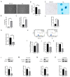
Figure 1
Figure 1
Cell shape, senescence-associated (SA)-β–gal assay, proliferation, and cell cycle. (A) Inverted microscopy image showing cell shape in normal and SA groups. (B) Comparison of cell size between normal and SA groups. (C) Elongation index for normal and SA groups is shown. (D) Comparison of cell viability between normal and SA groups. (E,F) Evaluation of senescence in normal and SA groups. The proportion of SA-β–gal stained cells was higher in the SA group. (G) Cell proliferation rate was measured by bromodeoxyuridine (BrdU) incorporation assay. (H) Cell cycle analysis was performed. (IK) The proportion of cells in the G0/G1, S and G2/M phase are shown. (L) TCF4 expression was decreased in the SA group. (M) β-catenin expression was decreased in the SA group. (N) Cyclin dependent kinase 1 (CDK1) expression was decreased in the SA group. (O) Cyclin D1 expression was not different between the normal and SA groups. All experiments were conducted in triplicate or quadruplicate. * for p < 0.05 using independent t-test.
Figure 2
Figure 2
Mitochondrial oxidative stress, elongation, and lysosome staining. (A) MitoSOX staining intensity of cells as analyzed using the Muse cell analyzer. (B) Mitochondrial oxidative stress levels were compared between the normal and SA groups. (C) Fluorescence imaging using the MitoSOX probe shows mitochondrial oxidative stress in cells. (D) MitoTracker red was used for mitochondrial imaging of cells from normal and SA groups. Mitochondrial elongation is shown in the SA group. (E) Mitochondrial elongation is greater in the SA group as compared to that in the normal group. (F,G) Lysosomal staining of cells from the normal and SA groups. (H,I) phospho- extracellular signal-regulated protein kinases 1 and 2 (pERK1/2) expression levels are shown. (J,K) SIRT1 expression levels are shown. All experiments were performed in triplicate or quadruplicate. * for p < 0.05 using independent t-test.
Figure 3
Figure 3
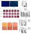
Animal study of SIRT1 activation using CRISPR/dCas9 in rat corneal endothelial cells (CECs). (A) Immunofluorescence staining of SIRT1 showing SIRT1a overexpression in SIRT1a group. (B) Real-time quantitative polymerase chain reaction (qRT-PCR) showing that relative SIRT1 mRNA expression was elevated to 246.7% of the control group. (C,D) Corneal opacity in SIRT1a group was decreased compared to control group on days 11 and 14. (EG) Alizarin red S staining showed higher density of CECs at the center in the SIRT1a group compared to the control group, as well as smaller cell size in the SIRT1a group compared to the control group. All experiments were performed in triplicate or quadruplicate. * for p < 0.05 using independent t-test.
Figure 4
Figure 4
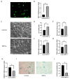
SIRT1 activation using CRISPR/dCas9 in cultured human corneal endothelial cells. (A) GFP-encoded plasmids were transfected into the cells. Green indicates GFP. (B) Relative SIRT1 mRNA expression in cultured hCECs of SIRT1a group was elevated to 247% of control. (C) Cell shape was polygonal in the SIRT1a group compared to the control. (D, E) Cell viability and proliferation rate were higher in SIRT1a group compared to control. (F, G) Relative mRNA expression levels of CCNA2 and PCNA were increased in SIRT1a group compared to control. (H) CDKN2A mRNA expression was lower in SIRT1a group compared to control. (I, J) The number of SA-β-gal positive cells were lower in SIRT1a group compared to control. All experiments were performed in triplicate or quadruplicate. * for p < 0.05 using independent t-test.
Figure 5
Figure 5
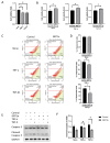
SIRT1 inhibits cytokine-induced cell death. (A) Cells were treated with tumor necrosis factor-alpha (TNF-α) and transforming growth factor-beta1 (TGF-β1). Cell viability was reduced when treated with TNF-α or TGF-β1. (B) Cell viability was increased in SIRT1a group compared to control in untreated or upon treatment with either TNF-α or TGF-β1. (C,D) Mitochondrial membrane potential was depolarized upon TNF-α or TGF-β1 treatment. The number of depolarized cells was increased in SIRT1a group compared to control upon treatment with TNF-α or TGF-β1. (E,F) Cleaved caspase 3 level was decreased in SIRT1a group compared to control upon treatment with TNF-α or TGF-β1. All experiments were performed in triplicate or quadruplicate * for p < 0.05 using independent t-test.
Figure 6
Figure 6
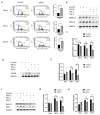
SIRT1 inhibits cytokine-induced cell cycle arrest. (A) Cell cycle analysis showed that the number of cells in S-phase was increased in SIRT1a group compared to control in untreated and upon treatment with either tumor necrosis factor-alpha (TNF-α) and transforming growth factor-beta1 (TGF-β1). (BH) phospho- extracellular signal-regulated protein kinases 1 and 2, glycogen synthase kinase 3 beta (GSK3β), Notch and HES1 levels were reduced in SIRT1a group compared to control upon treatment with TNF-α or TGF-β1. All experiments were performed in triplicate or quadruplicate. * for p < 0.05 using independent t-test.

References

    1. Treffers W.F. Human corneal endothelial wound repair. In vitro and in vivo. Ophthalmology. 1982;89:605–613. doi: 10.1016/S0161-6420(82)34757-0. - DOI - PubMed
    1. Nayak S.K., Binder P.S. The growth of endothelium from human corneal rims in tissue culture. Invest. Ophthalmol. Vis. Sci. 1984;25:1213–1216. - PubMed
    1. Joyce N.C. Proliferative capacity of the corneal endothelium. Prog. Retin. Eye Res. 2003;22:359–389. doi: 10.1016/S1350-9462(02)00065-4. - DOI - PubMed
    1. Senoo T., Joyce N.C. Cell cycle kinetics in corneal endothelium from old and young donors. Invest. Ophthalmol. Vis. Sci. 2000;41:660–667. - PubMed
    1. Ueno M., Asada K., Toda M., Hiraga A., Montoya M., Sotozono C., Kinoshita S., Hamuro J. MicroRNA Profiles Qualify Phenotypic Features of Cultured Human Corneal Endothelial Cells. Invest. Ophthalmol. Vis. Sci. 2016;57:5509–5517. doi: 10.1167/iovs.16-19804. - DOI - PubMed

LinkOut - more resources