. 2020 Nov 4;25(21):5139.
doi: 10.3390/molecules25215139.

A Photoalkylative Fluorogenic Probe of Guttiferone A for Live Cell Imaging and Proteome Labeling in Plasmodium falciparum

Affiliations

A Photoalkylative Fluorogenic Probe of Guttiferone A for Live Cell Imaging and Proteome Labeling in Plasmodium falciparum

Romain Duval et al. Molecules. .

Abstract

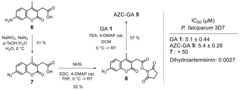
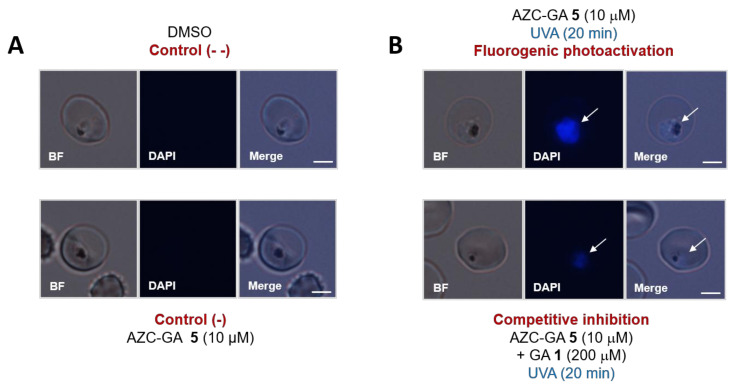
Guttiferone A (GA) 1, a polycyclic polyprenylated acylphloroglucinol (PPAP) isolated from the plant Symphonia globulifera (Clusiaceae), constitutes a novel hit in antimalarial drug discovery. PPAPs do not possess identified biochemical targets in malarial parasites up to now. Towards this aim, we designed and evaluated a natural product-derived photoactivatable probe AZC-GA 5, embedding a photoalkylative fluorogenic motif of the 7-azidocoumarin (AZC) type, devoted to studying the affinity proteins interacting with GA in Plasmodium falciparum. Probe 5 manifested a number of positive functional and biological features, such as (i) inhibitory activity in vitro against P. falciparum blood-stages that was superimposable to that of GA 1, dose-response photoalkylative fluorogenic properties (ii) in model conditions using bovine serum albumin (BSA) as an affinity protein surrogate, (iii) in live P. falciparum-infected erythrocytes, and (iv) in fresh P. falciparum cell lysate. Fluorogenic signals by photoactivated AZC-GA 5 in biological settings were markedly abolished in the presence of excess GA 1 as a competitor, indicating significant pharmacological specificity of the designed molecular probe relative to the native PPAP. These results open the way to identify the detected plasmodial proteins as putative drug targets for the natural product 1 by means of proteomic analysis.

Keywords: 7-azidocoumarin; Guttiferone A; Plasmodium falciparum; fluorogenesis; photoactivation.


Conflict of interest statement

The authors declare no conflict of interest nor competing financial interest.

Figures

Figure 1
Figure 1
Representative antimalarial PPAPs (IC50 = 0.2 to ca. 5 µM in vitro).
Figure 2
Figure 2
SARs in ester series [11] for the design of photoactivatable probe AZC-GA 5, and its principle of fluorogenic covalent capture of affinity proteins (star = protein).
Figure 3
Figure 3
Synthesis and in vitro antiplasmodial activity of AZC-GA 2.
Figure 4
Figure 4
Fluorimetric and SDS-PAGE analysis of the fluorogenic photoalkylation of BSA by AZC-GA 5. (A): 50 µM AZC-GA 5 in RIPA lysis buffer were used for the dose–response photoalkylation of BSA (0 to 8 mg/mL) at various irradiation times (left top panel). 2 mg/mL BSA in RIPA lysis buffer were used for the dose–response photoalkylation by AZC-GA 5 (0 to 100 µM) at various irradiation times (right top panel). Dose–response variation of AMC-type fluorescence was assessed using BSA (0 to 8 mg/mL, left bottom panel) and non-photoactivated 6 (0 to 100 µM (right bottom panel), taking 6 as exemplary of the AMC fluorophore present in AMC-GA-BSA photoadducts (Figure 2). Fluorimetric analysis was performed on a Xenius XML spectrofluorimeter (SAFAS, Monaco) at 23 °C using the following detection settings: λEX = 360 nm, λEM = 448 nm. Values represent the mean of three sequential measurements (interval time = 5 s). Standard deviations are systematically shown on the plots but are often invisible due to their close-to-zero values. (B): 2 mg/mL BSA in RIPA lysis buffer was used for the dose–response photoalkylation by AZC-GA 5 under UVA irradiation for 20 min. Noteworthy, BSA shows slight blue autofluorescence in the absence of AZC-GA 5 (lane 0 µM). Fluorescence revelation was performed using the UV bed of an E-Box VX2 scanner (Vilber-Lourmat, Collegien, France) and pictured as seen with the naked eye. All photoactivations were performed on ice (0–4 °C). MWM = molecular weight marker.
Figure 5
Figure 5
Photoalkylation of live 3D7 P. falciparum trophozoite blood-stages by AZC-GA 5 ± GA 1. (A): Negative controls consisted of parasites treated with dimethyle sulfoxide (DMSO) or with non-photoactivated AZC-GA 5. (B): Fluorogenic experiments consisted of parasites treated with photoactivated AZC-GA 5 in the absence or presence of excess GA 1. Images were obtained on an Olympus BX60F-3 microscope using DAPI excitation channel (λEX = 359 nm). All UVA photoactivations were performed on ice (0–4 °C) for 20 min. Fluorescence was recorded as seen with the naked eye. Images were taken using identical exposure times (132 ms) and are representative of several independent experiments (n = 2–4, see Figure S1 in the Supplementary Materials). White arrows indicate the parasites. Scale bars represent 2 µm.
Figure 6
Figure 6
Photoalkylation of asynchronous 3D7 P. falciparum cell lysate by AZC-GA 5 ± GA 1. (A): 0–1 mM range; lysate concentration was 1 mg/mL total protein. (B): 0–100 µM range; lysate concentration was 5 mg/mL total protein. Noteworthy, various plasmodial proteins show slight blue autofluorescence in the absence of AZC-GA 5 (lanes 0 µM). Fluorescence revelation was performed using the UV bed of an E-Box VX2 scanner (Vilber-Lourmat) and pictured as seen with the naked eye (A) or under limited exposure in a black chamber (B). All photoactivations were performed on ice (0–4 °C) for 20 min. MWM = molecular weight marker.

Similar articles

Cited by

References

    1. WHO . World Malaria Report 2019. World Health Organization; Geneva, Switzerland: 2019. pp. 1–232.
    1. Bhatt S., Weiss D.J., Cameron E., Bisanzio D., Mappin B., Dalrymple U., Battle K.E., Moyes C.L., Henry A., Eckhoff P.A., et al. The effect of malaria control on Plasmodium falciparum in Africa between 2000 and 2015. Nature. 2015;526:207–211. doi: 10.1038/nature15535. - DOI - PMC - PubMed
    1. Imwong M., Suwannasin K., Kunasol C., Sutawong K., Mayxay M., Rekol H., Smithuis F.M., Hlaing T.M., Tun K.M., Van Der Pluijm R.W., et al. The spread of artemisinin-resistant Plasmodium falciparum in the Greater Mekong subregion: A molecular epidemiology observational study. Lancet Infect. Dis. 2017;17:491–497. doi: 10.1016/S1473-3099(17)30048-8. - DOI - PMC - PubMed
    1. Conrad M.D., Rosenthal P.J. Antimalarial drug resistance in Africa: The calm before the storm? Lancet Infect. Dis. 2019;19:e338–e351. doi: 10.1016/S1473-3099(19)30261-0. - DOI - PubMed
    1. Tajuddeen N., Van Heerden F.R. Antiplasmodial natural products: An update. Malar. J. 2019;18:1–62. doi: 10.1186/s12936-019-3026-1. - DOI - PMC - PubMed

MeSH terms

LinkOut - more resources