. 2020 Nov:61:103104.
doi: 10.1016/j.ebiom.2020.103104. Epub 2020 Nov 3.

Persistence of viral RNA, pneumocyte syncytia and thrombosis are hallmarks of advanced COVID-19 pathology

Affiliations

Persistence of viral RNA, pneumocyte syncytia and thrombosis are hallmarks of advanced COVID-19 pathology

Rossana Bussani et al. EBioMedicine. 2020 Nov.

Abstract

Background: COVID-19 is a deadly pulmonary disease with peculiar characteristics, which include variable clinical course and thrombophilia. A thorough understanding of the pathological correlates of the disease is still missing.

Methods: Here we report the systematic analysis of 41 consecutive post-mortem samples from individuals who died of COVID-19. Histological analysis is complemented by immunohistochemistry for cellular and viral antigens and the detection of viral genomes by in situ RNA hybridization.

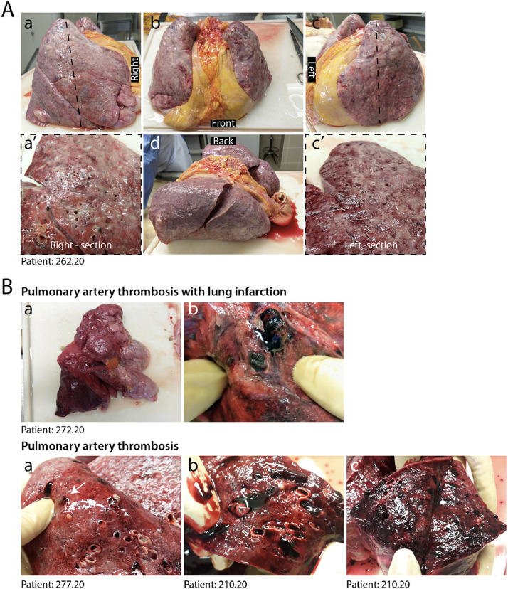
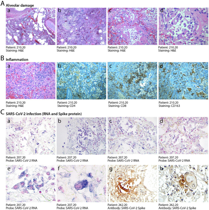
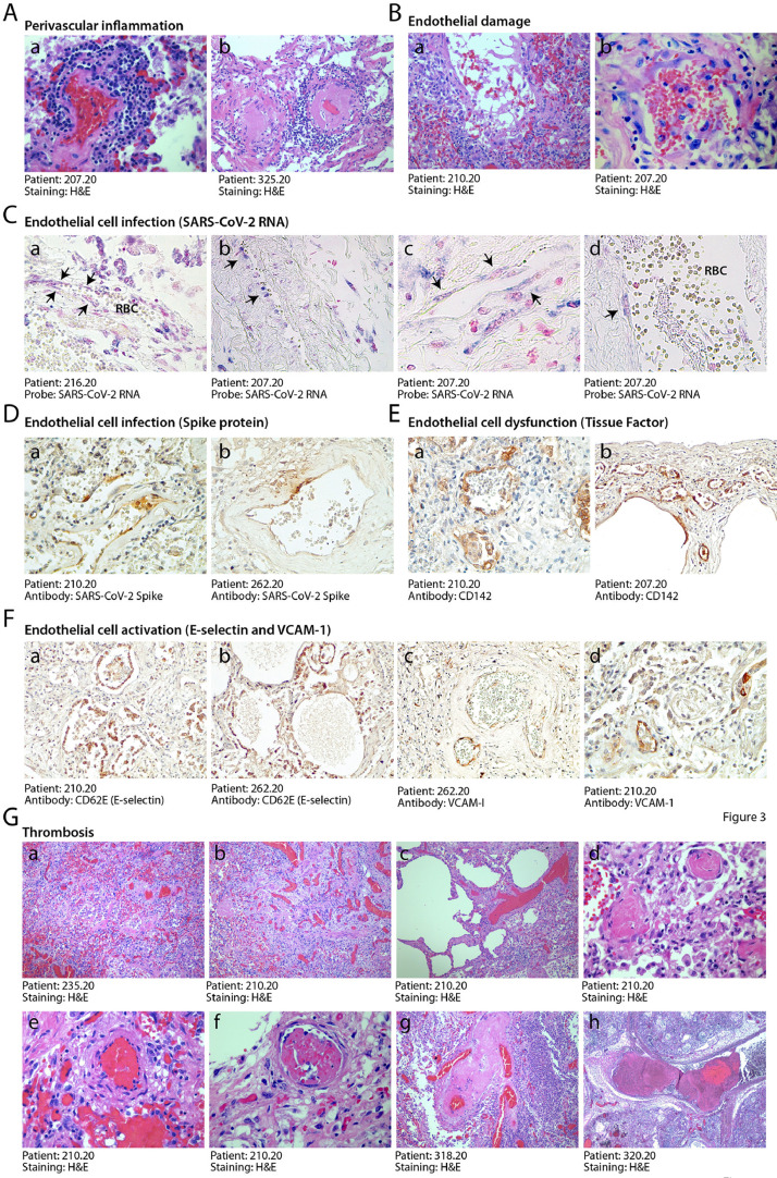
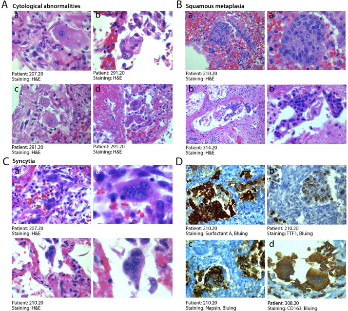
Findings: COVID-19 is characterized by extensive alveolar damage (41/41 of patients) and thrombosis of the lung micro- and macro-vasculature (29/41, 71%). Thrombi were in different stages of organization, consistent with their local origin. Pneumocytes and endothelial cells contained viral RNA even at the later stages of the disease. An additional feature was the common presence of a large number of dysmorphic pneumocytes, often forming syncytial elements (36/41, 87%). Despite occasional detection of virus-positive cells, no overt signs of viral infection were detected in other organs, which showed non-specific alterations.

Interpretation: COVID-19 is a unique disease characterized by extensive lung thrombosis, long-term persistence of viral RNA in pneumocytes and endothelial cells, along with the presence of infected cell syncytia. Several of COVID-19 features might be consequent to the persistence of virus-infected cells for the duration of the disease.

Funding: This work was supported by a King's Together Rapid COVID-19 Call grant from King's College London. MG is supported by the European Research Council (ERC) Advanced Grant 787971 "CuRE" and by Programme Grant RG/19/11/34633 from the British Heart Foundation.

Keywords: COVID-19; Endothelial dysfunction; Post-mortem analysis; SARS-CoV-2; Spike protein; Syncytia.


Conflict of interest statement

Declaration of Competing Interest The authors declare no conflict of interest.

Figures

Fig. 1
Fig. 1
Macroscopic appearance of COVID lungs. A. Gross appearance of en bloc resection of heart and lungs from a severely affected IC patient, shown in its left, anterior, posterior and right views, as indicated (a–d). Lungs appeared congested and firm. Cut sections of both right and left lungs revealed tan-grey solid parenchyma with numerous hemorrhagic areas and loss of air spaces (a’, c’). B. A large thrombus in the inferior branch of the right pulmonary artery determined a large infarct in the right lung. Most lungs presented multiple thrombi, with loss of alveolar spaces and extensive hemorrhages (c–e).
Fig. 2
Fig. 2
Histopathological evidence of alveolar damage, inflammation and SARS-CoV-2 infection in COVID-19 lungs. A. Severe and diffuse alveolar damage. Low magnification images (a, b, x2.5) show diffuse end-stage alveolar damage, with patchy inflammatory infiltrates. Higher magnification images better demonstrate massive disruption of alveolar structures and hyaline membranes (c, x20) and detachment of epithelial cells from the alveolar wall (d, x40). B. Moderate inflammatory infiltration. In most patients, lungs were sparsely infiltrated by clusters of inflammatory cells (a, 2.5x), largely composed of CD4+ helper and CD8+ suppressor lymphocytes (b, and c respectively, 2.5x), as well as of CD163+ histiocytes (d, 2.5x). C. Detection of SARS-CoV-2 RNA and Spike protein. In situ hybridization for SARS-CoV-2 RNA revealed extensive positivity in the cytoplasm of several alveolar cells (shown for one representative patient in a-d, x10; e, x20, f, x 40). Cuboidal pneumocytes (g, 20x) and several atypical cells (h, 40x, showing a bi-nucleated cell with a large inclusion body) were also positive for the expression of the Spike protein. Nuclei are stained with nuclear fast red.
Fig. 3
Fig. 3
Endothelial dysfunction and extensive thrombosis in COVID-19 lungs. A. Perivascular inflammation. Numerous patients show clusters of inflammatory cells around small and medium size vessels. In all cases, perivascular lymphocytic cuffs were particularly evident around small arterioles, which had thick walls, often undergoing fibrinoid necrosis (a,b). Other images are in Suppl. Fig. 2A. B. Endothelial damage. Medium and small vessels showed multiple vascular abnormalities, with massive liquefactive degeneration of endothelial cells (a) and their detachment from the vascular wall (b). C. Infection of endothelial cells by SARS-CoV-2. In situ hybridization showing SARS-CoV-2 signal in elongated cells along the luminal side of the vessel wall (arrows), compatible with their endothelial origin (a,b,x10; c, d, x40). RBC: red blood cells. Nuclei are stained with nuclear fast red. D. Expression of the viral Spike protein by endothelial cells. Cells lining blood vessels (shown for a small (a, 10x) and medium (b, 10x) size artery positive for Spike protein. E. Expression of Tissue Factor (CD142). This factor was expressed by the endothelium of several blood vessels of different caliber (a, b, 10x). F. Expression of endothelial cell activation markers. E-selectin (a, b, 10x) and VCAM-1 (c, d, 10x) were expressed in the endothelium of several lung vessels of different caliber. G. Multiple thrombi in different stages of organization. Histological analysis revealed the presence of diffuse micro-thrombi (a, b, x2.5), which sometimes appeared rich in fibrin and poorly organized, consistent with their recent formation (c, x10), but in most instances were largely organized (d, x20). High magnification images confirmed the co-existence of multiple thrombi in different stages of organization in the same lung (a recent thrombus and an organizing thrombus coexist in the lung of the same IC patient, shown in panels e and f, respectively, x20), as well as thrombotic lesions that have likely evolved structurally in multiple times, being composed of an older and a younger component (g, x10; h, x2.5).
Fig. 4
Fig. 4
Abnormal pneumocytes and cellular syncytia in COVID-19 lungs. A. Cytological features of SARS-CoV-2-infected lungs. A consistent and typical feature in COVID-19 lungs was the presence of major cytological abnormalities, including very large cells with dysmorphic phenotypes (a, x63; b, x40) often showing bi-o multinucleation (c,d, x40). B. Squamous metaplasia (pseudosyncytia). A common finding was the metaplasia of the alveolar epithelium, with a marked change in morphology of pneumocytes and their aggregation to form pseudosyncytia (a, b, x20; a’, b’, x63). C. Syncytia. In addition to squamous metaplasia, true syncytial elements were observed in numerous lungs, showing large cytoplasm and nuclear aggregation (a, b, x20; a’, b’, x63). D. Origin of syncytial elements. The giant and multinucleated cells forming either pseudo-syncytia or real syncytia scored positive for the pneumocyte markers Surfactant-A (a, x20), TTF1 (b, x20) and Napsin (c, x20), indicative of their epithelial origin. The COVID-19 lungs also showed the more occasional presence of CD163-positive syncytia of histiocytic origin (d, x20). In A-C, H&E: hematoxylin and eosin.

References

    1. Zhu N, Zhang D, Wang W. A Novel Coronavirus from Patients with Pneumonia in China, 2019. N Engl J Med. 2020;382(8):727–733. - PMC - PubMed
    1. Tang N, Bai H, Chen X, Gong J, Li D, Sun Z. Anticoagulant treatment is associated with decreased mortality in severe coronavirus disease 2019 patients with coagulopathy. J Thromb Haemost. 2020;18(5):1094–1099. - PMC - PubMed
    1. Buja LM, Wolf DA, Zhao B. The emerging spectrum of cardiopulmonary pathology of the coronavirus disease 2019 (COVID-19): report of 3 autopsies from Houston, Texas, and review of autopsy findings from other United States cities. Cardiovasc Pathol. 2020;48 - PMC - PubMed
    1. Barton LM, Duval EJ, Stroberg E, Ghosh S, Mukhopadhyay S. COVID-19 Autopsies, Oklahoma, USA. Am J Clin Pathol. 2020;153(6):725–733. - PMC - PubMed
    1. Fox SE, Akmatbekov A, Harbert JL, Li G, Quincy Brown J, Vander Heide RS. Pulmonary and cardiac pathology in African American patients with COVID-19: an autopsy series from New Orleans. Lancet Respir Med. 2020 - PMC - PubMed

MeSH terms