. 2021 Jan 13;95(3):e01969-20.
doi: 10.1128/JVI.01969-20. Print 2021 Jan 13.

The SARS-CoV-2 Conserved Macrodomain Is a Mono-ADP-Ribosylhydrolase

Affiliations

The SARS-CoV-2 Conserved Macrodomain Is a Mono-ADP-Ribosylhydrolase

Yousef M O Alhammad et al. J Virol. .

Abstract

Severe acute respiratory syndrome coronavirus 2 (SARS-CoV-2) and other SARS-related CoVs encode 3 tandem macrodomains within nonstructural protein 3 (nsp3). The first macrodomain, Mac1, is conserved throughout CoVs and binds to and hydrolyzes mono-ADP-ribose (MAR) from target proteins. Mac1 likely counters host-mediated antiviral ADP-ribosylation, a posttranslational modification that is part of the host response to viral infections. Mac1 is essential for pathogenesis in multiple animal models of CoV infection, implicating it as a virulence factor and potential therapeutic target. Here, we report the crystal structure of SARS-CoV-2 Mac1 in complex with ADP-ribose. SARS-CoV-2, SARS-CoV, and Middle East respiratory syndrome coronavirus (MERS-CoV) Mac1 domains exhibit similar structural folds, and all 3 proteins bound to ADP-ribose with affinities in the low micromolar range. Importantly, using ADP-ribose-detecting binding reagents in both a gel-based assay and novel enzyme-linked immunosorbent assays (ELISAs), we demonstrated de-MARylating activity for all 3 CoV Mac1 proteins, with the SARS-CoV-2 Mac1 protein leading to a more rapid loss of substrate than the others. In addition, none of these enzymes could hydrolyze poly-ADP-ribose. We conclude that the SARS-CoV-2 and other CoV Mac1 proteins are MAR-hydrolases with similar functions, indicating that compounds targeting CoV Mac1 proteins may have broad anti-CoV activity.IMPORTANCE SARS-CoV-2 has recently emerged into the human population and has led to a worldwide pandemic of COVID-19 that has caused more than 1.2 million deaths worldwide. With no currently approved treatments, novel therapeutic strategies are desperately needed. All coronaviruses encode a highly conserved macrodomain (Mac1) that binds to and removes ADP-ribose adducts from proteins in a dynamic posttranslational process that is increasingly being recognized as an important factor that regulates viral infection. The macrodomain is essential for CoV pathogenesis and may be a novel therapeutic target. Thus, understanding its biochemistry and enzyme activity are critical first steps for these efforts. Here, we report the crystal structure of SARS-CoV-2 Mac1 in complex with ADP-ribose and describe its ADP-ribose binding and hydrolysis activities in direct comparison to those of SARS-CoV and MERS-CoV Mac1 proteins. These results are an important first step for the design and testing of potential therapies targeting this unique protein domain.

Keywords: ADP-ribose; SARS-CoV-2; coronavirus; macrodomain; mono-ADP-ribose; poly-ADP-ribose.


Figures

FIG 1
FIG 1
The SARS-CoV-2 Mac1 is a small domain within nsp3 and is highly conserved between other human CoV Mac1 protein domains. (A) Cartoon schematic of the SARS-CoV-2 nonstructural protein 3. The conserved macrodomain (Mac1) is in yellow. (B) Sequence alignment of Mac1 from CoVs; SARS-CoV-2, SARS-CoV, MERS-CoV, and mouse hepatitis virus (MHV), from the alphaviruses Venezuelan equine encephalitis virus (VEEV) and Sindbis virus (SINV), and from hepatitis E virus (HEV). Sequences were aligned using the ClustalW method from Clustal Omega online tool with manual adjustment. Identical residues are boldface, shaded in gray, and marked with asterisks; semiconserved residues are shaded in gray and marked with two dots (one change among all viruses) or one dot (two changes or conserved within the CoV family).
FIG 2
FIG 2
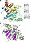
Structure of SARS-CoV-2 Mac1 complexed with ADP-ribose. (A) The structure was rendered as ribbons and colored using the visible spectrum from the N terminus (blue) to the C terminus (red). (B) The structure was colored by secondary structure showing sheets (magenta) and helices (green). The ADP-ribose is rendered as gray cylinders, with oxygens and nitrogens colored red and blue, respectively.
FIG 3
FIG 3
Extended residues at the C terminus of the SARS-CoV-2 Mac1 clash with symmetry-related molecules. (A) Comparison of the amino acid sequences of SARS-CoV-2 Mac1, 6W02, and 6WEY. (B) Superposition of SARS-CoV-2 Mac1 (magenta) subunit B onto subunit A of 6W02 reveals that the C terminus would clash with symmetry-related molecules (coral).
FIG 4
FIG 4
Binding mode of ADP-ribose in SARS-CoV-2 Mac1. (A) Fo-Fc Polder omit map (green mesh) contoured at 3σ. (B) Hydrogen bond interactions (dashed lines) between ADP-ribose and amino acids. (C) Interactions with water molecules. Direct hydrogen bond interactions are represented by dashed lines, and water-mediated contacts to amino acids are represented by solid lines.
FIG 5
FIG 5
Comparison of the SARS-CoV-2 Mac1 protein with homologous structures. (A and B) Superposition of SARS-CoV-2 Mac1 (magenta) with other recently determined homologous structures. (A) SARS-CoV-2 Mac1 apo structure (6WEN); (B) SARS-CoV-2 Mac1 complexed with ADP-ribose (6W02). The ADP-ribose molecule is in gray for SARS-CoV-2 and is represented as green cylinders for 6W02 in panel B. (C and D) Comparison of the residues in the ADP-ribose binding site. (C) SARS-CoV-2 Mac1 apo structure (blue; 6WEN); (D) SARS-CoV-2 Mac1 complexed with ADP-ribose (green; 6W02). The ADP-ribose of SARS-CoV-2 is rendered as gray cylinders and is represented as green cylinders for 6W02 in panel B.
FIG 6
FIG 6
Structural comparison of the SARS-CoV-2 Mac1 protein with the SARS-CoV and MERS-CoV Mac1 proteins. (A and B) Superposition of SARS-CoV-2 macrodomain (magenta) with coronavirus macrodomain structures. (A) SARS-CoV Mac1 with ADP-ribose (gold) (2FAV); (B) MERS-CoV Mac1 with ADP-ribose (teal) (5HOL). (C and D) Superposition of SARS-CoV-2 Mac1 (magenta) with other coronavirus Mac1 structures highlighting the ADP-ribose binding site. (C) SARS-CoV (gold); (D) MERS-CoV (teal). The ADP-ribose molecules are colored gray for SARS-CoV-2 Mac1 (A to D) and are rendered as green cylinders for SARS-CoV Mac1 (A and C) and MERS-CoV Mac1 (B and D).
FIG 7
FIG 7
Human CoVs bind to ADP-ribose with similar affinity. (A and B) ADP-ribose binding of human Mdo2 and SARS-CoV, MERS-CoV, and SARS-CoV-2 Mac1 proteins by ITC. Images in panel A are from one experiment and are representative of at least 2 independent experiments. Data in panel B are combined averages from multiple independent experiments for each protein. Mdo2, n = 2; SARS-CoV, n = 5; MERS-CoV, n = 6; SARS-CoV-2, n = 2. (C) The macrodomain proteins (10 μM) were incubated with increasing concentrations of ADP-ribose and measured by DSF as described in Materials and Methods. Mdo2, n = 4; SARS-CoV, n = 6; MERS-CoV, n = 5; SARS-CoV-2, n = 3.
FIG 8
FIG 8
The SARS-CoV-2 Mac1 protein is a mono-ADP-ribosylhydrolase. (A) Affinity of ADP-ribose binding antibodies for ADP-ribosylated PARP10 CD. MARylated PARP10 and non-MARylated PARP10 CD were detected by immunoblotting (IB) with anti-GST (MA4-004; Invitrogen), anti-ADP-ribose binding reagents, and anti-MAR (MAB1076; MilliporeSigma), anti-PAR (MABC547; MilliporeSigma), and anti-MAR/PAR (MABE1075; MilliporeSigma) antibodies. (B) The SARS-CoV-2 macrodomain was incubated with MARylated PARP10 CD in vitro at equimolar ratios (1 μM) for the indicated times at 37°C. ADP-ribosylated PARP10 CD was detected by IB with anti-ADP-ribose binding reagent (MAB1076; MilliporeSigma). Total PARP10 CD and macrodomain protein levels were determined by Coomassie blue (CB) staining. The reaction with PARP10 CD incubated alone at 37°C was stopped at 0 or 60 min. (C) The level of de-MARylation was measured by quantifying band intensity using ImageJ software. Intensity values were plotted and fitsted to a nonlinear regression curve; error bars represent standard deviations. Results in panel A are from representative experiments of two independent experiments, and data in panel B are the combined results of the two independent experiments.
FIG 9
FIG 9
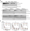
Comparison of the human Mdo2 and CoV Mac1 deMARylating activity. (A) The Mdo2, MERS-CoV, SARS-CoV, and SARS-CoV-2 macrodomains were incubated with MARylated PARP10 CD in vitro at [substrate]:[Mac1] ratios of 1:1 (1 μM), 5:1 (500 nM, 100 nM), and 10:1 (1 μM, 100 nM) for the indicated times at 37°C. ADP-ribosylated PARP10 CD was detected as described above, and total PARP10 CD and macrodomain protein levels were determined by Coomassie blue. (B) Time-dependent substrate concentrations were determined by quantifying band intensity using Image Studio software. The data were then analyzed using Mathematica 12, as described in Materials and Methods, to determine the initial rate (k) of substrate decay. Results in panel A are from representative experiments of three independent experiments, and data in panel B are the combined results of the three independent experiments.
FIG 10
FIG 10
Coronavirus Mac1 proteins do not hydrolyze poly-ADP-ribose (PAR). (A) Differential PARylation of PARP1 by various concentrations of NAD+. Recombinant human PARP1 was automodified in a reaction buffer supplemented with increasing concentrations of NAD+ to generate substrates for the PAR hydrolase assays. PAR was detected by immunoblot analysis of reaction products with the anti-PAR antibody 96-10. (B) PAR hydrolase assays were performed with PARP1 that was either extensively poly-ADP-ribosylated (500 μM NAD+) or partially poly-ADP-ribosylated (5 μM NAD+) to produce oligo-ADP-ribose. Macrodomains were incubated with both automodified PARP1 substrates for 1 h. PAR was detected by immunoblotting with the anti-PAR antibody 96-10. PARG (catalytically active 60-kDa fragment) was used as a positive control. The results are representative of 2 independent experiments.
FIG 11
FIG 11
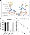
Development of an ELISA to detect de-MARylation. (A) Cartoon schematic of the ELISA. ELISA plates precoated with glutathione and preblocked were used to capture GST-tagged PARP10 proteins, which was used as a substrate for de-MARylation. The removal of MAR was detected by anti-MAR antibodies. (B) MARylated PARP10 (MAR+) and non-MARylated PARP10 (MAR-) with no SARS-CoV-2 Mac1 as controls were detected with anti-mono-ADP-ribose binding reagent (α-MAR) (MAB1076; MilliporeSigma) or with anti-GST (α-GST) (MA4-004; Invitrogen). (C) Starting at 12.5 nM, 2-fold serial dilutions of the SARS-CoV-2 Mac1 protein were incubated in individual wells with MARylated PARP10 CD for 60 min at 37°C. The data are the combined results of 2 independent experiments.

Update of

References

    1. Zhou P, Yang XL, Wang XG, Hu B, Zhang L, Zhang W, Si HR, Zhu Y, Li B, Huang CL, Chen HD, Chen J, Luo Y, Guo H, Jiang RD, Liu MQ, Chen Y, Shen XR, Wang X, Zheng XS, Zhao K, Chen QJ, Deng F, Liu LL, Yan B, Zhan FX, Wang YY, Xiao GF, Shi ZL. 2020. A pneumonia outbreak associated with a new coronavirus of probable bat origin. Nature 579:270–273. doi:10.1038/s41586-020-2012-7. - DOI - PMC - PubMed
    1. Coronaviridae Study Group of the International Committee on Taxonomy of Viruses. 2020. The species Severe acute respiratory syndrome-related coronavirus: classifying 2019-nCoV and naming it SARS-CoV-2. Nat Microbiol 5:536–544. doi:10.1038/s41564-020-0695-z. - DOI - PMC - PubMed
    1. Srinivasan S, Cui H, Gao Z, Liu M, Lu S, Mkandawire W, Narykov O, Sun M, Korkin D. 2020. Structural genomics of SARS-CoV-2 indicates evolutionary conserved functional regions of viral proteins. Viruses 12:360. doi:10.3390/v12040360. - DOI - PMC - PubMed
    1. Wu C, Liu Y, Yang Y, Zhang P, Zhong W, Wang Y, Wang Q, Xu Y, Li M, Li X, Zheng M, Chen L, Li H. 2020. Analysis of therapeutic targets for SARS-CoV-2 and discovery of potential drugs by computational methods. Acta Pharm Sin B 10:766–788. doi:10.1016/j.apsb.2020.02.008. - DOI - PMC - PubMed
    1. Fehr AR, Perlman S. 2015. Coronaviruses: an overview of their replication and pathogenesis, Methods Mol Biol 1282:1–23. doi:10.1007/978-1-4939-2438-7_1. - DOI - PMC - PubMed

Publication types

MeSH terms

Substances

LinkOut - more resources