. 2021 Jan-Jun:296:100039.
doi: 10.1074/jbc.RA120.015883. Epub 2020 Nov 23.

The nucleocytosolic O-fucosyltransferase SPINDLY affects protein expression and virulence in Toxoplasma gondii

Affiliations

The nucleocytosolic O-fucosyltransferase SPINDLY affects protein expression and virulence in Toxoplasma gondii

Giulia Bandini et al. J Biol Chem. 2021 Jan-Jun.

Abstract

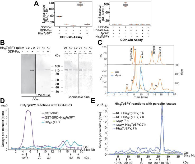
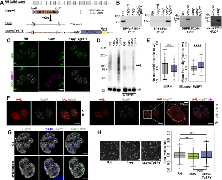
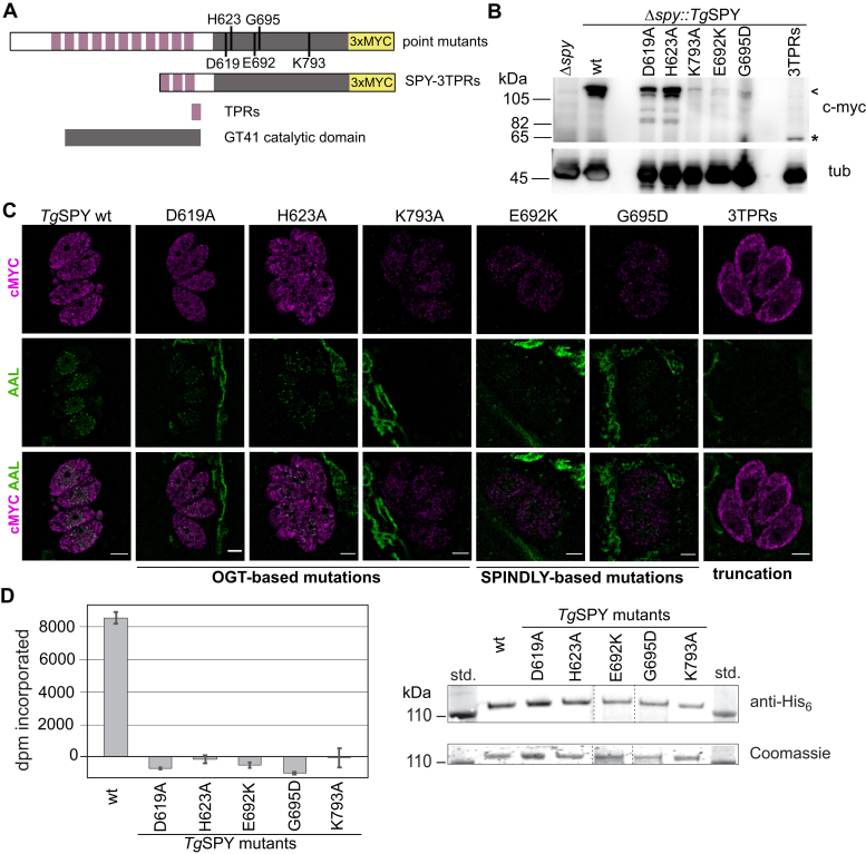
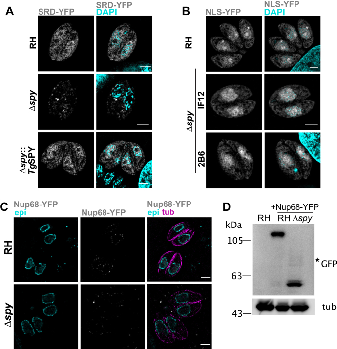
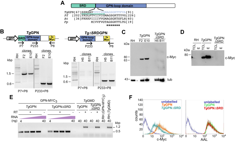
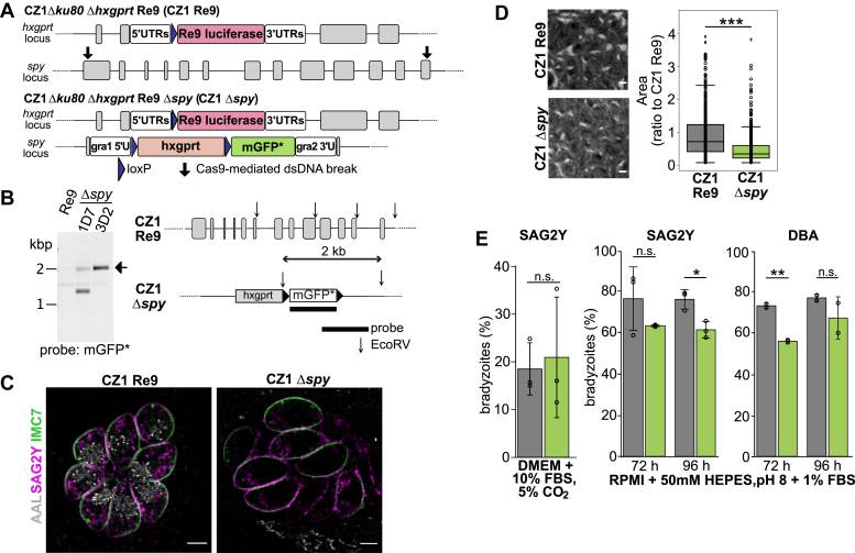
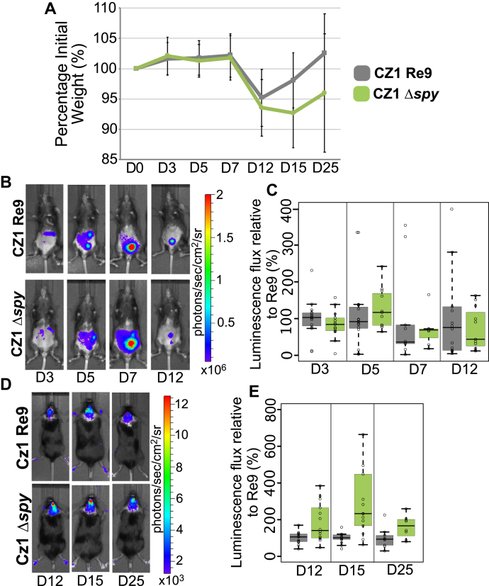
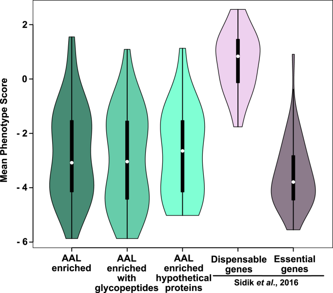
Once considered unusual, nucleocytoplasmic glycosylation is now recognized as a conserved feature of eukaryotes. While in animals, O-GlcNAc transferase (OGT) modifies thousands of intracellular proteins, the human pathogen Toxoplasma gondii transfers a different sugar, fucose, to proteins involved in transcription, mRNA processing, and signaling. Knockout experiments showed that TgSPY, an ortholog of plant SPINDLY and paralog of host OGT, is required for nuclear O-fucosylation. Here we verify that TgSPY is the nucleocytoplasmic O-fucosyltransferase (OFT) by 1) complementation with TgSPY-MYC3, 2) its functional dependence on amino acids critical for OGT activity, and 3) its ability to O-fucosylate itself and a model substrate and to specifically hydrolyze GDP-Fuc. While many of the endogenous proteins modified by O-Fuc are important for tachyzoite fitness, O-fucosylation by TgSPY is not essential. Growth of Δspy tachyzoites in fibroblasts is modestly affected, despite marked reductions in the levels of ectopically expressed proteins normally modified with O-fucose. Intact TgSPY-MYC3 localizes to the nucleus and cytoplasm, whereas catalytic mutants often displayed reduced abundance. Δspy tachyzoites of a luciferase-expressing type II strain exhibited infection kinetics in mice similar to wild-type but increased persistence in the chronic brain phase, potentially due to an imbalance of regulatory protein levels. The modest changes in parasite fitness in vitro and in mice, despite profound effects on reporter protein accumulation, and the characteristic punctate localization of O-fucosylated proteins suggest that TgSPY controls the levels of proteins to be held in reserve for response to novel stresses.

Keywords: Apicomplexa; Toxoplasma gondii; fucosyltransferase; glycosylation; nucleus; posttranslational modification; protein stability; structured illumination microscopy.


Conflict of interest statement

Conflict of interest The authors declare that they have no conflicts of interest with the contents of this article.

Figures

Figure 1
Figure 1
Recombinant His6TgSPY can hydrolyze GDP-Fuc and is active against protein substrates including itself. A, sugar nucleotide hydrolysis. Acceptor-independent consumption of the indicated sugar nucleotide donor substrates was assayed based on quantitation of GDP or UDP reaction products using GDP-Glo (right panel) or UDP-Glo (left panel) assays. Reactions were conducted in the presence or absence of either His6TgSPY or TgGat1, an α-galactose transferase that serves as a positive control for UDP-Gal hydrolysis. The high level of GDP in the absence of enzyme is due to the intrinsic instability of GDP-Fuc. Box plots: the box defines the interquartile range with the black line marking the median, while the whiskers mark maximum and minimum values. Data shown are the average of three technical replicates ±standard deviation (SD), of a representative trial. B, the same preparation of highly purified recombinant His6TgSPY was incubated in the presence or absence of 2 μM GDP-Fuc. The indicated volumes were subjected to SDS-PAGE and western blotted with biotinylated-AAL to detect fucosylation (left panel). A parallel blot was incubated with biotinylated-AAL in the presence of α-methyl fucopyranoside (Me-αFuc) as a competitive inhibitor (middle panel). The blotted gel was stained with Coomassie blue to confirm equal loading and indicated the purity of the His6TgSPY preparation (right panel). C, a parallel His6TgSPY reaction that was autofucosylated in the presence of 2 μM GDP-[3H]Fuc was subjected to SDS-PAGE and electroblotting onto a PVDF membrane. The SPY band was subjected to conditions of reductive β-elimination and analyzed by HPAEC on a CarboPac MA-1 column. Top: separation of a panel of sugar alcohols detected electrochemically. Bottom: separation of the reaction product spiked with the sugar alcohol sample, in a trial in which fractions were collected for analysis of radioactivity. The asterisk (∗) denotes peaks of unknown origin. D, purified His6TgSPY was incubated in the presence of 2 μM GDP-[3H]Fuc and recombinantly prepared and purified GST-polySer as a potential acceptor substrate for 7 h. The controls omitted one or the other protein fraction. The reaction product was separated on an SDS-PAGE gel, which, after Coomassie blue staining and fixation, was cut into equal sized slices and counted in a scintillation counter. E, alternatively, the reaction was conducted in the presence of a desalted cytosolic extract of RH (parental) or Δspy parasites for 0 or 7 h. Similar results were obtained in a complete set of independent reactions (not shown).
Figure 2
Figure 2
Disruption of T. gondii spy has a modest effect on parasite growth in vitro that is rescued by complementation.A, schematic representation of the spy locus in wild-type RH, the spy knockout, and the cell line complemented with TgSPY-MYC3. B, PCR analysis verifying the absence of endogenous spy (SPYe) in the knockout and complemented strains. Additionally, the DHFR cassette has been removed from the spy locus in the knockout strain used here. Codon reassigned SPY (SPYc) was used to complement the knockout strains and can be selectively amplified by PCR. Primers used for diagnostic PCRs are indicated below each gel (P###) and the sequences can be found in Table S1. C, SIM shows that nuclear staining by AAL is lost in the knockout and rescued by complementation with the endogenous enzyme. D, the loss and rescue of O-Fuc are confirmed by lectin blotting of whole cells. E, box plot comparing the number of AAL-positive punctae (RH n = 86, Δspy::TgSPY n = 103, three biological repeats) and the mean intensity for the AAL signal from 3D projections (RH n = 111, Δspy::TgSPY n = 106, three biological repeats) in wild-type and complemented strains. The increase in total signal for the complemented cell line was significant (p = 2.6 × 10⁻¹¹). F, SIM shows colabeling of O-Fuc with AAL, tubulin with antitubulin, and Nup67-YFP with anti-GFP in RH parasites. The top row shows a maximum intensity projection (MIP), while the lower row shows a single plane. Nup67-YFP: an FG-Nup that has not been found to be O-fucosylated; epichr: epichromatin; tub: tubulin; scale bars: 2 μm, unless indicated otherwise. G, SIM shows that TgSPY-MYC3 localizes to the cytosol, nucleoplasm, and residual body (white arrow), but not the nucleolus (asterisks). Labeling of the parental RH strain was essentially negative at this level of exposure (data not shown). Scale bars: 0.2 cm. H, plaque assays comparing wild-type, knockout, and complemented cell lines show that complementation with the endogenous enzyme rescues the mild growth phenotype observed in the knockout. Data are from three biological repeats. p values: ∗ 0.01; ∗∗∗ 2.9 × 10⁻⁵.
Figure 3
Figure 3
Mutagenesis analysis shows that AAL staining is dependent on the catalytic activity of TgSPY and the full TPRs domain. A, schematic representation of the TgSPY constructs used in the mutagenesis studies. N-terminal TPRs are shown in purple and the CAZy GT41 catalytic domain in gray. The targeted residues are indicated. B, western blot showing apparent Mr and expression levels of the mutants in the clones selected for further analysis. The position of full-length TgSPY proteins is indicated by an arrowhead and that of the truncated version with an asterisk. Tubulin served as a loading control. C, SIM showed localization and AAL staining for all point mutants and 3TPRs truncation. Based on AAL, no activity was detected for either K793A or the truncated construct, while all other mutants showed a reduced but detectable AAL pattern. Most mutant TgSPY proteins appear to be less abundant than wild-type TgSPY. D, the mutant isoforms were expressed as N-terminally His6-tagged proteins in E. coli and partially purified on Talon-Co++ columns. Equivalent amounts of protein were assayed for transfer of 3H from GDP-[3H]Fuc to a Ser-rich 25-mer peptide from TgRINGF1. SDS-PAGE followed by Coomassie blue staining (lower right) shows the calibration for the protein amounts in each reaction and was confirmed by Western blotting using anti-His6 (upper right). Data are from a single representative trial conducted in triplicate, and error bars represent ±SD.
Figure 4
Figure 4
O-fucosylated reporter proteins’ stability is affected by lack of O-Fuc.A, RH, Δspy and Δspy::TgSPY parasites were electroporated with SRD-YFP, and expression was examined 24 h later by SIM. SRD-YFP was markedly reduced in Δspy versus parental strain or complemented parasites (Δspy::TgSPY). B, a control chimera, NLS-YFP, that is not O-fucosylated in wild-type, was stably expressed in RH, and two Δspy clones showed no decrease in NLS-YFP expression versus parental strain. C, SIM shows Nup68-YFP, which was stably expressed under a tubulin promoter, was decreased in Δspy versus parental. Colocalization was performed using antibodies against epichromatin (epi) and tubulin (tub). D, Nup68-YFP was detected at a lower molecular weight compared with wild-type based on western blotting with anti-GFP, suggesting it is degraded.
Figure 5
Figure 5
O-fucosylated reporter proteins’ stability is affected by lack of SRD.A, schematic representation of the domains of TgGPN. Residues 1–79 were defined as SRD and TgGPNΔSRD starts at aa 80. The residues marked with asterisks are part of the active site of GPNs (59, 60). B, genotyping by PCR of the clones expressing either full-length or truncated GPN. All primers are listed in Table S1. Pf: Plasmodium falciparum, Pp: Pyrococcus abyssi. C, C-terminally MYC3-tagged TgGPN and TgGPNΔSRD were ectopically expressed in RH, and two pairs of clonal populations were analyzed by western blot with anti-cMYC. The mobility of full-length TgGPN-MYC3 suggested an apparent Mr higher than theoretical (46,600), possibly due to the O-fucosylation. No clear band was observed for TgGPNΔSRD. D, when AAL-enriched proteins were probed with anti-cMYC, full-length TgGPN was detected in both total cell lysate (TCL) and enriched fraction (E), whereas TgGPNΔSRD was not detected in either fraction. E, semiquantitative RT-PCR indicated comparable mRNA levels for full-length and truncated isoforms. T. gondii GDP-mannose 4,6-dehydratase (TgGMD) served as a positive control. F, flow cytometry showed a decrease in mAb 9E10 binding to TgGPNΔSRD-MYC3 compared with full-length TgGPN. Scale bars: 2 μm. Epi: epichromatin; tub: tubulin.
Figure 6
Figure 6
Generation and of spy knockout in Toxoplasma type II strain CZ1 in vitro analysis.A, schematic representation of the strategy used to knockout TgSPY in the type II strain CZ1. The parental strain was transformed with plasmid expressing Cas9 and gRNAs directed to first and last SPY exon as detailed in the methods. Additionally, a linear fragment containing homologous sequences to the dsDNA breaks regions was used to replace spy with an hpt resistance cassette, allowing for selection of the mutant parasites. B, southern blot analysis was used to verify single integration of the mGFP cassette in the spy locus of CZ1 Δku80 Δhpt Re9. Of the two clones (ID7 and 3D2) analyzed, 1D7 showed an additional GFP positive band (∼1.4 kbp) together with the expected one (2 kbp) suggesting that the recombination cassette was integrated in a second locus and not only the targeted one. Consequently, 3D2 was chosen for characterization experiments. C, the CZ1 Re9 (parental strain) and the derivative CZ1 Δspy strains were immunostained with SAG2Y, which confirmed their differentiation to bradyzoites. AAL indicates O-Fuc status, while IMC7 outlines the cells owing to its localization to the inner membrane complex near the plasma membrane. Scale bar: 5 μm. D, clonal plaque assays indicated a stronger growth defect in Δspy in type II CZ1 than in type I RH (∗∗∗ p value = 2 × 10⁻¹⁶) (Fig. 2F). Three biological repeats. Scale bar: 0.2 cm. E, spontaneous differentiation (left) from tachyzoites to bradyzoites in DMEM +10% FBS was not affected by Δspy as determined by SAG2Y labeling. In contrast, differentiation into bradyzoites by incubation in alkaline induction media (pH 8 and 1%FBS) lowered the percentage of bradyzoites at 72 h (measured by DBA labeling) or 96 h (based on SAG2Y antibody) (∗ p < 0.01; ∗∗ p < 0.005). Quantification performed on 100 vacuoles/cell line/experiment, average ±S.D. shown, the circles represent each replicate value; SAG2Y, three biological replicates, DBA two biological replicates).
Figure 7
Figure 7
Infection with type II strain spy-deficient parasites results in higher luminescence in vivo.A, parental (CZ1 Re9) and Δspy strains were injected with 104 tachyzoites (sublethal infection) and weight loss and recovery were followed during the acute phase and establishment of the chronic one. The graph shows the average weight with error bars representing ±S.D. B, luminescence in the abdominal cavity (acute phase) was measured at days 3, 5, 7, and 12. Representative examples are shown C, boxplot shows that luminescence flux was comparable between parental and knockout strain. D, representative examples of luminescence measured during brain infection on days 12, 15, and 25. E, increased parasite-dependent luminescence was observed in the brains of the Δspy compared with parental strain. Panels A–E show the averages of three biological repeats, with five mice per repeat.
Figure 8
Figure 8
Phenotype scores indicate O-fucosylated proteins are important for tachyzoites fitness. Violin plots comparing the mean phenotype score distributions of the complete set of AAL enriched proteins (n = 69), the subset for which glycopeptides were identified (n = 33) and the subset composed of hypothetical conserved proteins (n = 29) with known dispensable and essential genes for tachyzoites survival in culture (text ref. 41). Violin plots were used to better represent the distribution of phenotype scores in the various groups and were generated using R Studio. The white dot represents the median and the thick black line the interquartile range.

References

    1. Torrey E.F., Yolken R.H. Toxoplasma oocysts as a public health problem. Trends Parasitol. 2013;29:380–384. - PubMed
    1. Lykins J., Li X., Levigne P., Zhou Y., El Bissati K., Clouser F., Wallon M., Morel F., Leahy K., El Mansouri B., Siddiqui M., Leong N., Michalowski M., Irwin E., Goodall P. Rapid, inexpensive, fingerstick, whole-blood, sensitive, specific, point-of-care test for anti-Toxoplasma antibodies. PLoS Negl. Trop. Dis. 2018;12:e0006536. - PMC - PubMed
    1. Torgerson P.R., Mastroiacovo P. The global burden of congenital toxoplasmosis: a systematic review. Bull. World Health Organ. 2013;91:501–508. - PMC - PubMed
    1. Guo M., Dubey J.P., Hill D., Buchanan R.L., Gamble H.R., Jones J.L., Pradhan A.K. Prevalence and risk factors for Toxoplasma gondii infection in meat animals and meat products destined for human consumption. J. Food Prot. 2015;78:457–476. - PubMed
    1. Pleyer U., Schlüter D., Mänz M. Ocular toxoplasmosis: recent aspects of pathophysiology and clinical implications. Ophthalmic Res. 2014;52:116–123. - PubMed

Publication types

LinkOut - more resources