. 2021 Jan-Jun:296:100042.
doi: 10.1074/jbc.RA120.015306. Epub 2020 Nov 22.

Structures and mechanism of human glycosyltransferase β1,3-N-acetylglucosaminyltransferase 2 (B3GNT2), an important player in immune homeostasis

Affiliations

Structures and mechanism of human glycosyltransferase β1,3-N-acetylglucosaminyltransferase 2 (B3GNT2), an important player in immune homeostasis

Yue Hao et al. J Biol Chem. 2021 Jan-Jun.

Abstract

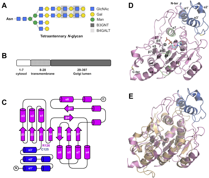
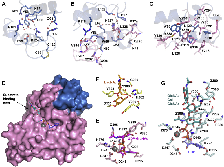
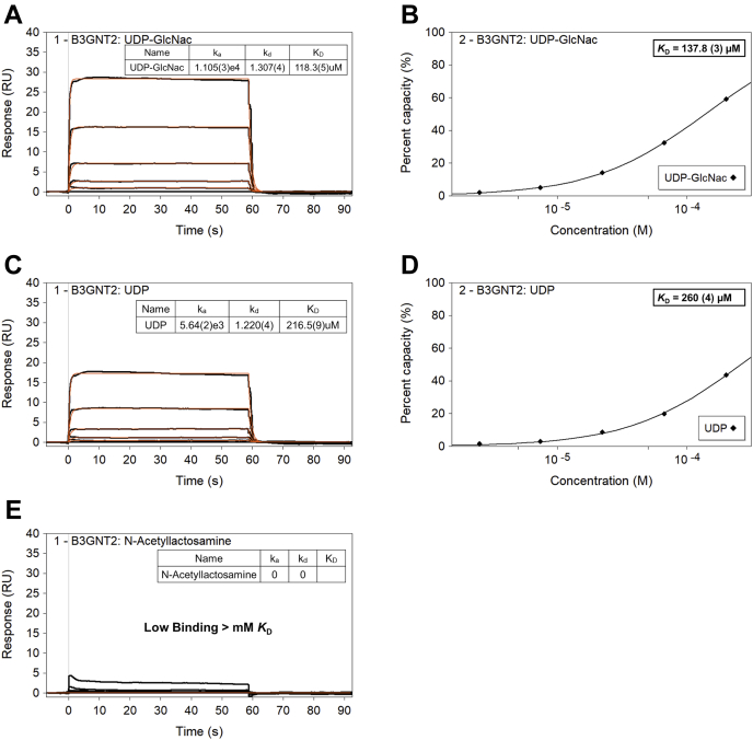
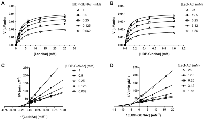
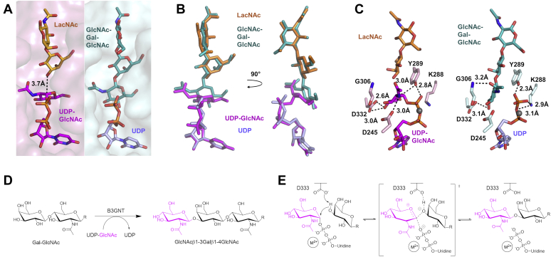
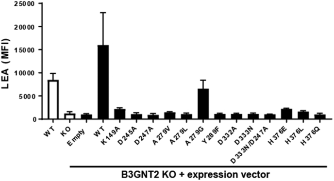
β1,3-N-acetylglucosaminyltransferases (B3GNTs) are Golgi-resident glycosyltransferases involved in the biosynthesis of poly-N-acetyl-lactosamine chains. They catalyze the addition of the N-acetylglucosamine to the N-acetyl-lactosamine repeat as a key step of the chain elongation process. Poly-N-acetyl-lactosamine is involved in the immune system in many ways. Particularly, its long chain has been demonstrated to suppress excessive immune responses. Among the characterized B3GNTs, B3GNT2 is the major poly-N-acetyl-lactosamine synthase, and deletion of its coding gene dramatically reduced the cell surface poly-N-acetyl-lactosamine and led to hypersensitive and hyperresponsive immunocytes. Despite the extensive functional studies, no structural information is available to understand the molecular mechanism of B3GNT2, as well as other B3GNTs. Here we present the structural and kinetic studies of the human B3GNT2. Five crystal structures of B3GNT2 have been determined in the unliganded, donor substrate-bound, acceptor substrate-bound, and product(s)-bound states at resolutions ranging from 1.85 to 2.35 Å. Kinetic study shows that the transglycosylation reaction follows a sequential mechanism. Critical residues involved in recognition of both donor and acceptor substrates as well as catalysis are identified. Mutations of these invariant residues impair B3GNT2 activity in cell assays. Structural comparison with other glycosyltransferases such as mouse Fringe reveals a novel N-terminal helical domain of B3GNTs that may stabilize the catalytic domain and distinguish among different acceptor substrates.

Keywords: enzyme mechanism; glycobiology; glycosyltransferase; poly-N-acetyl-lactosamine; structural biology; β1,3-N-acetylglucosaminyltransferase 2.


Conflict of interest statement

Conflict of interest The authors declare that they have no conflicts of interest with the contents of this article.

Figures

Figure 1
Figure 1
Overall structure of human B3GNT2 luminal domain.A, synthesis of polylactosamine on tetraantennary N-glycan catalyzed by B3GNT and B4GALT enzymes. B, B3GNT2 is a type II transmembrane protein with a short N-terminal cytosolic segment, a transmembrane region, and a C-terminal domain in Golgi lumen. C, secondary structure topology of human B3GNT2 luminal domain. N-terminal domain is colored in blue, and GT-A domain is colored in magenta. Figure was prepared with TopDraw (48). D, overall structure of B3GNT2_UDP (chain A). N-terminal domain is colored in blue, and GT-A domain is colored in pink. The three common sequence motifs in B3GT family are colored in green. Magnesium ion is shown as a gray sphere, and UDP is shown as cyan sticks. Disulfide bonds are highlighted in yellow. E, superposition of human B3GNT2 luminal domain (blue and pink) and the catalytic domain (GT-A domain) of mouse Manic Fringe (wheat).
Figure 2
Figure 2
N-terminal domain and substrate-binding cleft in GT-A domain.A, hydrogen-bonding interactions (dashed lines) within the N-terminal domain. Side chains of residues involved are shown as sticks. Water molecules are shown as red spheres. The Cys96-Cys125 disulfide bond is highlighted in yellow. B, hydrogen-bonding interactions (dashed lines) between the N-terminal domain (blue) and the GT-A domain (pink). Side chains and backbones of residues involved are shown as sticks. Water molecules are shown as red spheres. C, hydrophobic interactions between the N-terminal domain (blue) and the GT-A domain (pink). Side chains of residues involved are shown as sticks. D, surface representation of B3GNT2 structure, showing the substrate-binding cleft in GT-A domain. N-terminal domain is colored in blue and GT-A domain is colored in pink. Substrate-binding cleft is indicated with an arrow. GlcNAcβ1-3Galβ1-4GlcNAc (teal) and UDP (purple), as well as magnesium ion (gray sphere), are shown in the substrate-binding cleft. E, donor substrate UDP-GlcNAc (magenta) is coordinated by residues (pink) in the substrate-binding cleft through hydrogen-bonding interactions (in structure B3GNT2_UDPGlcNAc). Magnesium ion is shown as a gray sphere, and water molecules are shown as red spheres. F, acceptor substrate LacNAc (orange) is coordinated by residues (yellow) in the substrate-binding cleft through hydrogen-bonding interactions (in structure B3GNT2_LacNAc). Water molecules are shown as red spheres. G, products GlcNAcβ1-3Galβ1-4GlcNAc (teal) and UDP (purple) are coordinated by residues (cyan) in the substrate-binding cleft through hydrogen-bonding interactions (in structure B3GNT2_tri_UDP). Magnesium ion is shown as a gray sphere, and water molecules are shown as red spheres.
Figure 3
Figure 3
SPR binding characterization of UDP-GlcNAc, UDP, and LacNAc. Steady-state (equilibrium) affinity binding data of UDP-GlcNAc (B) and UDP (D) to B3GNT2 coupled on SA sensor. The kinetic binding model fit (A and C) shows 1:1 binding of test analytes with rapid equilibrium and fast dissociation.
Figure 4
Figure 4
Steady-state kinetic experiment for B3GNT2-catalyzed transglycosylation.A, Michaelis–Menten plots of the initial velocities versus LacNAc concentrations for each fixed concentration of UDP-GlcNAc. The data set was globally fit to Equation 1 that describes the rapid-equilibrium random-order sequential mechanism, and the best fit is represented by the solid lines. B, Michaelis–Menten plots of the initial velocities versus UDP-GlcNAc concentrations for each fixed concentration of LacNAc. The data set was globally fit to Equation 1 that describes the rapid-equilibrium random-order sequential mechanism, and the best fit is represented by the solid lines. C, double-reciprocal plots of initial velocities versus LacNAc concentrations for each fixed concentration of UDP-GlcNAc. The solid lines show the linear regression fit. D, double-reciprocal plots of initial velocities versus UDP-GlcNAc concentrations for each fixed concentration of LacNAc. The solid lines show the linear regression fit.
Figure 5
Figure 5
Mechanism of B3GNT2 glycosyltransferase reaction. (A) Left panel, superposition of B3GNT2_UDPGlcNAc and B3GNT2_LacNAc at the substrate-binding cleft. The surface representation of substrate-binding cleft of B3GNT2_UDPGlcNAc is shown in pink. The distance between the C1 of the GlcNAc in UDP-GlcNAc (magenta) and the hydroxyl group on C3 of the Gal in LacNAc (orange) is marked by dashed line. Right panel, the substrate-binding cleft (cyan surface) in B3GNT2_tri_UDP is shown in the same orientation as the left panel and the two products (GlcNAcβ1-3Galβ1-4GlcNAc in teal and UDP in purple) are shown as sticks. (B) Superposition of the substrates and products in the substrate-binding cleft. UDP-GlcNAc, magenta; LacNAc, orange; GlcNAcβ1-3Galβ1-4GlcNAc, teal; UDP, purple. (C) Change of the hydrogen-bonding interactions in the substrate-binding cleft before (left panel) and after (right panel) the reaction takes place. UDP-GlcNAc, magenta; LacNAc, orange; GlcNAcβ1-3Galβ1-4GlcNAc, teal; UDP, purple; magnesium ion, gray sphere. (D) Glycosyltransferase reaction catalyzed by B3GNT2. (E) Mechanism of a divalent-metal-dependent inverting glycosyltransferase reaction. Asp333 is proposed to be the active site base. The GlcNAc to be transferred is proposed to undergo an oxocarbenium ion-like transition state.
Figure 6
Figure 6
Assessment of B3GNT2 point mutants. Flow cytometry analysis of LEA lectin binding was performed on WT, B3GNT2 KO, or B3GNT2 KO cells expressing the indicated B3GNT2 point mutation. Data shown are the average cell surface LEA mean fluorescence intensity (MFI) from two independent experiments.

References

    1. Brewer C.F., Miceli M.C., Baum L.G. Clusters, bundles, arrays and lattices: novel mechanisms for lectin-saccharide-mediated cellular interactions. Curr. Opin. Struct. Biol. 2002;12:616–623. - PubMed
    1. Camby I., Le Mercier M., Lefranc F., Kiss R. Galectin-1: a small protein with major functions. Glycobiology. 2006;16:137R–157R. - PubMed
    1. Pace K.E., Lee C., Stewart P.L., Baum L.G. Restricted receptor segregation into membrane microdomains occurs on human T cells during apoptosis induced by galectin-1. J. Immunol. 1999;163:3801–3811. - PubMed
    1. Pillai S., Netravali I.A., Cariappa A., Mattoo H. Siglecs and immune regulation. Annu. Rev. Immunol. 2012;30:357–392. - PMC - PubMed
    1. Togayachi A., Kozono Y., Ishida H., Abe S., Suzuki N., Tsunoda Y., Hagiwara K., Kuno A., Ohkura T., Sato N., Sato T., Hirabayashi J., Ikehara Y., Tachibana K., Narimatsu H. Polylactosamine on glycoproteins influences basal levels of lymphocyte and macrophage activation. Proc. Natl. Acad. Sci. U. S. A. 2007;104:15829–15834. - PMC - PubMed

Substances

LinkOut - more resources