. 2020 Nov 6;11(1):5625.
doi: 10.1038/s41467-020-19444-y.

Sub-nanoliter metabolomics via mass spectrometry to characterize volume-limited samples

Affiliations

Sub-nanoliter metabolomics via mass spectrometry to characterize volume-limited samples

Yafeng Li et al. Nat Commun. .

Abstract

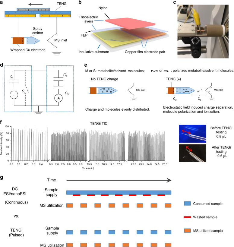
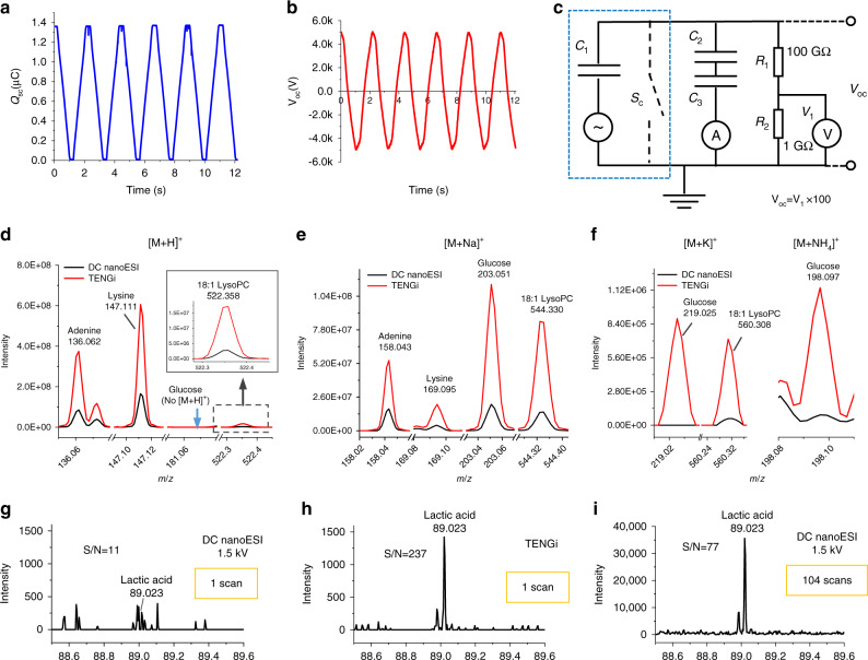
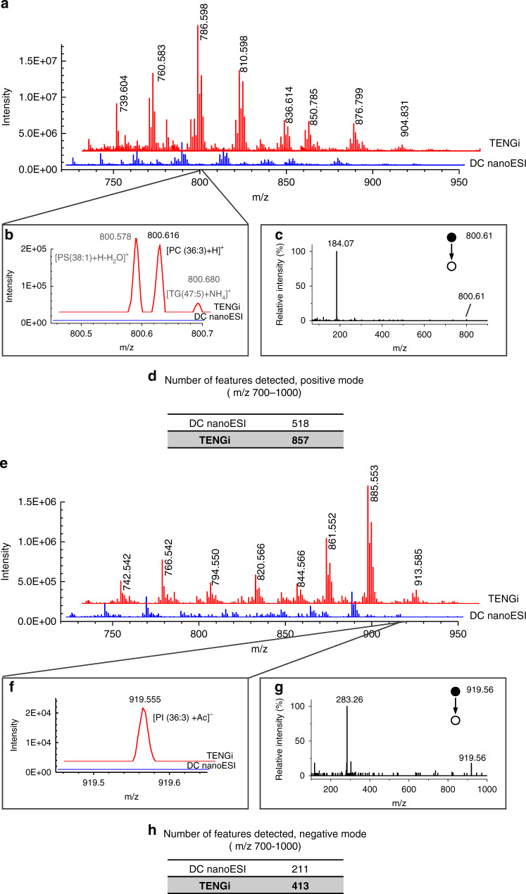
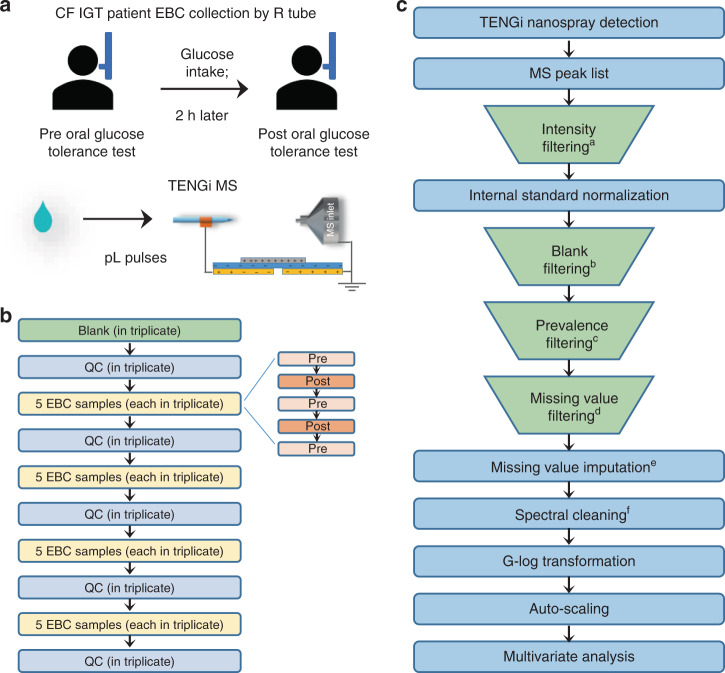
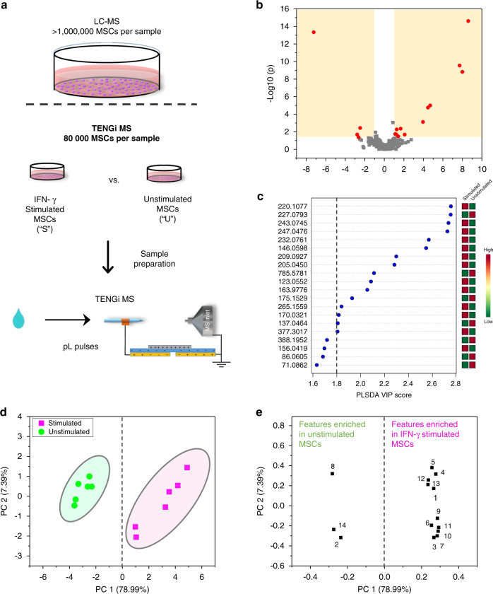
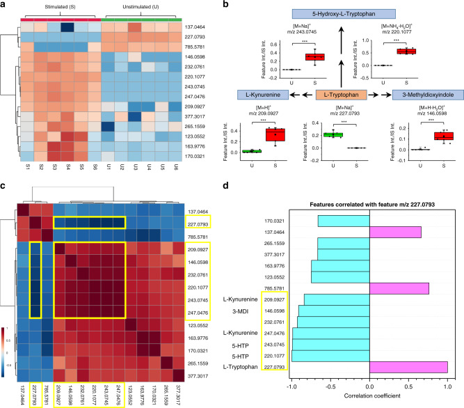
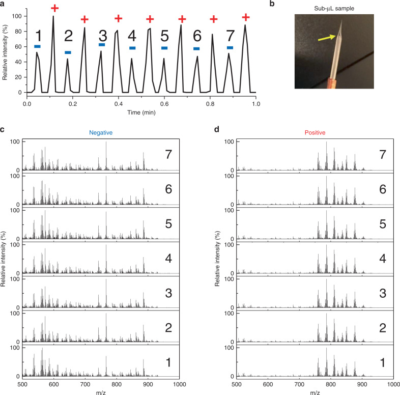
The human metabolome provides a window into the mechanisms and biomarkers of various diseases. However, because of limited availability, many sample types are still difficult to study by metabolomic analyses. Here, we present a mass spectrometry (MS)-based metabolomics strategy that only consumes sub-nanoliter sample volumes. The approach consists of combining a customized metabolomics workflow with a pulsed MS ion generation method, known as triboelectric nanogenerator inductive nanoelectrospray ionization (TENGi nanoESI) MS. Samples tested with this approach include exhaled breath condensate collected from cystic fibrosis patients as well as in vitro-cultured human mesenchymal stromal cells. Both test samples are only available in minimum amounts. Experiments show that picoliter-volume spray pulses suffice to generate high-quality spectral fingerprints, which increase the information density produced per unit sample volume. This TENGi nanoESI strategy has the potential to fill in the gap in metabolomics where liquid chromatography-MS-based analyses cannot be applied. Our method opens up avenues for future investigations into understanding metabolic changes caused by diseases or external stimuli.


Conflict of interest statement

The authors declare no competing interests.

Figures

Fig. 1
Fig. 1. TENGi nanoESI illustration, mechanism and characteristics.
a Schematic of the TENGi nanoESI system. b Schematic of the SF TENG device used in this study. c Photo of the custom cartridge used to secure nanoESI emitters in TENGi experiments. d Equivalent electric circuit (C1–C3 depict capacitors; Sc represents a short-circuit switch). e Charge generation mechanism in TENGi. M and S represent metabolite and solvent molecule, respectively; “+” and “−” represent positive and negative charges, respectively; C2 and C3, as also included in d, represent two capacitors formed during TENGi nESI process. f Total ion chronogram (TIC) produced by TENGi nanoESI of 0.8 µL of a 1 : 9 v/v mixture of MeOH : H2O together with photos of the solution contained in the emitter before and after analysis. The red arrows in the photos indicate the volume of sample solution in the nanoESI emitter. As a pulsed ionization method, the TIC appears as pulses instead of continuous signals. By sealing the larger end of the spray emitter to prevent evaporation, 0.8 µL of sample can pulse-spray for more than 25 min. g Illustration of sample utilization in conventional DC ESI/nanoESI and TENGi. As the mass spectrometer intermittently traps and scans ions, TENGi can be synchronized with these events so as to achieve higher sample utilization.
Fig. 2
Fig. 2. TENG device and TENGi nanoESI characterization.
a Measured short-circuit charge (Qsc) for the TENG device used in this work. The charge generated per cycle is 1.37 µC. b Measured TENG output voltage (Voc). c Equivalent circuit for TENG output voltage measurements, using an external load resistance of 100 GΩ to bring the voltage to the voltmeter measuring range. The blue square indicates the equivalent circuit of the TENG device. df The behavior of four typical metabolite standards (adenine, lysine, glucose, and lysoPC) with TENGi and conventional DC nanoESI. gi The comparison of TENGi and DC nanoESI for the detection of lactic acid ([M-H] m/z = 89) in EBC of a healthy volunteer: g single scan conventional DC nanoESI mass spectrum, h single scan TENGi nanoESI mass spectrum, and i average mass spectrum generated from 104 nanoESI scans.
Fig. 3
Fig. 3. Dilute liver lipid extract (25 μg mL−1) analyzed by both conventional DC nanoESI and TENGi MS.
ad Positive-ion mode MS comparison: a full MS, b enlarged view of m/z 800.616 that was detected by TENGi but not by DC nanoESI, c TENGi MS/MS identification of the species with m/z 800.616; d number of features detected in positive-ion mode in the 700–1000 m/z range. eh Negative-ion mode MS comparison: e full MS, f enlarged view of m/z 919.555 that was detected by TENGi but not by DC nanoESI. g MS/MS identification of the species with m/z 919.555; h number of features detected in negative-ion mode in the 700–1000 m/z range.
Fig. 4
Fig. 4. Experimental and data processing workflow for TENGi MS EBC metabolomics.
a Illustration of EBC sample collection and TENGi MS analysis; b TENGi MS run order, including method blanks, quality control (QC) samples, and EBC sample replicates. Pre and Post samples were run in an interleaved fashion within each sample segment, and randomized in terms of their overall order. Three replicate tests were conducted for each sample. Outliers were discarded based on the extracted ion chronogram pulse area of the spiked IS; c TENGi MS data processing protocol. aSpectral features with abundances lower than 2000 were removed from the dataset. bBlank filtering was applied to remove features with normalized abundances lower than 3 times that of the blank. cA spectral feature had to be detected in at least 80% of the EBC samples, otherwise it was removed. dSpectral features in the QCs with 80% missing values or not detected were filtered out and sample(s) with more than 20% missing values were deemed as outlier(s) and removed (Supplementary Fig. 10). eMissing values were replaced by half of the minimum positive value in the original data. fQC features with median relative SD (RSDQC) > 30% were removed. The order of the four filtering processes is interchangeable.
Fig. 5
Fig. 5. Reproducibility study of triboelectric ion source for tiny metabolomics.
TENGi MS reproducibility using the same nanoESI emitter (a, b) and between three different nanoESI emitters (c, d). In all cases, the sample sprayed was a QC sample made up with pooled EBC. a, c Extracted ion chronograms for the IS. The label displayed over each individual pulse (7, 8, 9, etc.) represents the integrated area of the IS, 13C tyrosine. Different colors represent different time regions. b, d The corresponding averaged mass spectra for the TENGi pulses shown in regions A, B, C and 1, 2, 3, respectively. The colors match those for the regions shown in a and c. The median intra and inter-emitter RSDQC values shown in b and d were 12.8% and 17.2%, respectively. Source data are provided as a Source Data file.
Fig. 6
Fig. 6. TENGi metabolomics results for the comparison of CF IGT patients pre- and post-oral glucose tolerance test.
a PLS-DA score plot using all features. b PLS-DA VIP score plot. The vertical dashed line indicates the VIP score > 1.8 cutoff. Five metabolic features were selected based on this cutoff. cg Box plots of IS-normalized MS peak abundances for these five features. For the Pre group, n = 10 biologically independent samples were tested; for Post group, n = 9 biologically independent samples were tested. Differences between the two groups were evaluated using the two-tailed Student’s t-test (***p-value < 0.001, *p-value < 0.05, detailed p-values are: 0.00086, 0.014, 0.019, 0.022, 0.026, respectively. These were calculated based on the final dataset after G-log transformation and autoscaling through MetaboAnalyst). Source data are provided as Source Data files. The lower, middle and upper lines in box plots cg correspond to 25th, 50th, and 75th percentiles. The whiskers extend to the most extreme data point within 1.5 interquartile range (IQR).
Fig. 7
Fig. 7. TENGi sub-nanoliter metabolomic studies of IFN-γ-stimulated MSCs.
a TENGi MS workflow; b volcano plot for IFN-γ stimulated (S) vs. unstimulated (U) MSCs. c PLS-DA VIP score plot with the top 20 features ranked by their PLS-DA VIP scores. d Principal component analysis score plot for IFN-γ stimulated vs. unstimulated MSCs using 14 significant features selected by volcano plot criteria and PLS-DA VIP score > 1.8; e corresponding principal component analysis score loadings plot identifying features that discriminate stimulated vs. non-stimulated MSCs.
Fig. 8
Fig. 8. IFN-γ stimulation affects tryptophan metabolism in MSCs through three catabolic pathways.
a Heat map of the top 14 most important features. b Pathway analysis results, showing three catabolic pathways are affected. Box plots shows the alterations in these tryptophan-related metabolites. Differences between the two groups were evaluated using the two-tailed Student’s t-test (***p-value < 0.001, detailed p-values of each feature: top left to right: 2.9E − 10, 2.5E − 15; bottom left to right: 7.6E − 04, 4.6E − 14, and 1.8E − 05. These are also provided in Table 1. P-values were calculated on the final dataset after G-log transformation and autoscaling through MetaboAnalyst). Source data are provided as Source Data files. The lower, middle, and upper lines in the box plots (in b) correspond to 25th, 50th, and 75th percentile. The whiskers extend to the most extreme data point within 1.5 IQR. n = 6 biologically independent samples were included in each group. c Pearson’s correlation map for the 14 feature set. d Pearson’s correlation coefficient between the 14 features of interest relative to m/z 227.0793 (tryptophan). Plots c and d show that there are six features closely correlated with m/z 227.0793 (tryptophan), five of which are from tryptophan catabolic products shown in b pathway analysis. Yellow squares indicate the metabolites that are highly correlated. Note 5-HTP has two ion forms as does l-kynurenine. Due to space limitations, b only shows the box plot of [M + H]+ (m/z 209.0927), the [M + K]+ (m/z 247.0476) ion is shown in Supplementary Fig. 11.
Fig. 9
Fig. 9. Polarity switching TENGi MS to obtain both positive and negative signals in the same experiment with sub-nanoliter sample consumption.
The sample tested was a 50 μg mL−1 liver lipid extract. a TIC of TENGi coupled with polarity switching, Negative (blue “−” sign) and positive (red “+” sign) signals are detected alternatively. b Detail of emitter with 0.8 μL sample loaded. Yellow arrows indicate the position of the sample solution in the emitter. c Mass spectra for negative pulses; d mass spectra for positive pulses.

References

    1. Griffiths WJ, et al. Targeted metabolomics for biomarker discovery. Angew. Chem. Int. Ed. Engl. 2010;49:5426–5445. doi: 10.1002/anie.200905579. - DOI - PubMed
    1. Spratlin JL, Serkova NJ, Eckhardt SG. Clinical applications of metabolomics in oncology: a review. Clin. Cancer Res. 2009;15:431–440. doi: 10.1158/1078-0432.CCR-08-1059. - DOI - PMC - PubMed
    1. Dettmer K, Aronov PA, Hammock BD. Mass spectrometry-based metabolomics. Mass Spectrom. Rev. 2007;26:51–78. doi: 10.1002/mas.20108. - DOI - PMC - PubMed
    1. Agathocleous M, et al. Ascorbate regulates haematopoietic stem cell function and leukaemogenesis. Nature. 2017;549:476–481. doi: 10.1038/nature23876. - DOI - PMC - PubMed
    1. Antonucci R, Atzori L, Barberini L, Fanos V. Metabolomics: the new clinical chemistry for personalized neonatal medicine. Minerva Pediatr. 2010;62:145–148. - PubMed

Publication types

MeSH terms