. 2020 Nov 6;11(1):5628.
doi: 10.1038/s41467-020-19361-0.

A single-cell atlas and lineage analysis of the adult Drosophila ovary

Affiliations

A single-cell atlas and lineage analysis of the adult Drosophila ovary

Katja Rust et al. Nat Commun. .

Erratum in

Abstract

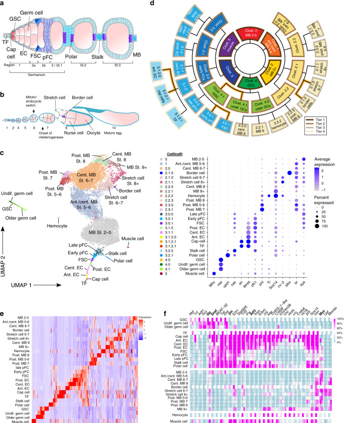
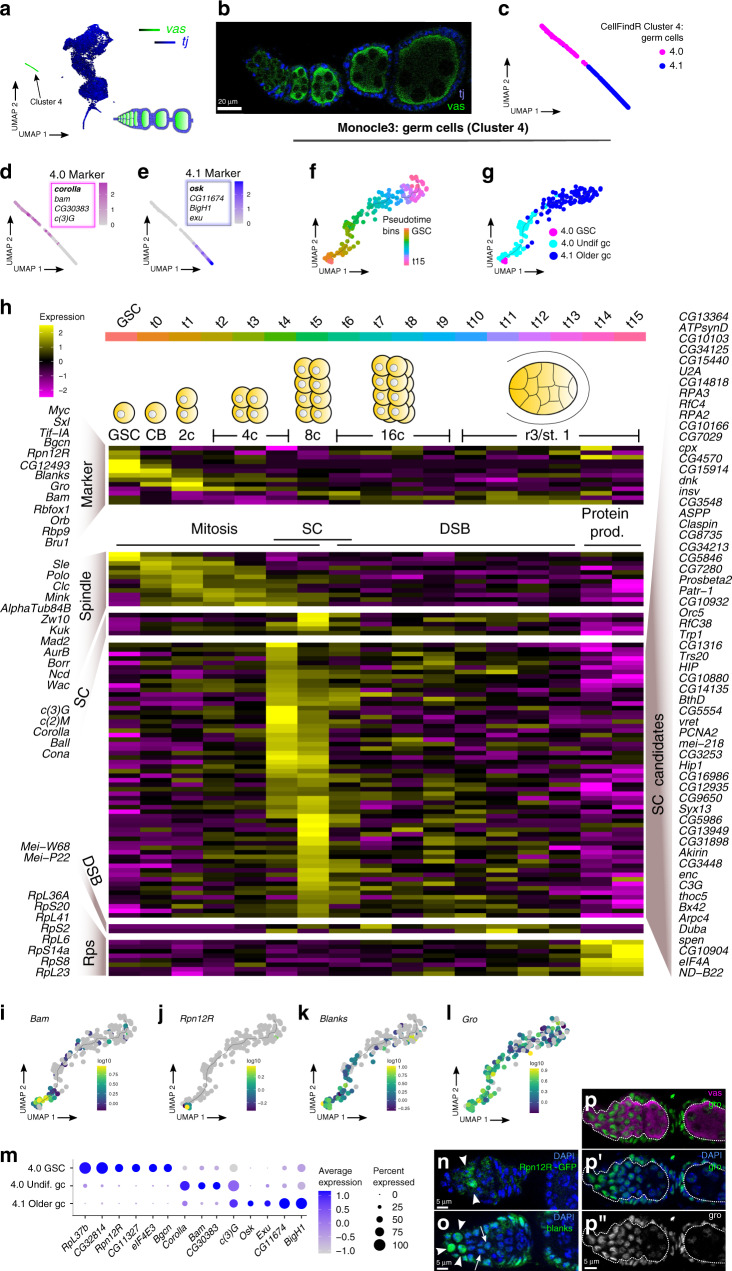
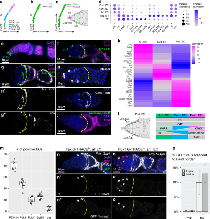
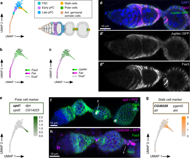
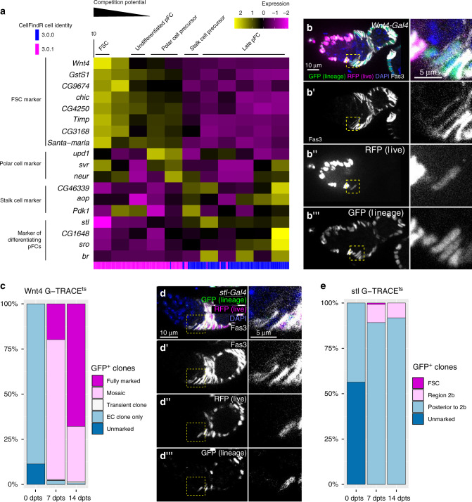
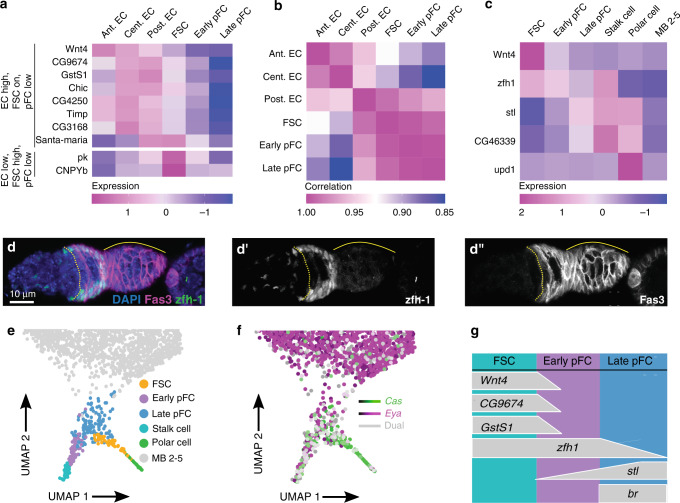
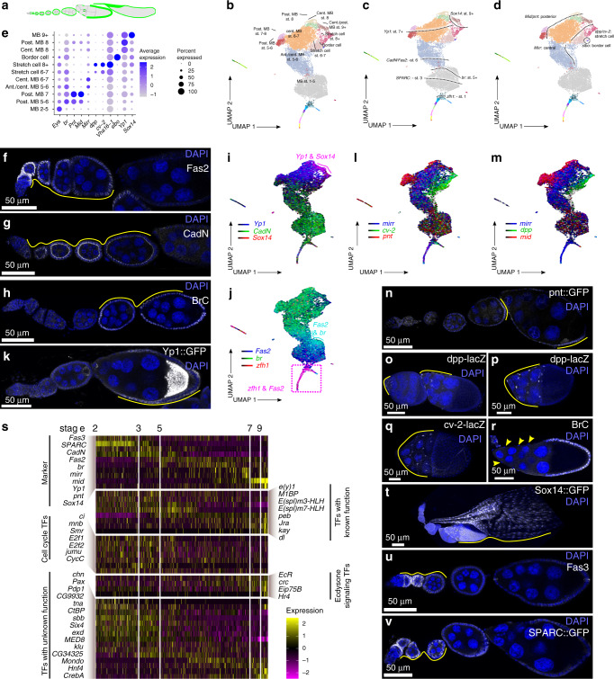
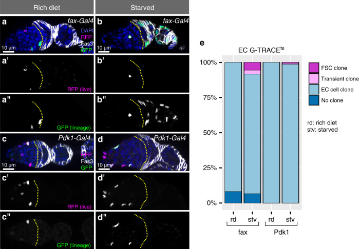
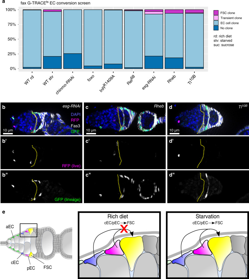
The Drosophila ovary is a widely used model for germ cell and somatic tissue biology. Here we use single-cell RNA-sequencing (scRNA-seq) to build a comprehensive cell atlas of the adult Drosophila ovary that contains transcriptional profiles for every major cell type in the ovary, including the germline stem cells and their niche cells, follicle stem cells, and previously undescribed subpopulations of escort cells. In addition, we identify Gal4 lines with specific expression patterns and perform lineage tracing of subpopulations of escort cells and follicle cells. We discover that a distinct subpopulation of escort cells is able to convert to follicle stem cells in response to starvation or upon genetic manipulation, including knockdown of escargot, or overactivation of mTor or Toll signalling.


Conflict of interest statement

The authors declare no competing interests.

Figures

Fig. 1
Fig. 1. CellFindR identifies distinct populations of cells in the ovary.
a Diagram of the anterior tip of the ovariole, including the germarium and two budded follicles. b Diagram of the entire ovariole. c UMAP plot of the merged dataset and gene expression profiles of selected markers. d Hierarchy of CellFindR clusters. Tier 1 (dark brown outlines), Tier 2 (brown outlines), Tier 3 (taupe outlines) and Tier 4 (beige outlines) clusters were produced by the first, second, third and fourth round of CellFindR clustering, respectively. Orange branch lines indicate subclusters of a single terminal cluster that were identified through additional analysis. e Heat map showing the expression of the top 10 most unique genes for each cluster across the entire dataset. f Heat map showing the activity for selected regulons identified by SCENIC in each cluster. The first column shows regulon activity in dataset2 and the second column shows regulon activity in dataset3 for each regulon respectively. Scale bar shows percent of regulon activity. TF: terminal filament; GSC: germline stem cells; EC: escort cells; FSC: follicle stem cells; pFC: prefollicle cells; polar: polar cell; stalk: stalk cell; MB: main body follicle cells; St.: Stage; ant.: anterior; cent.: central; post.: posterior; undif.: undifferentiated.
Fig. 2
Fig. 2. Germ cells.
a SCope expression plot of vas (green) and tj (blue) on UMAP plot and a diagram of an ovariole showing cell types in the corresponding colors. b Early stages of Drosophila ovariole stained for tj (blue) and vas (green). ce UMAP plots showing the distribution of the two germ cell clusters initially identified by CellFindR (c), and the expression pattern of a marker for each cluster. Expression of the marker in bold text is shown on the plot and additional markers are listed below (de). fg monocle3 analysis of germ cells orders cells into a linear trajectory (f) that distributes the cells from the two germ cell clusters onto opposite ends of the pseudotime trajectory and identifies GSCs (g). h Heat map showing transcriptional changes across pseudotime identifies markers of each stage of germ cell differentiation from the GSC to the Region 3/Stage 1 follicle, including stages that are enriched for the expression of mitosis genes, synaptonemal complex genes, double-stranded break genes, and protein production genes. Genes with a similar expression profile as known synaptonemal complex genes are presented as novel synaptonemal complex candidate genes. il The expression profile in pseudotime of representative markers of different stages of germ cell differentiation. m Dot plot showing the specificity of selected markers for GSCs, undifferentiated germ cells, and older germ cells. np Rpn12R-GFP germarium stained for GFP (n), or wildtype germaria stained for blanks (o) or gro (p, p’), shown in the green channel or in white (p”), and for DAPI (blue, n, o, p’) and vasa (magenta, p) as indicated. Arrowheads in n and o point at positive cells. Arrows in o point at germ cells with lower expression of blanks. White line in p demarks the border between germ cells and somatic cells. GSC: germline stem cell; undif.: undifferentiated; gc: germ cell; protein prod.: protein production; SC: synaptonemal complex; DSB: double-strand break; Rps: ribosomal proteins.
Fig. 3
Fig. 3. Anterior germarial somatic cells.
ac UMAP plots of the five clusters that contain somatic cells in the anterior half of the germarium (a) and markers that distinguish apical cells (cap cells and terminal filament cells) from escort cells (bc). d Dot plot showing the expression of selected markers in each of the five clusters. ej Wildtype germarium stained for en (e), or germaria from enhancer traps or protein traps stained for GFP, RFP, or LacZ as indicated (fj). fax::GFP germaria were also stained for cas (white, g”), the GFP channel is shown separately (white, g’). Staining for en, GFP or RFP (green), LacZ (white), Fas3 or vasa (magenta) and DAPI (blue). Yellow dotted line demarks the Region 2a/2b border. kl Heat map (k) and summary diagram (l) showing markers distinguishing aECs, cECs, and pECs. m Quantification of the number of ECs per germarium that express the indicated marker genes. Each dot is a germarium. n = 10, 15, 15, 17, 17 germaria for PZ1444, Pdk1, Pdk1, GstS1, and cas, respectively. In the box plots, the midline corresponds to the median; the lower and upper hinges correspond to the first and third quartiles; and the whiskers span the smallest and largest values within 1.5 of the interquartile range. no Germaria from flies with fax-Gal4 (n) or Pdk1-Gal4 (o) combined with G-TRACEts raised at 18 °C, shifted to 29 °C upon eclosion, and well-fed for 14 days before dissection, stained for GFP (green), RFP (magenta), Fas3 (white), and DAPI (blue). n’, o’ RFP channel (white). n”, o” GFP channel (white). Yellow dotted line demarks the Region 2a/2b border. p Quantification of the percent of germaria with GFP+ ECs adjacent to the Region 2a/2b border (Fas3 expression boundary) in germaria with fax-Gal4 or Pdk1-Gal4 driving G-TRACEts after 7 or 14 days at 29 °C. n = 380, 865, 81, and 187 GFP+ cells adjacent to Fas3 border for Pdk1 7dpts, Pdk1 14 dpts, fax 7dpts, and fax 14dpts, respectively. Error bars indicate S.E.M. TF: terminal filament; EC: escort cell; ant.: anterior; cent.: central; post.: posterior.
Fig. 4
Fig. 4. Posterior germarial somatic cells, polar cells, and stalk cells.
ac UMAP plots of the clusters that contain the cells in the early FSC lineage, polar cells, and stalk cells showing the distribution of clusters (a), and the expression patterns of Fas3 (b) and Jupiter (c), which are strongly expressed in these clusters, relative to fax, which is a marker of ECs. d Germarium with Jupiter::GFP stained for GFP (green), Fas3 (magenta), and DAPI (blue). d’ GFP channel shown in white. d” Fas3 channel shown in white. eh UMAP plots showing the specificity of upd1 expression in the polar cell cluster (e) and CG46339 expression in the stalk cell cluster (g) and germaria with upd1-Gal4 (f) or CG46339-Gal4 (h) driving RFP expression stained for RFP (white), Fas3 (green), and DAPI (blue) shown in maximum intensity projections. The expression patterns of these enhancer trap lines are consistent with the prediction that upd1 is expressed specifically in polar cells (p) and CG46339 is expressed specifically in stalk cells (s). FSC: follicle stem cell; pFC: prefollicle cell; ant.: anterior.
Fig. 5
Fig. 5. The early FSC lineage.
a Heat map showing gene expression across pseudotime in the FSC and pFCs clusters. Blue and magenta lines indicate the original CellFindR identity. b Germarium with Wnt4-Gal4 driving G-TRACEts stained for RFP (magenta), GFP (green), Fas3 (white), and DAPI (blue). Inset shows Wnt4-Gal4low Fas3+ cell at the Fas3 boundary. Fas3 (b’), RFP (b”) and GFP (b”’) are also shown in white. c Quantification of ovarioles with Wnt4-Gal4 driving G-TRACEts without GFP-positive cells (unmarked), with only EC clones, transient follicle cell clones, mosaic labeling of the follicle epithelium or a fully marked follicle epithelium at 0, 7, or 14 days post temperature shift (dpts). The presence of ovarioles with EC clones at the 0 dpts time point is likely because this is where Wnt4-Gal4 activity is strongest and Gal4 may not be fully repressed by Gal80 in these cells. Notably, we never observed GFP+ follicle cell clones at 0 dpts, consistent with lower expression of Wnt4-Gal4 in FSCs. n = 149, 156 and 120 ovarioles for 0, 7 or 14 dpts respectively. d Germarium with stl-Gal4 driving G-TRACEts stained for RFP (magenta), GFP (green), Fas3 (white), and DAPI (blue). stl-Gal4 drives RFP expression sparsely in pFCs in the 2b Region and is consistently expressed in Region 3. GFP+ clones typically include pFCs in region 2b (inset) but not FSCs or pFCs at the 2a/2b border. Fas3 (d’), RFP (d”) and GFP (d”’) are shown separately in white. e Quantification of ovarioles with stl-Gal4 driving G-TRACEts without GFP-positive cells (unmarked), with FSC clones, transient follicle cell clones located in Region 2b, or transient follicle cell clones posterior to Region 2b at 0, 7 or 14 dpts. Ovarioles at the 0 dpts frequently contained small GFP+ clones of up to 4 cells posterior to region 3. These clones were usually confined to stalk cells where stl-Gal4 activity is strongest. n = 140, 135 and 128 ovarioles for 0, 7 or 14 dpts respectively. FSC: follicle stem cell; pFC: prefollicle cell; EC: escort cell; dpts: days post temperature shift.
Fig. 6
Fig. 6. Distinct expression patterns in cell types of the early follicle cell lineage.
a Heat map of gene expression in EC populations, FSCs and pFCs. Most stem cell markers identified by pseudotime analysis of the early FSC lineage (Fig. 5a) are also expressed in ECs. In contrast, pk and CNPYb show high expression on FSCs but not ECs and pFCs. b Heat map showing the correlation in the gene expression profiles of EC, FSC and pFC clusters. We observe a high degree of similarity between cell types of the same lineage, as expected, but also find that the gene expression profile of pECs has substantial overlap with FSCs and pFCs. c Heat map showing the expression of markers that can be used to distinguish FSCs, early pFCs, late pFCs and stalk and polar cells from each other. d Maximum intensity projection of a wildtype germarium stained for zfh1 (green), Fas3 (magenta), and DAPI (blue). d’ zfh1 staining (white). d” Fas3 staining (white). ef UMAP plots showing FSC, pFC, stalk and polar cell clusters (e) and the expression patterns of cas and eya (f). g Summary of markers that can be used to distinguish FSCs, early pFCs, and late pFCs from each other. FSC: follicle stem cell; pFC: prefollicle cell; EC: escort cell; ant.: anterior; cent.: central; post.: posterior; MB: main body follicle cell.
Fig. 7
Fig. 7. Distinct stages of main body follicle cells.
ad A diagram of the ovariole highlighting the main body follicle cells in green (a), and UMAP plots showing main body follicle cell cluster identities (b). The spatial arrangement of cells on the plot positions cells according to the stage of oogenesis (c) and, starting in mid-oogenesis, anterior-posterior position on the follicle (c). e A dot plot showing the expression patterns of genes that are expressed in different types of main body follicle cells and their derivatives. fr The expression patterns of selected markers on UMAP plots or in ovarioles, as indicated. In all images of ovarioles, the selected marker is shown in white and DAPI is shown in blue. The images in (f, g, h and r) show wildtype ovarioles stained for Fas2 (f), CadN (g), and BrC (h and r). The remaining images are of germaria from enhancer trap or protein trap lines, as indicated. Maximum intensity projections are shown in (oq). s Heat map showing the changes in gene expression across pseudotime in main body follicle cells. Genes that serve as markers of different stages of pseudotime as well as transcription factors with predicted functions, such as cell cycle genes at the early stages before the mitosis-to-endocycle switch, and ecdysone responsive genes at late stages are shown. Transcription factors with unknown functions in oogenesis that have dynamic expression patterns across pseudotime are also shown. tv Ovarioles from Sox14::GFP (t); wildtype (u) or SPARC::GFP (v) flies stained for GFP or Fas3 (white) and DAPI (blue). MB: main body follicle cell; ant.: anterior; cent.: central; post.: posterior; TFs: transcription factors.
Fig. 8
Fig. 8. Posterior ECs convert to FSCs in response to nutrient deprivation.
ad Germaria from flies with fax-Gal4 (a, b) or Pdk1-Gal4 (c, d) driving G-TRACEts stained for DAPI (blue), Fas3 (white), RFP (magenta), and GFP (green). a’d’ RFP channel in white. a”d” GFP channel in white. Yellow line outlines the 2a/2b border. fax-Gal4 and Pdk1-Gal4 do not produce GFP + FSC clones on a rich diet (0 out of 5 flies with follicle cell clones) (a, c), whereas, following exposure to 24 h of starvation, fax-Gal4-expressing cells produce GFP + FSC clones (4 out of 5 flies contained follicle cell clones) (b) but Pdk1-Gal4-expressing cells almost never do (d) (2 out of 5 flies with follicle cell clones, we observed 1 ovariole with a transient clone and 1 ovariole with an FSC clone out of 138 ovarioles total). e Quantification of GFP + clone types in flies with fax-Gal4 or Pdk1-Gal4 driving G-TRACEts exposed to a rich diet or starvation. n = 170, 211, 134, and 138 ovarioles for fax-Gal4 rich diet, fax-Gal4 starved, Pdk1-Gal4 rich diet, and Pdk1-Gal4 starved, respectively. p-values from two-sided Student’s T-test for comparisons of the frequency of follicle cell clones (FSC + transient): fax-Gal4 rich vs starved: p = 0.002; Pdk1-Gal4 rich vs starved: p = 0.57. EC: escort cell; rd: rich diet; stv: starved; FSC: follicle stem cell.
Fig. 9
Fig. 9. EC conversion to FSCs is genetically controlled.
a Quantification of GFP + clone types in flies with fax-Gal4 driving G-TRACEts alone on a rich diet (rd) for 14 days (WT rich diet); starved for 24 hrs within the 14 dpts (WT starved); or on a rich diet for 14 days in combination with chinmo or escargot RNAi knockdown, or overexpression of a dominant-negative allele of the insulin receptor (InRK1409A), a constitutively active allele of Relish (Rel68) or Toll (Tl10B) or a wildtype allele of Rheb or foxo. p-values from two-sided Student’s T-test for comparisons between the frequency of follicle cell clones (FSC + transient) in WT rich diet 14d and mutant condition that have a p < 0.05: esg-RNAi: p = 0.008; Rheb: p = 0.03; Tl10B: p = 0.03. n = 204 (WT rich diet), 213 (WT starved), 113 (chinmo-RNAi), 135 (foxo), 127 (InRK1409A), 125 (Rel68), 154 (esg-RNAi), 143 (Rheb), 118 (Tl10B) ovarioles. bd Germaria with fax-Gal4 driving expression of G-TRACEts and esg-RNAi (h), Rheb (i), or Tl10B (j) stained for GFP (green), RFP (magenta), Fas3 (white) and DAPI (blue). b’d’ RFP channel show in white. b”d” GFP channel shown in white. GFP + follicle cell clones are present even though flies were maintained on well-fed conditions. Yellow line outlines the 2a/2b border. e Model summarizing our observations that starvation, but not well-fed conditions, induces central ECs and/or posterior ECs to convert to FSCs. EC: escort cell; rd: rich diet; stv: starved; suc: sucrose; aEC: anterior escort cell; cEC: central escort cell; pEC: posterior escort cell; FSC: follicle stem cell.

References

    1. Xie T, Spradling A. C. decapentaplegic is essential for the maintenance and division of germline stem cells in the Drosophila ovary. Cell. 1998;94:251–260. - PubMed
    1. Margolis J, Spradling A. Identification and behavior of epithelial stem cells in the Drosophila ovary. Development. 1995;121:3797–3807. - PubMed
    1. Fadiga, J. & Nystul, T. G. The follicle epithelium in the Drosophila ovary is maintained by a small number of stem cells. Elife8, (2019). - PMC - PubMed
    1. Nystul TG, Spradling A. Regulation of epithelial stem cell replacement and follicle formation in the Drosophila ovary. Genetics. 2010;184:503–515. - PMC - PubMed
    1. Reilein A, et al. Alternative direct stem cell derivatives defined by stem cell location and graded Wnt signalling. Nat. Cell Biol. 2017;19:433–444. - PMC - PubMed

Publication types

Substances