. 2020 Nov 6;3(1):647.
doi: 10.1038/s42003-020-01381-7.

Ediacaran Doushantuo-type biota discovered in Laurentia

Affiliations

Ediacaran Doushantuo-type biota discovered in Laurentia

Sebastian Willman et al. Commun Biol. .

Abstract

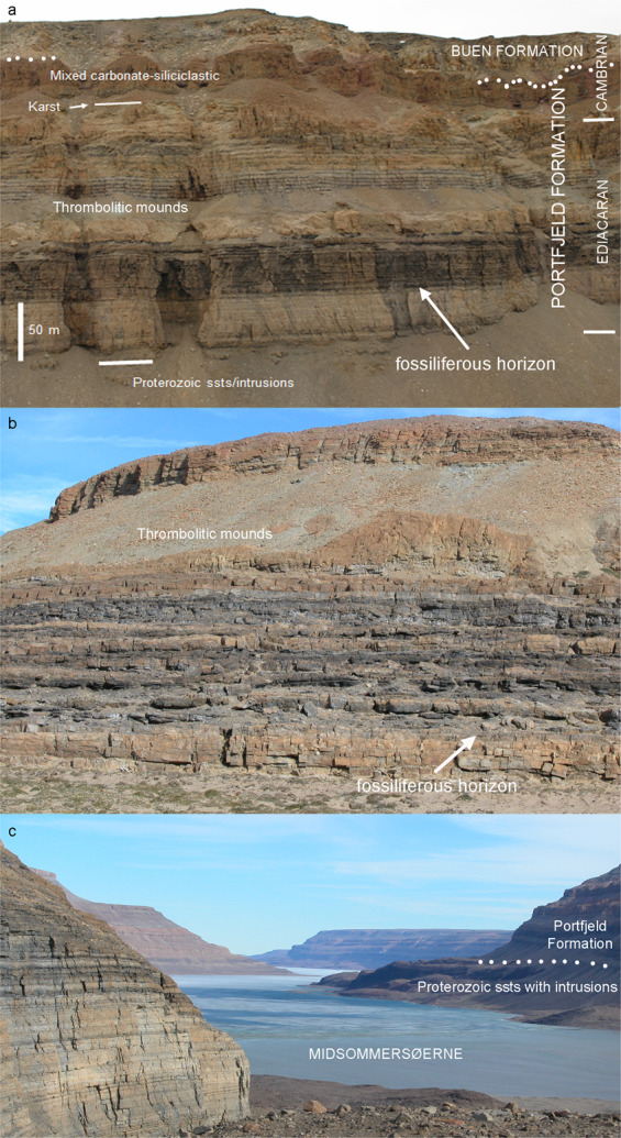
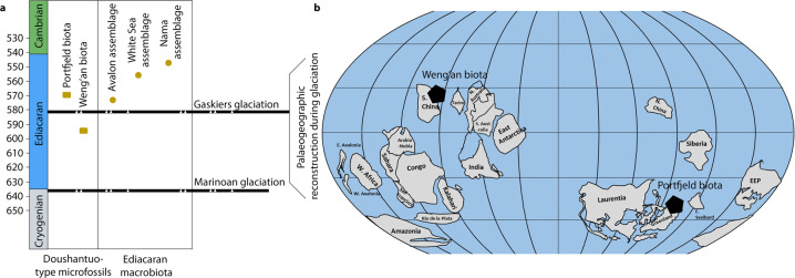
The Ediacaran period (635-541 Ma) was a time of major environmental change, accompanied by a transition from a microbial world to the animal world we know today. Multicellular, macroscopic organisms preserved as casts and molds in Ediacaran siliciclastic rocks are preserved worldwide and provide snapshots of early organismal, including animal, evolution. Remarkable evolutionary advances are also witnessed by diverse cellular and subcellular phosphatized microfossils described from the Doushantuo Formation in China, the only source showing a diversified assemblage of microfossils. Here, we greatly extend the known distribution of this Doushantuo-type biota in reporting an Ediacaran Lagerstätte from Laurentia (Portfjeld Formation, North Greenland), with phosphatized animal-like eggs, embryos, acritarchs, and cyanobacteria, the age of which is constrained by the Shuram-Wonoka anomaly (c. 570-560 Ma). The discovery of these Ediacaran phosphatized microfossils from outside East Asia extends the distribution of the remarkable biota to a second palaeocontinent in the other hemisphere of the Ediacaran world, considerably expanding our understanding of the temporal and environmental distribution of organisms immediately prior to the Cambrian explosion.


Conflict of interest statement

The authors declare no competing interests.

Figures

Fig. 1
Fig. 1. Field photographs of locality of the Portfjeld biota.
a Portfjeld Formation—basal Buen sandstones at Midsommersøer, notice the conspicuous band of dark cherty dolomites. b Detail of Portfjeld Formation, west of Midsommersøer, with the same darky cherty dolomites overlain by thrombolitic mounds, showing the lithostratigraphic horizon yielding the Portfjeld biota. c View looking east along Wandel Dal with Midsommersøer, taken from the fossil locality (off shot left). Scale bar valid for a.
Fig. 2
Fig. 2. Geography, geology, and stratigraphy of the study area, North Greenland.
a Geological map showing the sample locality at Midsommersøer, North Greenland. b Stratigraphic schemes in northern Ellesmere Island and North Greenland. c Stratigraphic section through the Portfjeld Formation at the western end of Midsommersøer compared with δ13Ccarb (o/oo PDB) values indicating the Shuram–Wonoka anomaly (about 570–560 Ma), “F” indicates the sample locality.
Fig. 3
Fig. 3. Putative eggs, embryos, and red algae from the Portfjeld Formation.
a–f Putative cleavage embryos. b, d, f Enlarged to show the detail of polygonal cell junctions. g Red algal thalli similar to Gremiphyca corymbiata. h–m Putative eggs showing various degrees of taphonomic degradation (h, k ductile; j, m brittle). i Shows a possible peanut-shaped cell division. j, m Showing breakage of vesicle wall, the shape of the breakage, its size and location on the specimen is similar in many specimens and may therefore be interpreted as a biological feature rather than random breakage. n Shows a comparably large unidentified acritarch showing polygonal shrinkage and a golf ball-like vesicle surface structure. Scale bar 100 µm, unless where individually stated. Accession numbers; a, b PMU 36863/1. c, d PMU 36864/1. e, f PMU 36865/1. f PMU 36865/1. g PMU 36866/1. h PMU 36867/1. i PMU 36868/1. j PMU 36869/1. k PMU 36869/2. l PMU 36870/1. m PMU 36869/3. n PMU 36864/2.
Fig. 4
Fig. 4. Acanthomorphic acritarchs, helically coiled spheroids, microbial mat fragment.
a, b Cavaspina acuminata, showing nature of sparsely separated, tapering processes. c, d Asterocapsoides wenganensis showing densely arranged, conical processes. Box in d shows the conical shape of mostly unbroken processes. e, f Helically anti-clockwise coiling, spheroidal microfossils with closed termination. g Putative egg shell broken during preparation displaying internal contents (indicated by arrow). h Microbial mat displaying community structures with intertwined filamentous structures. i, j Coiled cyanobacterium Jiangispirellus groenlandicus and close up of individual cell walls. k Spirellus shankari cyanobacterium showing the nature of the coiling helix. Scale bar 100 µm, unless where individually stated. Accession numbers; a, b PMU 36871/1. c, d PMU 36872/1. e PMU 36866/2. f PMU 36866/3. g PMU 36872/2. h PMU 36873/1. i, j PMU 36873/2. k PMU 36874/1.
Fig. 5
Fig. 5. Putative eggs and embryos at various stages of taphonomic degradation.
a Putative cleavage embryo displaying individual cells. b–s Putative eggs/embryos in various states of degradation; internal contents are often preserved and seen as external enveloping layer is broken or peeled off (b–d, g–i, q) but absent when compressed (p); surface structures vary from golf ball-like (f), wrinkled (b), pitted (j) to smooth (h), and from thin-, (s) to thick-walled (g). Some specimens have surface structures that may represent grooves (arrow in n) or polar invaginations (arrows in g). t Probable tightly coiled cyanobacterium (but see also Fig. 6 in Yin et al. for similar pseudo-uncoiling in Helicoforamina). Scale bar 100 µm. Accession numbers; a PMU 36868/2. b PMU 36869/4. c PMU 36874/2. d PMU 36875/1. e PMU 36876/1. f PMU 36876/2. g PMU 36877/1. h PMU 36875/2. i PMU 36877/2. j PMU 36877/3. k PMU 36874/3. l PMU 36869/5. m PMU 36876/3. n PMU 36872/2. o PMU 36878/1. p PMU 36864/3. q PMU 36865/2. r PMU 36879/1. s PMU 36877/4. t PMU 36868/2.
Fig. 6
Fig. 6. Stratigraphic and palaeogeographic distribution of evolutionary important Ediacaran assemblages.
a Stratigraphic distribution of the Portfjeld biota and the Weng’an biota (representative of Doushantuo Formation), the latter of which predates the classical macroscopic “Ediacaran biota” and probably also predating the Gaskiers glaciation. The potential stratigraphic range of the biotas is indicated by the vertical line but detailed stratigraphic correlation and relationships to other biotas remain to be resolved. b Palaeogeographic reconstruction at ca. 580 Ma, black polygons show location of the two taphonomic windows of this type known from the Ediacaran and their spatial separation (adapted from,).

References

    1. Barghoorn ES, Tyler SA. Microorganisms from the Gunflint chert. Science. 1965;147:563–577. doi: 10.1126/science.147.3658.563. - DOI - PubMed
    1. Schopf JW. Microflora of the Bitter Springs formation, late Precambrian, central Australia. J. Paleontol. 1968;42:651–688.
    1. Gehling, J. G., Droser, M. L., Jensen, S. R. & Runnegar, B. N. Ediacara organisms: relating form to function in Evolving Form and Function: Fossils and Development (ed. Briggs, D. E. G.) 43–66 (Yale Peabody Museum of Natural History, New Haven, 2005).
    1. Bengtson S, Zhao Y. Fossilized metazoan embryos from the earliest Cambrian. Science. 1997;277:1645–1648. doi: 10.1126/science.277.5332.1645. - DOI
    1. Xiao S, Zhang Y, Knoll AH. Three-dimensional preservation of algae and animal embryos in a Neoproterozoic phosphorite. Nature. 1998;391:553–558. doi: 10.1038/35318. - DOI

Publication types