Multicenter Study
. 2020 Dec:62:103083.
doi: 10.1016/j.ebiom.2020.103083. Epub 2020 Nov 5.

Discovery, validation and sequencing of urinary peptides for diagnosis of liver fibrosis-A multicentre study

Affiliations
Multicenter Study

Discovery, validation and sequencing of urinary peptides for diagnosis of liver fibrosis-A multicentre study

Ayman S Bannaga et al. EBioMedicine. 2020 Dec.

Abstract

Background: Liver fibrosis is a consequence of chronic inflammation and is associated with protein changes within the hepatocytes structure. In this study, we aimed to investigate if this is reflected by the urinary proteome and can be explored to diagnose liver fibrosis in patients with chronic liver disease.

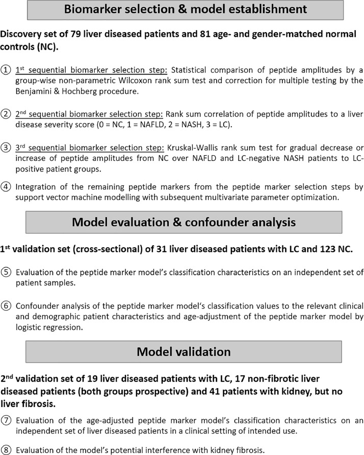
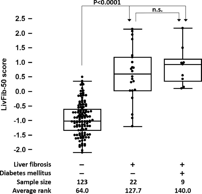
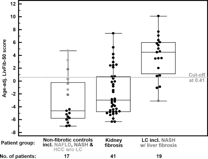
Methods: In a multicentre combined cross-sectional and prospective diagnostic test validation study, 129 patients with varying degrees of liver fibrosis and 223 controls without liver fibrosis were recruited. Additionally, 41 patients with no liver, but kidney fibrosis were included to evaluate interference with expressions of kidney fibrosis. Urinary low molecular weight proteome was analysed by capillary electrophoresis coupled to mass spectrometry (CE-MS) and a support vector machine marker model was established by integration of peptide markers for liver fibrosis.

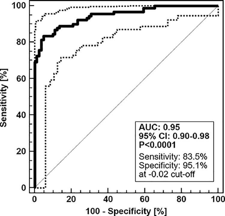
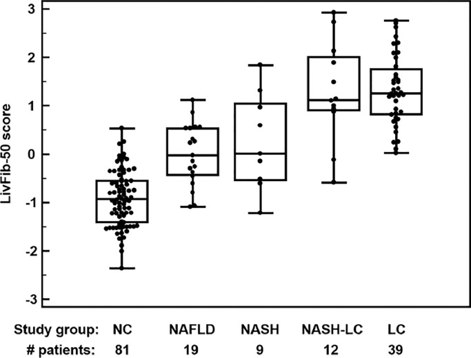
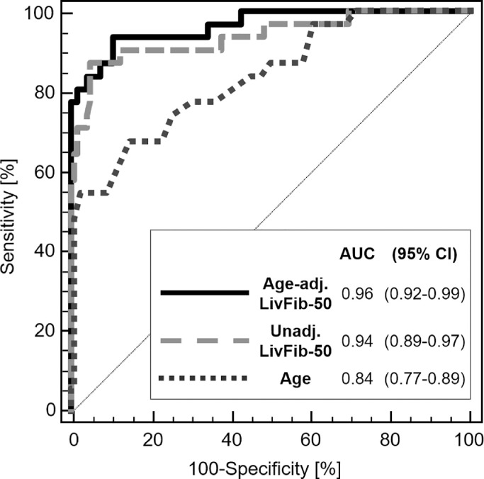
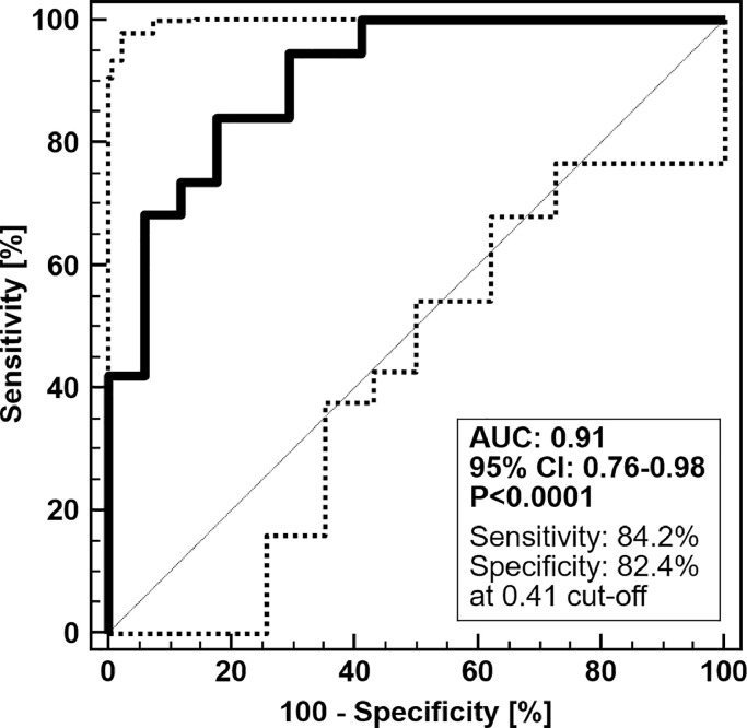
Findings: CE-MS enabled identification of 50 urinary peptides associated with liver fibrosis. When combined into a classifier, LivFib-50, it separated patients with liver fibrosis (N = 31) from non-liver disease controls (N = 123) in cross-sectional diagnostic phase II evaluation with an area under the curve (AUC) of 0.94 (95% confidence intervals (CI): 0.89-0.97, p<0.0001). When adjusted for age, LivFib-50 demonstrated an AUC of 0.94 (95% CI: 0.89-0.97, p<0.0001) in chronic liver disease patients with (N = 19) or without (N = 17) liver fibrosis progression. In this prospective diagnostic phase III validation set, age-adjusted LivFib-50 showed 84.2% sensitivity (95% CI: 60.4-96.6) and 82.4% specificity (95% CI: 56.6-96.2) for detection of liver fibrosis. The sequence-identified peptides are mainly fragments of collagen chains, uromodulin and Na/K-transporting ATPase subunit γ. We also identified ten putative proteolytic cleavage sites, eight were specific for matrix metallopeptidases and two for cathepsins.

Interpretation: In liver fibrosis, urinary peptides profiling offers potential diagnostic markers and leads to discovery of proteolytic sites that could be targets for developing anti-fibrotic therapy.

Keywords: Capillary electrophoresis mass spectrometry; Diagnosis; Liver fibrosis; Urinary peptide marker.


Conflict of interest statement

Declaration of Competing Interest Harald Mischak is founder and co-owner of Mosaiques Diagnostics GmbH, which developed the CE-MS technology. Jochen Metzger, Agnieszka Latosinska and Martin Pejchinovski are employees of Mosaiques Diagnostics GmbH. All other authors declare no conflict of interest.

Figures

Fig. 1
Fig. 1
Study flow chart. A total of 391 patients was included in the discovery and validation phases of the LivFib-50 peptide marker model for the detection of progressive liver fibrosis. Biomarker selection was carried out in three sequential steps resulting in 50 urinary peptides with differential and graded expression ranging from disease-free normal individuals. over NAFLD and NASH patients without LC to patients with well-established LC. Combining the peptide markers to the LivFib-50 model was followed by a first evaluation of the model's classification performance and confounder analysis in patients with LC and normal controls. Since the classification factors significantly correlate with the age of the patients, the LivFib-50 classification model was adjusted for age on these patient groups by logistic regression. In a final validation phase the age-adjusted LivFib-50 classification model was validated in an independent group of patients with liver disease, with or without LC and interference of classification was tested in a set of patients with renal fibrosis, but no liver fibrosis.
Fig. 2A
Fig. 2A
ROC curve and ROC characteristics for the discovery set. Patients with NAFLD, NASH, LC or HCC were treated as case group (N = 79) and were compared to non-diseased age- and gender-matched normal controls (N = 81). Bootstrapping of the classification results was performed by leave one out total cross-validation. Dotted lines represent the upper and lower bounds of the confidence interval.
Fig. 2B
Fig. 2B
Box and Whisker distribution plots for classification of the different patient groups of the discovery set with the LivFib-50 model. A Kruskal-Wallis test was performed for rank sum differences in the LivFib-50 classification scores and revealed significant differences in post-hoc analysis between normal controls to all liver diseased patient groups (p < 0.0001) as well as between patients with combined NASH and LC (NASH-LC) compared to NASH without concomitant LC (p = 0.04).
Fig. 3A
Fig. 3A
Distribution of classification scores of the LivFib-50 marker pattern in normal liver and liver fibrosis groups of the first validation set of patients. The liver fibrosis group (N = 31) was further divided into those with (N = 9) or without (N = 22) concomitant diabetes mellitus in order to evaluate the impact of diabetes mellitus on the LivFib-50 classification results. A post hoc test was performed for average rank differences between the three different subgroups (each with p< 0.05) after a significant result in the global Kruskal-Wallis test. The abbreviation n.s. indicates a non-significant result.
Fig. 3B
Fig. 3B
Classification of normal controls without clinical signs of liver disease (NC, N = 123) and those with clinical manifestations of liver cirrhosis (LC, N = 31) with the age-adjusted LivFib-50 peptide marker model in comparison to the proteomic model without age adjustment and age alone. Age adjustment of the LivFib-50 peptide marker model was performed using logistic regression. Significance P values for each ROC curve were determined to be <0.0001.
Fig. 4A
Fig. 4A
ROC curve and ROC characteristics of the age-adjusted LivFib-50 peptide marker model for patients with LC in the absence or presence of HCC (N = 19) compared to non-fibrotic control patients (N = 17) in independent validation. Of note, the three HCC patients without LC manifestations were treated as controls.
Fig. 4B
Fig. 4B
Box and Whisker distribution plots for classification of the different patient groups of the validation set with the age-adjusted LivFib-50 classification model. The validation set consists of patients without clinical signs of liver fibrosis (N = 17), patients with kidney fibrosis (N = 41) and patients with LC or fibrosis (N = 19).

References

    1. Seki E., Schwabe R.F. Hepatic inflammation and fibrosis: functional links and key pathways. Hepatology. 2015;61:1066–1079. - PMC - PubMed
    1. Chalasani N., Younossi Z., Lavine J.E., Diehl A.M., Brunt E.M., Cusi K. The diagnosis and management of non-alcoholic fatty liver disease: practice guideline by the American association for the study of liver diseases, American college of gastroenterology, and the American gastroenterological association. Hepatology. 2012;55:2005–2023. - PubMed
    1. Nalbantoglu I.L., Brunt E.M. Role of liver biopsy in nonalcoholic fatty liver disease. World J Gastroenterol. 2014;20:9026–9037. - PMC - PubMed
    1. Bedossa P., Patel K. Biopsy and noninvasive methods to assess progression of nonalcoholic fatty liver disease. Gastroenterology. 2016;150 1811-22.e4. - PubMed
    1. Machado M.V., Cortez-Pinto H. Non-invasive diagnosis of non-alcoholic fatty liver disease. A critical appraisal. J Hepatol. 2013;58:1007–1019. - PubMed

Publication types

MeSH terms