. 2020 Nov 7;13(1):149.
doi: 10.1186/s13045-020-00985-0.

HMGB1 released from GSDME-mediated pyroptotic epithelial cells participates in the tumorigenesis of colitis-associated colorectal cancer through the ERK1/2 pathway

Affiliations

HMGB1 released from GSDME-mediated pyroptotic epithelial cells participates in the tumorigenesis of colitis-associated colorectal cancer through the ERK1/2 pathway

Gao Tan et al. J Hematol Oncol. .

Abstract

Background: Pyroptosis is a form of proinflammatory gasdermin-mediated programmed cell death. Abnormal mucosal inflammation in the intestine is a critical risk factor for colitis-associated colorectal cancer (CAC). However, it is unknown whether pyroptosis participates in the development of CAC.

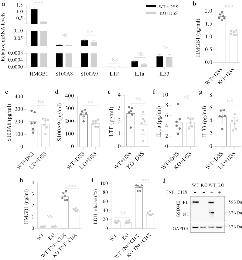
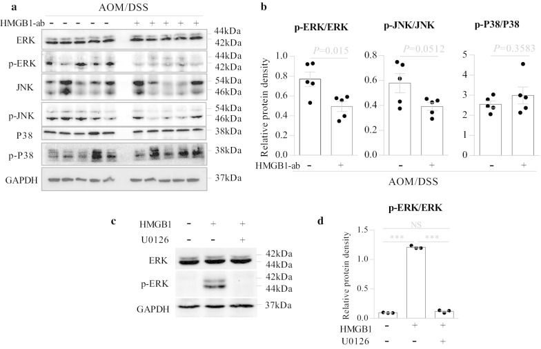
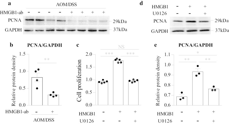
Methods: To investigate the role of gasdermin E (GSDME)-mediated pyroptosis in the development of CAC, Gsdme-/- mice and their wild-type (WT) littermate controls were challenged with azoxymethane (AOM) and dextran sodium sulfate (DSS) to induce a CAC model. Neutralizing antibodies against high-mobility group box protein 1 (HMGB1) were used to determine the role of HMGB1 in CAC. To identify the role of ERK1/2 in HMGB1-induced colon cancer cell proliferation, we performed western blotting and CCK8 assays using the ERK1/2-specific inhibitor U0126 in CT26 colon cancer cells.

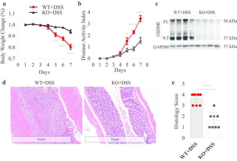
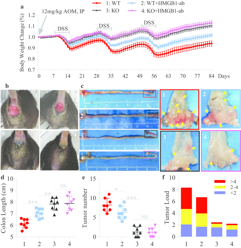
Results: In the CAC model, Gsdme-/- mice exhibited reduced weight loss and colon shortening, attenuated rectal prolapse, and reduced tumor numbers and sizes compared to WT littermates. Furthermore, treatment with neutralizing anti-HMGB1 antibodies decreased the numbers and sizes of tumors, ERK1/2 activation and proliferating cell nuclear antigen (PCNA) expression in AOM/DSS-challenged WT mice. In addition, our in vitro experiments demonstrated that HMGB1 induced proliferation and PCNA expression in CT26 colon cancer cells through the ERK1/2 pathway.

Conclusion: GSDME-mediated pyroptosis promotes the development of CAC by releasing HMGB1, which induces tumor cell proliferation and PCNA expression through the ERK1/2 pathway. This finding reveals a previously unrecognized link between pyroptosis and CAC tumorigenesis and offers new insight into CAC pathogenesis.


Conflict of interest statement

The authors declare that they have no competing interests.

Figures

Fig. 1
Fig. 1
The clinical correlation of GSDME in IBD patients. a IHC analyses of GSDME protein in the colonic mucosa from healthy controls (n = 40), UC patients (n = 42) and CD patients (n = 43). b UM un-inflamed mucosa, IM inflamed mucosa. Relative GSDME-NT levels (GSDME-NT/GAPDH) in the inflamed and un-inflamed colonic mucosa from UC patients (n = 15) and CD patients (n = 15) were further compared as determined by the paired t tests. c Representative immunoblot images of the colonic mucosa from UC and CD patients. The full length of GSDME (GSDME-FL) is shown in 56 kDa and GSDME-NT in 37 kDa. d Representative IHC images of the colonic mucosa from healthy controls, UC and CD patients. Scale bars: 400 μm. Yellow asterisks: epithelial cells. Yellow arrows: lymphocytes. NS, not significant; ***P < 0.001
Fig. 2
Fig. 2
GSDEM deletion mitigates DSS-induced colitis. Gsdme−/− (KO) mice and wild-type (WT) littermate controls were treated with 7-day DSS to induce acute colitis (n = 8 per group); then, body weight change (a) and disease activity index (b) were measured daily. c Immunoblot analyses of GSDME and GSDME-NT in the colonic mucosa of DSS mice. The full length of GSDME (GSDME-FL) is shown in 56 kDa and GSDME-NT in 37 kDa. d, e Representative HE-stained sections of middle colon tissues collected at day 7. Histopathology change reflected by semiquantitative scores (Scale bars: 1 mm). All data shown are representative of three independent experiments. *P < 0.05; **P < 0.01; ***P < 0.001
Fig. 3
Fig. 3
GSDME-mediated IEC pyroptosis increases the release of HMGB1. a IECs from Gsdme−/− (KO) mice and wild-type (WT) littermates treated with DSS for 7 days were isolated; then, total RNA was isolated and the mRNA levels of DAMPs were determined by qPCR and normalized to GAPDH. bg IECs from KO and WT littermates treated with DSS for 7 days were isolated, then washed with PBS containing Penicillin and Streptomycin for 3 times, and finally were plated in dishes with DMEM medium containing 10%FBS for 12 h. Cell medium was used for ELISA detection. hj Isolated IECs from KO and WT littermates were given TNF-α (50 ng/ml) plus CHX (20 µg/ml) treatment for 12 h. h, i Culture supernatant was collected for ELISA and LDH release assays. j Representative immunoblot images of IECs. Data are shown as means ± SEM from six mice in each group. Data shown are representative of three independent experiments. NS not significant; **P < 0.01; ***P < 0.001
Fig. 4
Fig. 4
Inhibition of HMGB1 alleviates AOM/DSS-induced CAC. Gsdme−/− (KO) mice and wild-type (WT) littermate controls were induced CAC with AOM/DSS as described above, and in this process, they were intraperitoneally injected with neutralizing anti-HMGB1 antibody (HMGB1-ab) at days 1, 3, 5 during DSS treatment (n = 8 per group). a Body weight change was measured daily. b Representative images of rectal prolapse at day 84 of the experimental procedure. c, d Representative images of the colon and the distal portion (arrows: tumor) and length values of the colon at day 84 of the experimental procedure. e, f The number of colonic tumors and the tumor size distribution according to tumor numbers (tumor load) were recorded. All data shown are representative of three independent experiments. NS not significant; *P < 0.05; **P < 0.01; ***P < 0.001
Fig. 5
Fig. 5
HMGB1 activates the ERK1/2 signaling pathway. a, b Wild-type (WT) mice were induced CAC with AOM/DSS as described above, and in this process, they were intraperitoneally injected with or without neutralizing anti-HMGB1 antibody (HMGB1-ab) at days 1, 3, 5 during DSS treatment. Tumor tissues in the colon were excised at day 84 of the experimental procedure, and whole tissue extracts from the tumors were prepared and analyzed for the phosphorylation of ERK1/2, JNK and P38 by western blotting. a The representative images. b Quantitative analyses of proteins. Data are shown as means ± SEM from five mice in each group. Data shown are representative of three independent experiments. c, d CT26 cells (1 × 105 cells/well) were seeded in 6-well plates. After 24 h, the cells were incubated for 1 h with or without U0126 (10 μM) before stimulation with HMGB1 (1 μg/ml) for 12 h; then, whole cell extracts were analyzed for the phosphorylation of ERK1/2 by western blotting. c The representative images. d Quantitative analyses of proteins. Data are shown as means ± SEM from three independent experiments. NS not significant; ***P < 0.001
Fig. 6
Fig. 6
HMGB1 induces tumor proliferation and PCNA expression through the ERK1/2 pathway. a, b Wild-type (WT) mice were induced CAC with AOM/DSS as described above, and in this process, they were intraperitoneally injected with or without neutralizing anti-HMGB1 antibody (HMGB1-ab) at days 1, 3, 5 during DSS treatment. Tumor tissues in the colon were excised at day 84 of the experimental procedure, and whole tissue extracts from the tumors were prepared and analyzed for PCNA expression by western blotting. a The representative images. b Quantitative analyses of proteins. Data are shown as means ± SEM from four mice in each group. Data shown are representative of three independent experiments. c CT26 cells (1 × 103 cells/well) were seeded in 96-well plates. After 24 h, the cells were incubated for 1 h with or without U0126 (10 μM) before stimulation with HMGB1 (1 μg/ml) for 48 h, and then, the cell proliferation activity was measured by CCK-8 assay. Data are shown as means ± SD from five wells in each group. Data shown are representative of three independent experiments. d, e CT26 cells (1 × 105 cells/well) were seeded in 6-well plates. After 24 h, the cells were incubated for 1 h with or without U0126 (10 μM) before stimulation with HMGB1 (1 μg/ml) for 48 h, and then, whole cell extracts were analyzed for PCNA expression by western blotting. d The representative images. e Quantitative analyses of proteins. Data are shown as means ± SEM from three independent experiments. NS not significant; *P < 0.05; **P < 0.01; ***P < 0.001

References

    1. Cassidy S, Syed BA. Colorectal cancer drugs market. Nat Rev Drug Discov. 2017;16:525–526. doi: 10.1038/nrd.2017.59. - DOI - PubMed
    1. Siegel RL, Miller KD, Jemal A. Cancer statistics. CA Cancer J Clin. 2020;70:7–30. doi: 10.3322/caac.21590. - DOI - PubMed
    1. Siegel RL, Miller KD, Goding Sauer A, Fedewa SA, Butterly LF, Anderson JC, et al. Colorectal cancer statistics. CA Cancer J Clin. 2020;7:145–164. doi: 10.3322/caac.21601. - DOI - PubMed
    1. Zhou CB, Fang JY. The role of pyroptosis in gastrointestinal cancer and immune responses to intestinal microbial infection. Biochim Biophys Acta. 2019;1872:1–10. - PubMed
    1. Aggarwal BB, Vijayalekshmi RV, Sung B. Targeting inflammatory pathways for prevention and therapy of cancer: short-term friend, long-term foe. Clin Cancer Res. 2009;15:425–430. doi: 10.1158/1078-0432.CCR-08-0149. - DOI - PubMed

Publication types