. 2021 Jan:162:450-460.
doi: 10.1016/j.freeradbiomed.2020.11.001. Epub 2020 Nov 6.

Selective cysteines oxidation in soluble guanylyl cyclase catalytic domain is involved in NO activation

Affiliations

Selective cysteines oxidation in soluble guanylyl cyclase catalytic domain is involved in NO activation

Maryam Alapa et al. Free Radic Biol Med. 2021 Jan.

Abstract

Nitric oxide (NO) binds to soluble guanylyl cyclase (GC1) and stimulates its catalytic activity to produce cGMP. Despite the key role of the NO-cGMP signaling in cardiovascular physiology, the mechanisms of GC1 activation remain ill-defined. It is believed that conserved cysteines (Cys) in GC1 modulate the enzyme's activity through thiol-redox modifications. We previously showed that GC1 activity is modulated via mixed-disulfide bond by protein disulfide isomerase and thioredoxin 1. Herein we investigated the novel concept that NO-stimulated GC1 activity is mediated by thiol/disulfide switches and aimed to map the specific Cys that are involved. First, we showed that the dithiol reducing agent Tris (2-carboxyethyl)-phosphine reduces GC1 response to NO, indicating the significance of Cys oxidation in NO activation. Second, using dibromobimane, which fluoresces when crosslinking two vicinal Cys thiols, we demonstrated decreased fluorescence in NO-stimulated GC1 compared to unstimulated conditions. This suggested that NO-stimulated GC1 contained more bound Cys, potentially disulfide bonds. Third, to identify NO-regulated Cys oxidation using mass spectrometry, we compared the redox status of all Cys identified in tryptic peptides, among which, ten were oxidized and two were reduced in NO-stimulated GC1. Fourth, we resorted to computational modeling to narrow down the Cys candidates potentially involved in disulfide bond and identified Cys489 and Cys571. Fifth, our mutational studies showed that Cys489 and Cys571 were involved in GC1'response to NO, potentially as a thiol/disulfide switch. These findings imply that specific GC1 Cys sensitivity to redox environment is critical for NO signaling in cardiovascular physiology.

Keywords: Allosteric regulation; Crosslinking; Cyclic GMP (cGMP); Disulfide; Nitric oxide (NO); Soluble guanylyl cyclase (GC1); Thiol.


Conflict of interest statement

Conflict of interest

The authors declare that they have no conflicts of interest with the contents of this article. The content is solely the responsibility of the authors and does not necessarily represent the official views of the National Institutes of Health.

Figures

Figure 1:
Figure 1:
A. DEA-NO concentration-response curves of GC1 ± TCEP. GC1 activity was measured in response to increasing concentrations of DEA-NO without (●) or with (○) 0.5 mM TCEP. The results are expressed in nmol cGMP.mg−1.min−1 ± SEM. n=3 independent experiments. B. Fluorescent intensity of DiBrb-bound GC1 (400 ng) as a function of increasing concentrations of DiBrb. C. Representative spectra of GC1 (100ng) cross-linked with 50 μM DiBrb after being stimulated with DEA-NO (10 μM) or under basal condition; n=6.
Figure 2.
Figure 2.
NO-stimulated free thiols changes in GC1. Free thiols under basal and NO-stimulation were alkylated with NEM and quantified by MS. Levels of free thiols were determined from levels of NEM-alkylated Cys and normalized to basal values (set at 100%). The redox status of 7 Cys in the α subunit and 5 Cys in the β subunit were significantly modified upon NO-stimulation. The results ± SEM are expressed as percentage of change in free thiols in the presence of NO. n=3. *, p < 0.05 and **, p < 0.01.
Figure 3:
Figure 3:
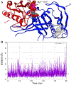
Computational Model of GC1 catalytic domain and simulation of β1 Cys489 and Cys571 distance. A. Modeling of the catalytic domain of GC1 from PDB file 4NI2 with docked GTP (sphere) and Mg2+ ions (see Experimental Procedures). A distance calculation predicts that the two Cys (β1 Cys489 and β1 Cys571) are 4.6 Å apart and oriented to form a possible disulfide. B. Molecular dynamics simulation to assess whether Cys489 and Cys571 are close enough to form a disulfide bond. The x-axis, scaled by 10 ps, represents the simulation duration and y-axis represents the distance between Cys489 and Cys571 at a time. The cutoff distance for disulfide formation is 4Å (indicated by a dotted line) between the sulfide groups of the two Cys. The distance stayed below and above 4Å for 51 % and 38 % of the time, respectively.
Figure 4.
Figure 4.
DEA-NO dose response curves of GC1 and mutants with and without TCEP. The activity of GC1 WT (A) and mutants αβ Cys489S (B), αβ Cys571A (C) and αβ CysDM (D) was measured with and without TCEP at different concentrations of DEA-NO. Results are expressed in nmol cGMP mg−1min−1 ± SEM.
Figure 5:
Figure 5:
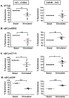
Purified GC1 WT and mutants show different fluorescent patterns under basal and NO-stimulation. Fluorescence of WT (A), αβCys489S (B), αβCys571A (C), and αβCysDM (D) cross-linked with 50 μM DiBrb, following (left panel) or preceding (right panel) 10 μM NO stimulation. The samples were excited at 385 nm and emission scan was done at a range of 400–600 nm. Analysis was done using Student’s T-test and p < 0.05 was considered statistically significant. * p < 0.05, ** p < 0.01, ***p < 0.001. To normalize the analysis, raw basal values were set at 100. It should be noted that the fluorescence spectra (raw values) were lower in the mutants compared to WT (except for C571A). For example, basal intensity for the WT was 1.37 ± 0.07 vs. 1.21 ± 0.03 for the CysDM.
Figure 6.
Figure 6.
DiBrb effect on NO-stimulated activity of purified GC1 WT and mutants. WT (A), αβCys489S (B) αβCys571A (C) or αβCysDM (D) were incubated with vehicle, 10 or 50 μM DiBrb prior to stimulation with 10μM of DEA-NO. The treated samples were normalized to the control (vehicle), which was set at 100 %. Statistical analysis was performed using one-way ANOVA and p< 0.05 was deemed significant (* p < 0.05, ** p < 0.01, ***p < 0.001).
Scheme. 1.
Scheme. 1.
DiBrb crosslinking design: (A) DiBrb was added after addition of vehicle or NO; (B) DiBrb was added before addition of vehicle or NO.

References

    1. Haase T, Haase N, Kraehling JR, Behrends S, Fluorescent fusion proteins of soluble guanylyl cyclase indicate proximity of the heme nitric oxide domain and catalytic domain, PLoS One 5(7) (2010) e11617. - PMC - PubMed
    1. Underbakke ES, Iavarone AT, Chalmers MJ, Pascal BD, Novick S, Griffin PR, Marletta MA, Nitric oxide-induced conformational changes in soluble guanylate cyclase, Structure 22(4) (2014) 602–11. - PMC - PubMed
    1. Fritz BG, Roberts SA, Ahmed A, Breci L, Molecular Model of a soluble guanylyl cyclase fragment determined by small-angle X-ray Scattering and Chemical Cross-Linking, Biochemistry 52(9) (2013) 1568–1582. - PMC - PubMed
    1. Campbell MG, Underbakke ES, Potter CS, Carragher B, Marletta MA, Single-particle EM reveals the higher-order domain architecture of soluble guanylate cyclase, Proc Natl Acad Sci U S A 111(8) (2014) 2960–5. - PMC - PubMed
    1. Horst BG, Yokom AL, Rosenberg DJ, Morris KL, Hammel M, Hurley JH, Marletta MA, Allosteric activation of the nitric oxide receptor soluble guanylate cyclase mapped by cryo-electron microscopy, eLife 8 (2019) e50634. - PMC - PubMed

Publication types

LinkOut - more resources