

Neuroimaging findings of brain MRI and CT in patients with COVID-19: A systematic review and meta-analysis
- PMID: 33161199
- PMCID: PMC7606068
- DOI: 10.1016/j.ejrad.2020.109393
Neuroimaging findings of brain MRI and CT in patients with COVID-19: A systematic review and meta-analysis
Abstract
Purpose: To comprehensively evaluate the incidences of abnormal neuroimaging findings in patients with COVID-19 via a systematic review and meta-analysis.
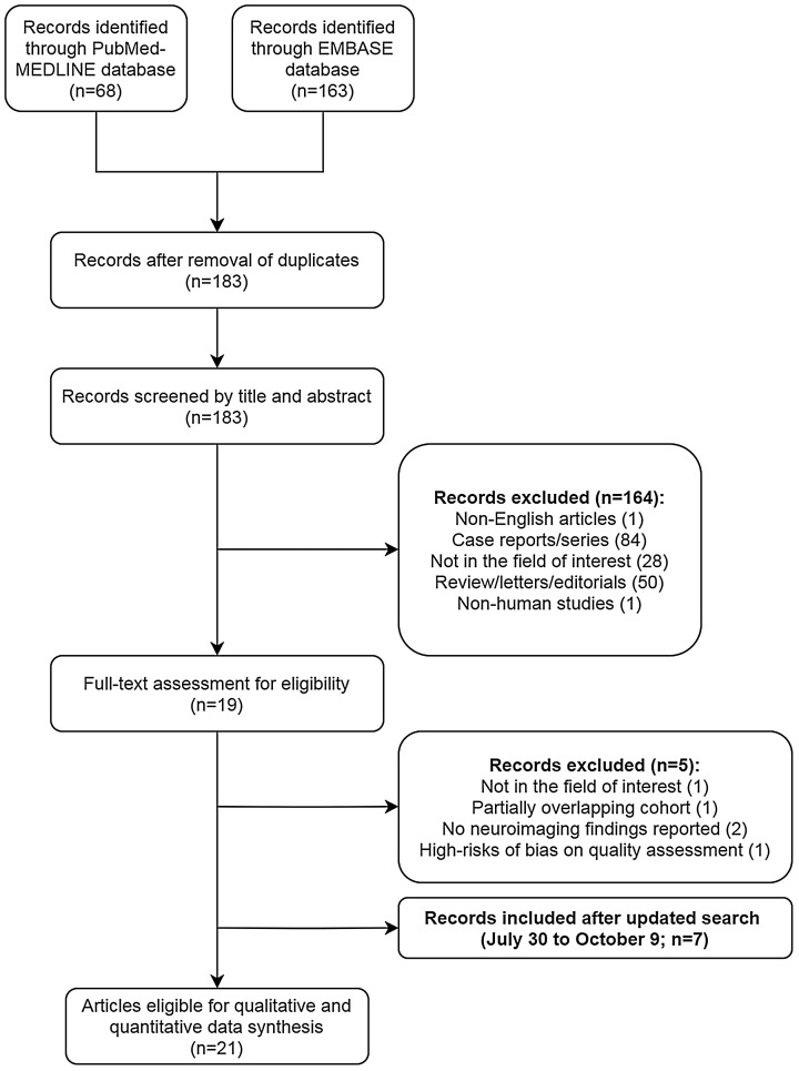
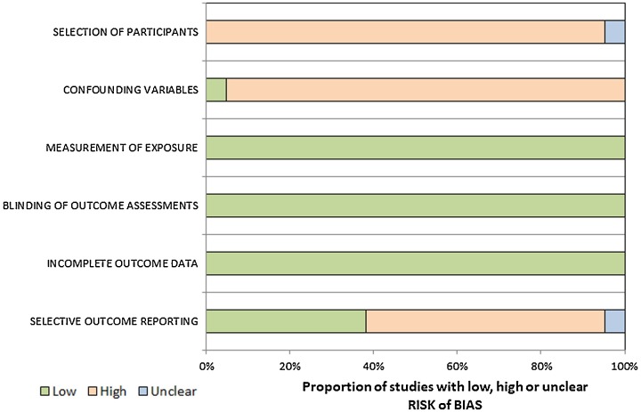
Method: PubMed-MEDLINE and EMBASE were searched for original articles reporting imaging findings of the brain in adult patients with COVID-19 between January 1, 2020 and October 9, 2020. Abnormal neuroimaging findings were categorized as (1) cerebral microhemorrhages, (2) acute spontaneous intracranial hemorrhage (ICH), (3) acute to subacute infarcts, and (4) encephalitis or encephalopathy. Pooled incidences of neuroimaging findings were assessed using random-effects modeling. Between-study heterogeneity was explored by using the χ2 statistic for pooled incidences and the inconsistency index I2. The quality of the studies was evaluated using the Risk of Bias Assessment Tool for Nonrandomized Studies. Subgroup meta-regression analysis was performed to identify potential sources of heterogeneity.
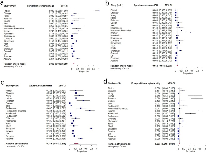
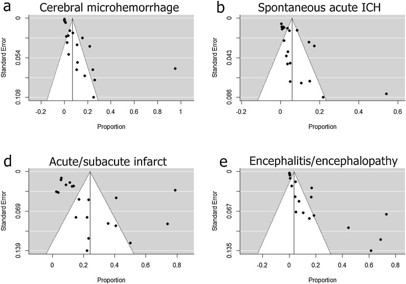
Results: Twenty-one eligible papers, including 2125 patients, were identified. The pooled incidences of cerebral microhemorrhages, acute spontaneous ICH, acute/subacute infarcts, and encephalitis/encephalopathy were 6.9 % (95 % confidence interval [CI], 4.9 %-8.9 %), 5.4 % (95 % CI, 3.1 %-7.6 %), 24.0 % (95 % CI, 16.1 %-31.8 %), and 3.3 % (95 % CI, 1.9 %-4.7 %), respectively. Substantial heterogeneities were noted for all neuroimaging findings (I2 = 87 %-97 %). Significant publication biases were present in the pooled incidences. In the subgroup meta-regression analysis, patients with mean or median ages over 65 years showed a significantly lower incidence of encephalitis/encephalopathy (P < 0.001). Furthermore, studies reported that patients in ICU had significantly higher incidences of cerebral microhemorrhages (P < 0.001) and encephalitis/encephalopathy (P < 0.001).
Conclusions: Considerable incidences of abnormal neuroimaging findings have been reported in patients with COVID-19. Acute to subacute cerebral infarction was the most prevalent neuroimaging finding.
Keywords: Brain diseases; COVID-19; Neuroimaging; Prevalence; Severe acute respiratory syndrome coronavirus 2.
Copyright © 2020 Elsevier B.V. All rights reserved.
Conflict of interest statement
The authors report no declarations of interest.
Figures
Similar articles
-
The Spectrum of Neuroimaging Findings on CT and MRI in Adults With COVID-19.AJR Am J Roentgenol. 2021 Oct;217(4):959-974. doi: 10.2214/AJR.20.24839. Epub 2020 Nov 25. AJR Am J Roentgenol. 2021. PMID: 33236647 Review.
-
Special report of the RSNA COVID-19 task force: systematic review of outcomes associated with COVID-19 neuroimaging findings in hospitalized patients.Br J Radiol. 2021 Nov 1;94(1127):20210149. doi: 10.1259/bjr.20210149. Epub 2021 Apr 29. Br J Radiol. 2021. PMID: 33914618 Free PMC article.
-
Brain and Lung Imaging Correlation in Patients with COVID-19: Could the Severity of Lung Disease Reflect the Prevalence of Acute Abnormalities on Neuroimaging? A Global Multicenter Observational Study.AJNR Am J Neuroradiol. 2021 Jun;42(6):1008-1016. doi: 10.3174/ajnr.A7072. Epub 2021 Mar 11. AJNR Am J Neuroradiol. 2021. PMID: 33707278 Free PMC article.
-
Severity of Chest Imaging is Correlated with Risk of Acute Neuroimaging Findings among Patients with COVID-19.AJNR Am J Neuroradiol. 2021 May;42(5):831-837. doi: 10.3174/ajnr.A7032. Epub 2021 Feb 4. AJNR Am J Neuroradiol. 2021. PMID: 33541897 Free PMC article.
-
Neuroimaging findings in children with COVID-19 infection: a systematic review and meta-analysis.Sci Rep. 2024 Feb 27;14(1):4790. doi: 10.1038/s41598-024-55597-2. Sci Rep. 2024. PMID: 38413808 Free PMC article.
Cited by
-
Is a high chest CT severity score a risk factor for an increased incidence of long-term neuroimaging findings after COVID-19?J Stroke Cerebrovasc Dis. 2023 Feb;32(2):106920. doi: 10.1016/j.jstrokecerebrovasdis.2022.106920. Epub 2022 Nov 30. J Stroke Cerebrovasc Dis. 2023. PMID: 36516593 Free PMC article.
-
Neurological pathophysiology of SARS-CoV-2 and pandemic potential RNA viruses: a comparative analysis.FEBS Lett. 2021 Dec;595(23):2854-2871. doi: 10.1002/1873-3468.14227. Epub 2021 Nov 22. FEBS Lett. 2021. PMID: 34757622 Free PMC article. Review.
-
Brain MRI findings in neurologically symptomatic COVID-19 patients: a systematic review and meta-analysis.J Neurol. 2023 Nov;270(11):5131-5154. doi: 10.1007/s00415-023-11914-9. Epub 2023 Aug 3. J Neurol. 2023. PMID: 37535100 Review.
-
Abnormalities of brain imaging in COVID-19 patients with neurological symptoms.Curr J Neurol. 2023 Jul 6;22(3):162-169. doi: 10.18502/cjn.v22i3.13796. Curr J Neurol. 2023. PMID: 38011453 Free PMC article.
-
Construction of nanomaterials as contrast agents or probes for glioma imaging.J Nanobiotechnology. 2021 May 3;19(1):125. doi: 10.1186/s12951-021-00866-9. J Nanobiotechnology. 2021. PMID: 33941206 Free PMC article. Review.
References
-
- COVID-19 Map: Johns Hopkins Coronavirus Resource Center. https://coronavirus.jhu.edu/map.html. (Accessed October 21,2020.
