

Crosstalk between endoplasmic reticulum stress and oxidative stress in the progression of diabetic nephropathy
- PMID: 33161510
- PMCID: PMC7925747
- DOI: 10.1007/s12192-020-01176-z
Crosstalk between endoplasmic reticulum stress and oxidative stress in the progression of diabetic nephropathy
Abstract
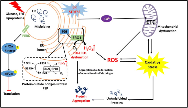
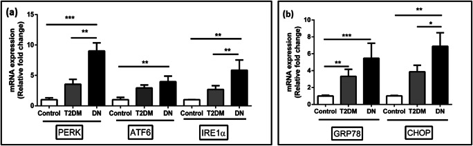
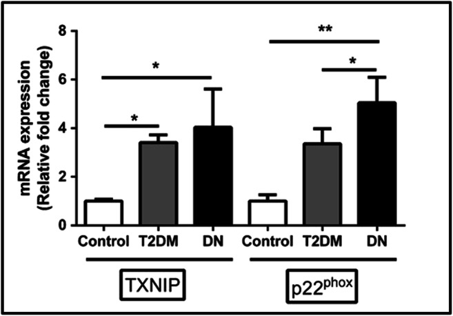
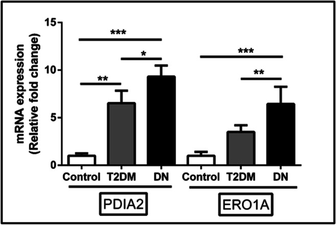
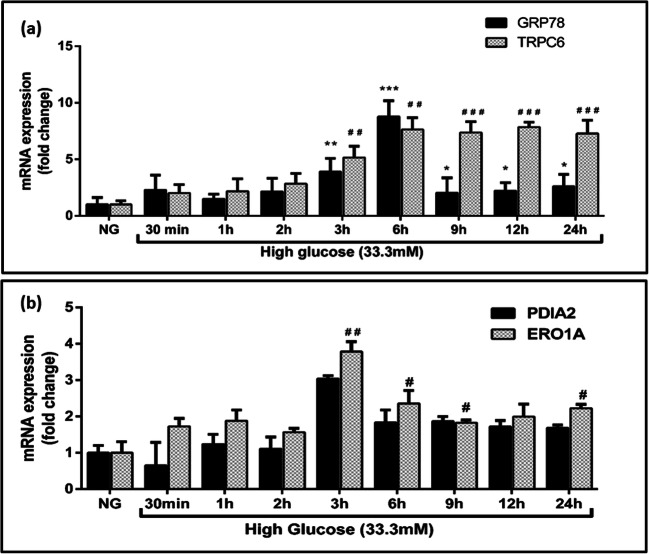
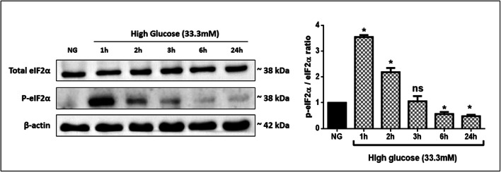
Increasing evidence in substantiating the roles of endoplasmic reticulum stress, oxidative stress, and inflammatory responses and their interplay is evident in various diseases. However, an in-depth mechanistic understanding of the crosstalk between the intracellular stress signaling pathways and inflammatory responses and their participation in disease progression has not yet been explored. Progress has been made in our understanding of the cross talk and integrated stress signaling network between endoplasmic reticulum stress and oxidative stress towards the pathogenesis of diabetic nephropathy. In this present study, we studied the crosstalk between the endoplasmic reticulum stress and oxidative stress by understanding the role of protein disulfide isomerase and endoplasmic reticulum oxidase 1α, a key player in redox protein folding in the endoplasmic reticulum. We had recruited a total of 90 subjects and divided into three groups (control (n = 30), type 2 diabetes mellitus (n = 30), and diabetic nephropathy (n = 30)). We found that endoplasmic reticulum stress markers, activating transcription factor 6, inositol-requiring enzyme 1α, protein kinase RNA-like endoplasmic reticulum kinase, C/EBP homologous protein, and glucose-regulated protein-78; oxidative stress markers, thioredoxin-interacting protein and cytochrome b-245 light chain; and the crosstalk markers, protein disulfide isomerase and endoplasmic reticulum oxidase-1α, were progressively elevated in type 2 diabetes mellitus and diabetic nephropathy subjects. The association between the crosstalk markers showed a positive correlation with endoplasmic reticulum stress and oxidative stress markers. Further, the interplay between endoplasmic reticulum stress and oxidative stress was investigated in vitro using a human leukemic monocytic cell line under a hyperglycemic environment and examined the expression of protein disulfide isomerase and endoplasmic reticulum oxidase-1α. DCFH-DA assay and flow cytometry were performed to detect the production of free radicals. Further, phosphorylation of eIF2α in high glucose-exposed cells was studied using western blot. In conclusion, our results shed light on the crosstalk between endoplasmic reticulum stress and oxidative stress and significantly contribute to the onset and progression of diabetic nephropathy and therefore represent the major therapeutic targets for alleviating micro- and macrovascular complications associated with this metabolic disturbance. Graphical abstract.
Keywords: Crosstalk; Diabetic nephropathy; ER oxidase 1α; Endoplasmic reticulum stress; Oxidative stress; Protein disulfide isomerase.
Conflict of interest statement
The authors declare that they have no conflict of interest.
Figures

Similar articles
-
Crosstalk between endoplasmic reticulum stress and oxidative stress: Focus on protein disulfide isomerase and endoplasmic reticulum oxidase 1.Eur J Pharmacol. 2021 Feb 5;892:173749. doi: 10.1016/j.ejphar.2020.173749. Epub 2020 Nov 25. Eur J Pharmacol. 2021. PMID: 33245896 Review.
-
Sodium-Glucose Cotransporter-2 Inhibitor Suppresses Endoplasmic Reticulum Stress and Oxidative Stress in Diabetic Nephropathy Through Nrf2 Signaling: A Clinical and Experimental Study.J Clin Pharmacol. 2024 Oct;64(10):1193-1203. doi: 10.1002/jcph.2465. Epub 2024 Jun 4. J Clin Pharmacol. 2024. PMID: 38831713
-
Superoxide dismutase 1 overexpression in mice abolishes maternal diabetes-induced endoplasmic reticulum stress in diabetic embryopathy.Am J Obstet Gynecol. 2013 Oct;209(4):345.e1-7. doi: 10.1016/j.ajog.2013.06.037. Epub 2013 Jun 20. Am J Obstet Gynecol. 2013. PMID: 23791840 Free PMC article.
-
Terpene glycoside component from Moutan Cortex ameliorates diabetic nephropathy by regulating endoplasmic reticulum stress-related inflammatory responses.J Ethnopharmacol. 2016 Dec 4;193:433-444. doi: 10.1016/j.jep.2016.09.043. Epub 2016 Sep 21. J Ethnopharmacol. 2016. PMID: 27664441
-
Stress in the kidney is the road to pERdition: is endoplasmic reticulum stress a pathogenic mediator of diabetic nephropathy?J Endocrinol. 2014 Sep;222(3):R97-111. doi: 10.1530/JOE-13-0517. Epub 2014 Jun 30. J Endocrinol. 2014. PMID: 24982467 Review.
Cited by
-
Single Nucleotide Polymorphisms of the RAC1 Gene as Novel Susceptibility Markers for Neuropathy and Microvascular Complications in Type 2 Diabetes.Biomedicines. 2023 Mar 22;11(3):981. doi: 10.3390/biomedicines11030981. Biomedicines. 2023. PMID: 36979960 Free PMC article.
-
The involvement of PDIA2 gene in the progression of renal cell carcinoma is potentially through regulation of JNK signaling pathway.Clin Transl Oncol. 2023 Oct;25(10):2938-2949. doi: 10.1007/s12094-023-03158-w. Epub 2023 Apr 5. Clin Transl Oncol. 2023. PMID: 37017923
-
Crosstalk of Hyperglycaemia and Cellular Mechanisms in the Pathogenesis of Diabetic Kidney Disease.Int J Mol Sci. 2024 Oct 10;25(20):10882. doi: 10.3390/ijms252010882. Int J Mol Sci. 2024. PMID: 39456664 Free PMC article. Review.
-
Exosomes as nanostructures deliver miR-204 in alleviation of mitochondrial dysfunction in diabetic nephropathy through suppressing methyltransferase-like 7A-mediated CIDEC N6-methyladenosine methylation.Aging (Albany NY). 2024 Feb 8;16(4):3302-3331. doi: 10.18632/aging.205535. Epub 2024 Feb 8. Aging (Albany NY). 2024. PMID: 38334961 Free PMC article.
-
Crocin protects against endoplasmic reticulum stress-related tubular injury in diabetic nephropathy via the activation of the PI3K/AKT/Nrf2 pathway.Iran J Basic Med Sci. 2024;27(4):439-446. doi: 10.22038/IJBMS.2023.73385.15942. Iran J Basic Med Sci. 2024. PMID: 38419890 Free PMC article.
References
-
- Back SH, Scheuner D, Han J, Song B, Ribick M, Wang J, Gildersleeve RD, Pennathur S, Kaufman RJ. Translation attenuation through eIF2alpha phosphorylation prevents oxidative stress and maintains the differentiated state in beta cells. Cell Metab. 2009;10:13–26. doi: 10.1016/j.cmet.2009.06.002. - DOI - PMC - PubMed
Publication types
MeSH terms
Substances
LinkOut - more resources
Full Text Sources
Medical
