. 2020 Oct 26:2020:1693730.
doi: 10.1155/2020/1693730. eCollection 2020.

Expression of AIM2 in Rheumatoid Arthritis and Its Role on Fibroblast-Like Synoviocytes

Affiliations

Expression of AIM2 in Rheumatoid Arthritis and Its Role on Fibroblast-Like Synoviocytes

Yong Chen et al. Mediators Inflamm. .

Abstract

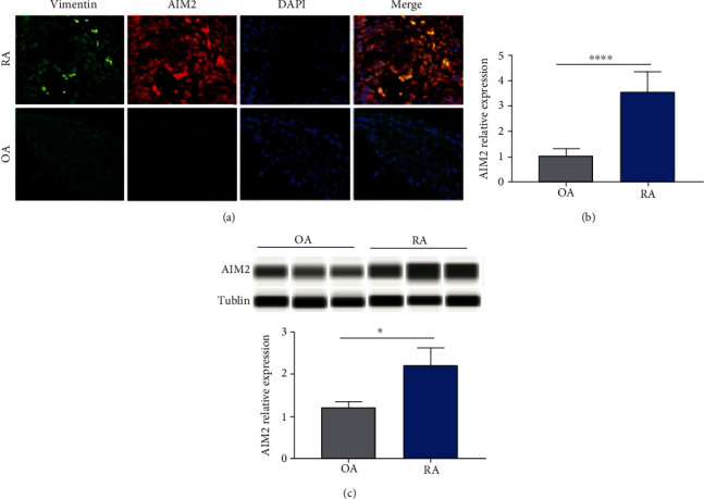
Objectives: To determine differences in AIM2 inflammasome expression levels between rheumatoid arthritis (RA) and osteoarthritis (OA) and to investigate the role of AIM2 in RA fibroblast-like synoviocytes (RA-FLS).

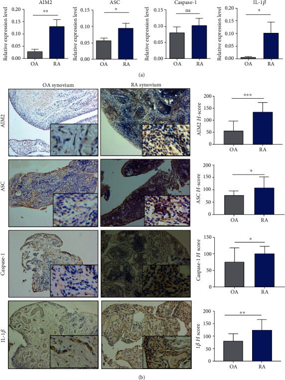
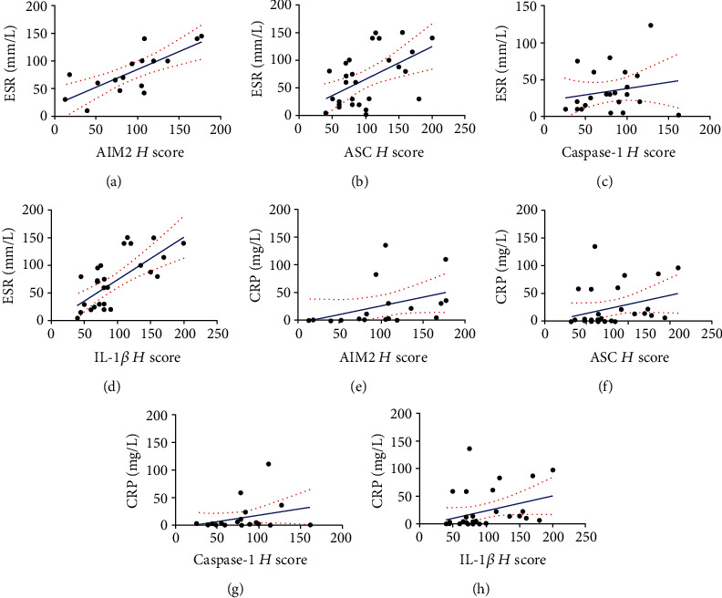
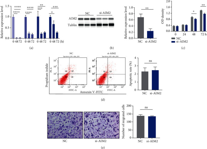
Methods: Serum AIM2 levels among health controls (HC, n = 20), OA (n = 25), and RA (n =49) patients were compared via ELISA. The different expression levels of AIM2, ASC, caspase-1, and IL-1β between RA and OA synovium were semiquantified by qRT-PCR and immunohistochemical (IHC) staining. IHC staining was recorded by H scores, and its correlation with the ESR and CRP levels of RA patients was determined. SiRNA AIM2 was transferred to RA-FLS and its effects on the proliferation and migration via CCK-8 assay and Transwell test, respectively.

Results: In RA sera, the HC expressed higher level of AIM2 than OA and RA patients, and ASC, caspase-1, and IL-1β expressed higher in RA patients than HC; no significant differences were observed between sera of OA and RA patients. However, in affected knee synovium, AIM2, ASC, caspase-1, and IL-1β were expressed higher in RA than that of OA. Moreover, the H scores of AIM2, ASC, and IL-1β were positively correlated with the ESR and CRP levels in RA patients. The proliferation of FLS was significantly inhibited after transferring with AIM2 siRNA to FLS. There were no differences in apoptosis and migration assay between the si-AIM2 group and the control group.

Conclusion: AIM2 inflammasome pathway involves in the pathogenesis of RA. si-AIM2 inhibits the proliferation of RA-FLS, which may be a promising therapeutic strategy for the treatment of RA.


Conflict of interest statement

None declared.

Figures

Figure 1
Figure 1
The differences in AIM2 pathway-related molecules in sera among HCs, OA, and RA patients. The HCs expressed obviously higher level of AIM2 than OA and RA patients (a), and ASC, caspase-1, and IL-1β expressed obviously higher in RA patients than HCs (b–d). No statistical differences of these molecules between OA and RA (a–d). p < 0.05, ∗∗∗p < 0.001, and ∗∗∗∗p < 0.0001.
Figure 2
Figure 2
The differences in AIM2 pathway-related molecules in synovium among OA and RA. mRNA AIM2 and AIM2 pathway-related proteins including ASC and IL-1β were expressed higher in RA than in OA; caspase-1 was higher in RA than in OA without statistical significance (a). H score for the synovium of RA, AIM2, and AIM2 pathway-related proteins including ASC, caspase-1, and IL-1β was more expressed than that of OA (b). p < 0.05, ∗∗p < 0.01, and ∗∗∗∗p < 0.0001.
Figure 3
Figure 3
H scores of AIM2, ASC, and IL-1β were positively correlated with ESR and CRP. AIM2 demonstrated a positive correlation with ESR (r = 0.74, p = 0.001, 95% CI: 0.38-0.9) and CRP (r = 0.65, p = 0.003, 95% CI: 0.25-0.86) (a, e). The positive correlations between ASC and ESR (r = 0.5, p = 0.005, 95% CI: 0.16-0.74) as well as CRP (r = 0.42, p = 0.02, 95% CI: 0.05-0.69) were detected (b, f). The positive correlations between IL-1β and ESR (r = 0.62, p = 0.0004, 95% CI: 0.31-0.81) and CRP (r = 0.41, p = 0.02, 95% CI: 0.05-0.67) were detected. The correlations between CASPSE-1 and ESR as well as CRP were not statistically significant (p > 0.05) (c, d).
Figure 4
Figure 4
AIM2 is expressed more in RA-FLS than in OA. AIM2 expressed in the cytoplasm of FLS, which is higher in RA-FLS than in OA-FLS ((a) vimentin was labelled as green, AIM2 as red, and nuclei as blue by immunofluorescent staining). The relative expression of AIM2 in mRNA and protein levels was higher in RA-FLS than in OA (b, c). p < 0.05 and ∗∗∗∗p < 0.0001.
Figure 5
Figure 5
Proliferation, apoptosis, and migration assay of RA-FLS after si-AIM2 transfection. AIM2 and its downstream proteins including ASC, caspase-1, and IL-1β were successfully silenced after 24 h of transfection (a). Western blot confirmed the successful silencing of the AIM2 expression by si-AIM2 (b). The proliferation of RA-FLS was inhibited after 48 h of transfection (c). There were no significant differences between the NC and si-AIM2 groups in apoptosis and migration assay (d, e). p < 0.05, ∗∗p < 0.01, ∗∗∗p < 0.001, and ∗∗∗∗p < 0.0001.

Similar articles

Cited by

References

    1. Calabresi E., Petrelli F., Bonifacio A. F., Puxeddu I., Alunno A. One year in review 2018: pathogenesis of rheumatoid arthritis. Clinical and Experimental Rheumatology. 2018;36(2):175–184. - PubMed
    1. Mahajan T. D., Mikuls T. R. Recent advances in the treatment of rheumatoid arthritis. Current Opinion in Rheumatology. 2018;30(3):231–237. doi: 10.1097/BOR.0000000000000496. - DOI - PMC - PubMed
    1. The RACI consortium, the GARNET consortium, Okada Y., et al. Genetics of rheumatoid arthritis contributes to biology and drug discovery. Nature. 2014;506(7488):376–381. doi: 10.1038/nature12873. - DOI - PMC - PubMed
    1. Smith S., Jefferies C. Role of DNA/RNA sensors and contribution to autoimmunity. Cytokine & Growth Factor Reviews. 2014;25(6):745–757. doi: 10.1016/j.cytogfr.2014.07.019. - DOI - PubMed
    1. Li W. C., Bai D. L., Xu Y., et al. Identification of differentially expressed genes in synovial tissue of rheumatoid arthritis and osteoarthritis in patients. Journal of Cellular Biochemistry. 2018;120(3):4533–4544. doi: 10.1002/jcb.27741. - DOI - PubMed