. 2020 Oct 9:11:2026.
doi: 10.3389/fimmu.2020.02026. eCollection 2020.

Cancer-Associated Fibroblasts-Derived Exosomes Suppress Immune Cell Function in Breast Cancer via the miR-92/PD-L1 Pathway

Affiliations

Cancer-Associated Fibroblasts-Derived Exosomes Suppress Immune Cell Function in Breast Cancer via the miR-92/PD-L1 Pathway

Dongwei Dou et al. Front Immunol. .

Retraction in

Abstract

Cancer-associated fibroblasts (CAFs) are an essential component in the tumor microenvironment and have been reported to contribute to tumor progression through many mechanisms; however, the detailed mechanism underlying the immune-suppression effect of CAFs is not clearly defined. In this study, human breast cancer-derived CAFs were cultured, and CAF-derived exosomes in a culture medium were isolated. Using a miRNA profiles assay, we identify a significantly higher level of microRNA-92 isolated in CAFs exosomes. After treatment by CAF-derived exosomes, breast cancer cells express higher programmed cell death receptor ligand 1 (PD-L1), accompanied with increased miR-92 expression. Increased PD-L1 expression, which was induced by CAF-derived exosomes, significantly promotes apoptosis and impaired proliferation of T cells. The underlying mechanism of this effect was studied, proliferation and migration of breast cancer cells were increased after the transfection of miR-92, LATS2 was recognized as a target gene of miR-92, and further confirmed by a luciferase assay. Immunoprecipitation showed that LATS2 can interact with YAP1, chromatin immunoprecipitation confirmed that after nuclear translocation YAP1 could bind to the enhancer region of PD-L1 to promotes transcription activity. Furthermore, the animal study confirmed that CAFs significantly promoted tumor progression and impaired the function of tumor-infiltrated immune cells in vivo. Our data revealed a novel mechanism that can induce immune suppression in the tumor microenvironment.

Keywords: CAF; PD-L1; YAP1; breast cancer; miR-92.


Figures

FIGURE 1
FIGURE 1
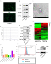
Cancer associated fibroblast-derived exosomes promote miR-92 and PD-L1 expression in breast cancer. (A) Immunofluorescence image of FSP and vimentin in breast cancer-derived cancer associated fibroblasts. (B) Western blot analysis of FSP, vimentin, α-SMA expression in cancer-associated fibroblasts and normal fibroblasts. (C) Transmission electron microscopy image of exosomes. (D) TRPS analysis of exosomes confirming the expected size range of 30–150 nm in diameter. (E) Western blot analysis of exosome markers in CAF-derived exosomes and cell lysates. (F) A clustergram performed hierarchical clustering of the upregulated and downregulated miRNAs in exosomes isolated from CAFs compared with normal fibroblasts cells. The data display a heat map with an indication of co-regulated genes across groups or individual samples. The magnitude of gene expression is presented by colors as indicated by the color bar shown on the right. (G) micro-RNA expression after exosomes treatment (H) flow cytometry analysis of PD-L1 expression in breast cancer cells. (I) PD-L1 expression analyzed by Western blot. Error bars represent mean ± SD; ****P < 0.0001; n.s., not significant; by paired two-sided Student’s t-test.
FIGURE 2
FIGURE 2
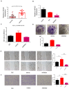
miR-92 promotes migration and proliferation of breast cancer cells. (A) miR-92 expression was examined by qRT-PCR in human breast cancer tissue and normal tissue. In total, 32 cancer tissues and 34 normal tissues were analyzed. (B) miR-92 expression after transfection of miR-92 mimic or inhibitor was analyzed by qRT-PCR (n = 3). (C) miR-92 overexpression promotes cell proliferation. Cancer cells were transfected with a miR-92 mimic or inhibitor and the absorption (A450 nm) was detected at 0, 24, 48, and 72 h, respectively. (D) Cancer cells transfected with the miR-92 mimic and NC control were assayed for clonogenicity in adherent cultures. (E) A wound-healing closure assay for cancer cells which transfected with the miR-92 mimic or inhibitor (magnification, ×50, scale bar: 500 μm). (F) Transwell assay for cancer cells which transfected with the miR-92 mimic or inhibitor (magnification, ×100). Error bars represent mean ± SD; ****P < 0.0001; n.s., not significant; by paired two-sided Student’s t-test.
FIGURE 3
FIGURE 3
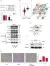
miR-92 targeting LATS2 and enhance nuclear translocation of YAP1. (A) Schematic representation of the miR-92 targeting sequences within the 3′UTR of LATS2. A luciferase reporter assay was conducted in MCF7 cells following transfection with the miR-92 mimic or NC, and together with WT or Mut LATS2 3′UTR luciferase reporter plasmid. (B) LATS2 expression was examined in normal and breast cancer tissue from a public dataset. (C) The protein–protein network view from the STRING database showing the networks of LATS2. (D) Western blot shows the YAP1, LATS2 expression in MCF7 which transfected with the miR-92 mimic or inhibitor. (E) Co-IP analysis with anti-YAP1 antibody or IgG in MCF7. (F) Western blot analysis of YAP expression after two shRNA target YAP was transfected. (G) Transwell assay for breast cancer cells which transfected with the miR-92 mimic and/or shRNA targeting YAP (magnification, ×100). Error bars represent mean ± SD; ****P < 0.0001; n.s., not significant; by paired two-sided Student’s t-test.
FIGURE 4
FIGURE 4
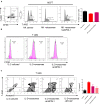
miR-92 promotes PD-L1 transcription activity by enhancing the nuclear translocation of YAP1. (A) Western blot shows the YAP1, LATS2, and PD-L1 expression in MCF7 which was transfected with the miR-92 mimic or inhibitor. (B) Flow-cytometry analysis for PD-L1 expression in MCF7 which was transfected with NC or miR-92. Blank is an unstained group indicating negative fluorescence intensity. (C) ChIP assay was performed with MCF7 which is shown by gel bands of the RT-PCR products with 30 cycle. (D) Western blot shows the YAP1 and PD-L1 expression in MCF7 which was transfected with the miR-92 mimic or inhibitor or/and shRNA targeting YAP.
FIGURE 5
FIGURE 5
Exosomes induced PD-L1 upregulation suppresses immune cell function in breast cancer. (A) An NK cell cytotoxicity assay was performed, MCF7 treated or untreated by exosomes were used as target cells. The quantification of cytotoxicity of NK cells were shown in right panel. (B) CFSE was used to evaluate the proliferation of T cells. T cells were cultured directly with MCF7 treated or untreated by exosomes for 48 h. (C) T cells were cultured directly with MCF7 treated or untreated by exosomes for 48 h, the apoptosis rate was evaluated by an Annexin V stain. Error bars represent mean ± SD; ****P < 0.0001; n.s., not significant; by paired two-sided Student’s t-test.
FIGURE 6
FIGURE 6
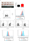
Cancer associated fibroblasts promote tumor progression and suppress immune cell function in vivo. (A) The weight of tumors isolated from the mice with MA-782 cells co-injected with or without MSC (n = 8). (B) PD-L1 expression of MA-782 which gated as mCherry+. (C,D) CD107a expression of cytotoxicity T cells and NK cells. Error bars represent mean ± SD; ****P < 0.0001; n.s., not significant; by paired two-sided Student’s t-test.

References

    1. Hutchinson L. Challenges, controversies, breakthroughs. Nat Rev Clin Oncol. (2010) 7:669–70. 10.1038/nrclinonc.2010.192 - DOI - PubMed
    1. Woolston C. Breast cancer. Nature. (2015) 527:S101–101. 10.1038/527S101a - DOI - PubMed
    1. Sabatier R, Finetti P, Mamessier E, Adelaide J, Chaffanet M, Ali HR, et al. Prognostic and predictive value of PDL1 expression in breast cancer. Oncotarget. (2015) 6:5449–64. 10.18632/oncotarget.3216 - DOI - PMC - PubMed
    1. Samanta D, Park Y, Ni X, Li H, Zahnow CA, Gabrielson E, et al. Chemotherapy induces enrichment of CD47 +/CD73 +/PDL1 + immune evasive triple-negative breast cancer cells. Proc Natl Acad Sci USA. (2018) 115:E1239–48. 10.1073/pnas.1718197115 - DOI - PMC - PubMed
    1. Houthuijzen JM, Jonkers J. Cancer-associated fibroblasts as key regulators of the breast cancer tumor microenvironment. Cancer Metastasis Rev. (2018) 37:577–97. 10.1007/s10555-018-9768-3 - DOI - PubMed

Publication types

MeSH terms