. 2020 Oct 1;10(10):3248-3266.
eCollection 2020.

Blockade of IGF/IGF-1R signaling axis with soluble IGF-1R mutants suppresses the cell proliferation and tumor growth of human osteosarcoma

Affiliations

Blockade of IGF/IGF-1R signaling axis with soluble IGF-1R mutants suppresses the cell proliferation and tumor growth of human osteosarcoma

Daigui Cao et al. Am J Cancer Res. .

Abstract

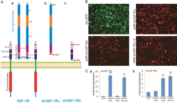
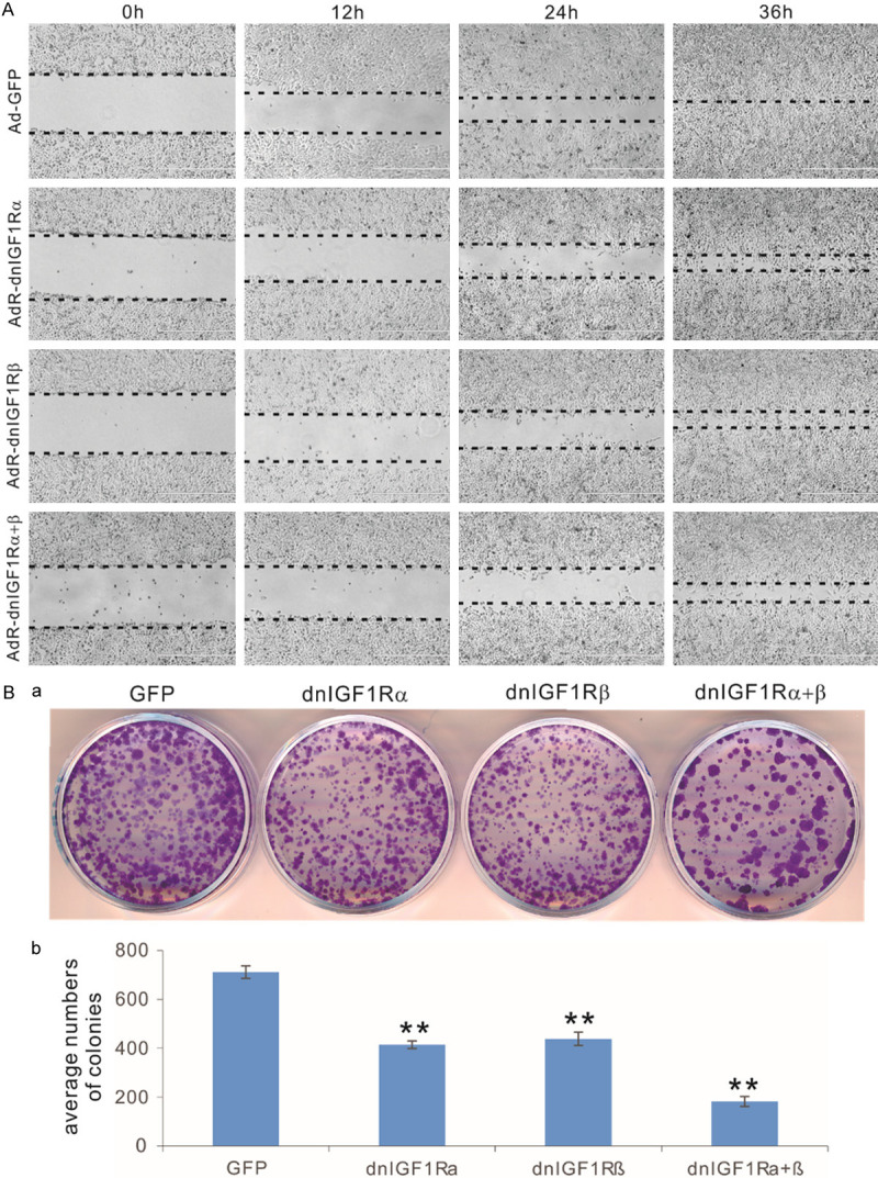
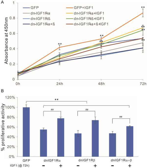
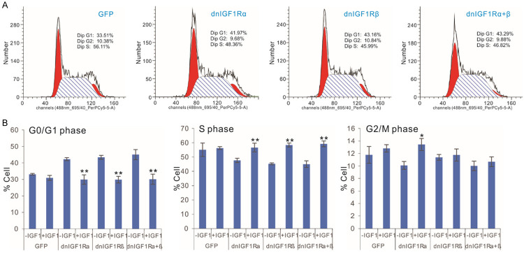
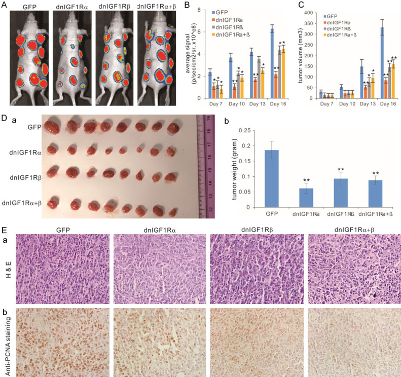
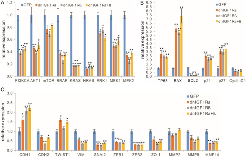
Primary bone tumor, also known as osteosarcoma (OS), is the most common primary malignancy of bone in children and young adults. Current treatment protocols yield a 5-year survival rate of near 70% although approximately 80% of patients have metastatic disease at the time of diagnosis. However, long-term survival rates have remained virtually unchanged for nearly four decades, largely due to our limited understanding of the disease process. One major signaling pathway that has been implicated in human OS tumorigenesis is the insulin-like growth factor (IGF)/insulin-like growth factor-1 receptor (IGF1R) signaling axis. IGF1R is a heterotetrameric α2β2 receptor, in which the α subunits comprise the ligand binding site, whereas the β subunits are transmembrane proteins containing intracellular tyrosine kinase domains. Although numerous strategies have been devised to target IGF/IGF1R axis, most of them have failed in clinical trials due to the lack of specificity and/or limited efficacy. Here, we investigated whether a more effective and specific blockade of IGF1R activity in human OS cells can be accomplished by employing dominant-negative IGF1R (dnIGF1R) mutants. We engineered the recombinant adenoviruses expressing two IGF1R mutants derived from the α (aa 1-524) and β (aa 741-936) subunits, and found that either dnIGF1Rα and/or dnIGF1Rβ effectively inhibited cell migration, colony formation, and cell cycle progression of human OS cells, which could be reversed by exogenous IGF1. Furthermore, dnIGF1Rα and/or dnIGF1Rβ inhibited OS xenograft tumor growth in vivo, with the greatest inhibition of tumor growth shown by dnIGF1Rα. Mechanistically, the dnIGF1R mutants down-regulated the expression of PI3K/AKT and RAS/RAF/MAPK, BCL2, Cyclin D1 and most EMT regulators, while up-regulating pro-apoptotic genes in human OS cells. Collectively, these findings strongly suggest that the dnIGF1R mutants, especially dnIGF1Rα, may be further developed as novel anticancer agents that target IGF signaling axis with high specificity and efficacy.

Keywords: IGF signaling; IGF-1R; Osteosarcoma; bone tumor; dominant-negative mutants; targeted therapy.


Conflict of interest statement

None.

Figures

Figure 1
Figure 1
Construction of dominant-negative IGF-1R (dnIGF1R) mutants as decoy receptors to specifically target IGF-1R signaling activity. A. Schematic depictions of the hetero-tetrameric structure and domains of the wild type IGF-1R and two dnIGF1R mutants, dnIGF-1Rα and dnIGF-1Rβ. L1, ligand binding domain 1; L2, ligand binding domain 2; CR, cysteine rich domain; ID-a, insert domain a; ID-b, insert domain b; FnIII-1, fibronectin type III domain 1; FnIII-2a, fibronectin type III domain 2a; FnIII-2b, fibronectin type III domain 2b; FnIII-3, fibronectin type III domain 3. The schematic diagrams are not drawn to scale. B. The dnIGF1R mutant expressing adenoviral vectors effectively transduce human osteosarcoma (OS) cells. Subconfluent OS line 143B cells were infected with Ad-GFP control virus, AdR-IGF1Rα, or AdR-IGF1Rβ, or co-infected with AdR-IGF1Rα and AdR-IGF1Rβ (AdR-dnIGF1Rα+β). Fluorescent signals were recorded at 48 h post infection. Representative images are shown. C. Recombinant adenovirus-mediated expression of the dnIGF1R mutants in human OS cells. Subconfluent OS line 143B cells were infected with Ad-GFP control virus, AdR-IGF1Rα, or AdR-IGF1Rβ, or co-infected with AdR-dnIGF1Rα+β. Total RNA was isolated at 48 h post infection and subjected to reverse transcription and qPCR analyses with the primers for the coding regions of the dnIGF1R mutants. GAPDH was used as a reference gene. “**”, P<0.01 compared with that of the GFP group’s.
Figure 2
Figure 2
The dominant-negative IGF-1R (dnIGF1R) mutants effectively inhibit cell migration and colony formation of human OS cells. (A) The dnIGF1R mutants inhibit cell wound healing in OS cells. Subconfluent 143B cells were infected with the indicated adenoviral vectors for 16 h and replated at near confluence. The well attached cells were injured with fine pipette tips, and the injured gaps were recorded and monitored for 36 h. Images at approximately same locations were taken under a bright field microscope. The wounding front lines are indicated with the dotted lines. (B) The dnIGF1R mutants inhibit colony formation from human OS cells. Subconfluent 143B cells were infected with the indicated adenoviral vectors for 16 h. The same numbers of infected OS cells were reseeded in 12-well cell culture plates in triplicate, and cultured for 10 days with medium changes every 2-3 days. At 10 days post replating, the cells were fixed and stained with crystal violet. Representative images are shown (a), and the established colonies were further quantitatively determined (b). “**”, P<0.01 compared with that of the GFP group’s.
Figure 3
Figure 3
The dominant-negative IGF-1R (dnIGF1R) mutants compete with IGF1-induced cell proliferative activity in human OS cells. Exponentially growing 143B cells were infected with the indicated adenoviral vectors for 16 h and replated into 96-well cell culture plates, with or without IGF1 stimulation (30 ng/mL). At the indicated time points, the cells were subjected to WST-1 assays (A), and the WST-1 assay results at 72 h were further quantitatively analyzed (B). “**”, P<0.01 compared with that of the GFP group’s; “##”, P<0.01 compared with that of the -IGF1 group’s.
Figure 4
Figure 4
The dominant-negative IGF-1R (dnIGF1R) mutants inhibit OS cell cycle progression by increasing G0/G1 phase and reducing S phase. A. Subconfluent 143B cells were infected with the indicated adenoviral vectors for 48 h and subjected to FACS analysis. The assays were done in triplicate, and representative results are shown. B. Subconfluent 143B cells were infected with the indicated adenoviral vectors and cultured with or without IGF1 (30 ng/mL). At 48 h post infection, the cells were collected and subjected to FACS analysis. “**”, P<0.01 compared with that of the -IGF1 group’s.
Figure 5
Figure 5
The dominant-negative IGF-1R (dnIGF1R) mutants suppress tumor growth in the xenograft tumor model of human OS. (A) Subconfluent Firefly luciferase-tagged 143B cells were infected with the indicated adenoviral vectors for 24 h, and collected for subcutaneous injection into the flanks of athymic nude mice. The mice were subjected to Xenogen imaging at days 7, 10, 13 and 16. The representative results for day 16 are shown. (B) Quantitative analysis of the average signal from the xenogeny imaging. “*”, P<0.05, “**”, P<0.01, compared with that of the GFP group’s. (C) Tumor volumes monitored with clipper measurement. “*”, P<0.05, “**”, P<0.01, compared with that of the GFP group’s. (D) Gross images of the retrieved tumor masses. Representative images of the retrieved tumor masses (a) and average tumor weight of the retrieved masses (b). (E) Histologic and immunohistochemical staining. The retrieved tumor samples were paraffin-embedded and subjected to H & E staining (a) and immunohistochemical staining using a PCNA antibody (b). Minus primary antibody and control IgG were used as negative controls (data not shown). Representative images are shown.
Figure 6
Figure 6
The dominant-negative IGF-1R (dnIGF1R) mutants effectively inhibit the downstream mediators of the IGF-1R signaling pathway in OS cells. Subconfluent 143B cells were infected with the indicated adenoviral vectors for 48 h. Total RNA was isolated and subjected to qPCR analyses of the IGF-1R signal downstream mediators (A), the apoptosis and cell cycle related genes (B), and EMT related genes (C). “*”, P<0.05, “**”, P<0.01, compared with that of the GFP group’s.

References

    1. Tang N, Song WX, Luo J, Haydon RC, He TC. Osteosarcoma development and stem cell differentiation. Clin Orthop Relat Res. 2008;466:2114–2130. - PMC - PubMed
    1. Haydon RC, Luu HH, He TC. Osteosarcoma and osteoblastic differentiation: a new perspective on oncogenesis. Clin Orthop Relat Res. 2007;454:237–246. - PubMed
    1. Wagner ER, He BC, Chen L, Zuo GW, Zhang W, Shi Q, Luo Q, Luo X, Liu B, Luo J, Rastegar F, He CJ, Hu Y, Boody B, Luu HH, He TC, Deng ZL, Haydon RC. Therapeutic implications of PPARgamma in human osteosarcoma. PPAR Res. 2010;2010:956427. - PMC - PubMed
    1. Wagner ER, Luther G, Zhu G, Luo Q, Shi Q, Kim SH, Gao JL, Huang E, Gao Y, Yang K, Wang L, Teven C, Luo X, Liu X, Li M, Hu N, Su Y, Bi Y, He BC, Tang N, Luo J, Chen L, Zuo G, Rames R, Haydon RC, Luu HH, He TC. Defective osteogenic differentiation in the development of osteosarcoma. Sarcoma. 2011;2011:325238. - PMC - PubMed
    1. Whelan JS. Osteosarcoma. Eur J Cancer. 1997;33:1611–1618. discussion 1618-1619. - PubMed

LinkOut - more resources