Review
. 2020 Oct 9:8:587685.
doi: 10.3389/fcell.2020.587685. eCollection 2020.

Emerging Functions of Actins and Actin Binding Proteins in Trypanosomatids

Affiliations
Review

Emerging Functions of Actins and Actin Binding Proteins in Trypanosomatids

Chhitar M Gupta et al. Front Cell Dev Biol. .

Abstract

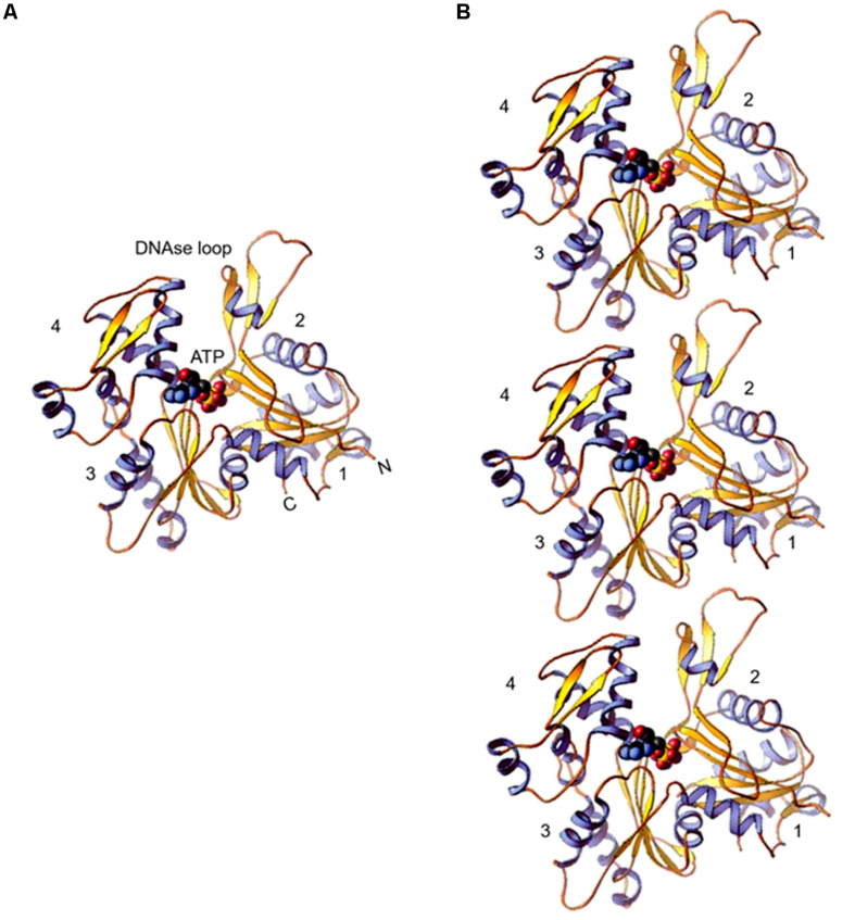
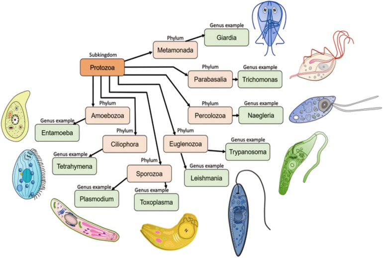
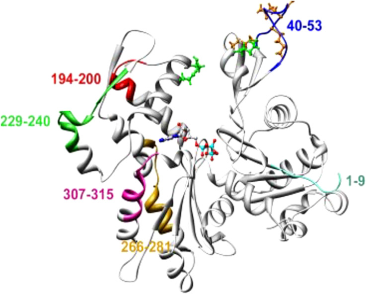
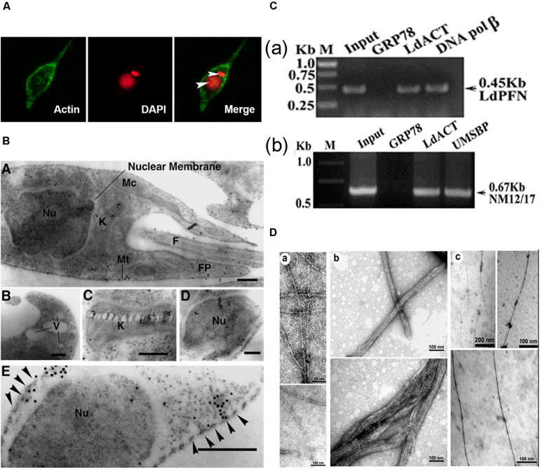
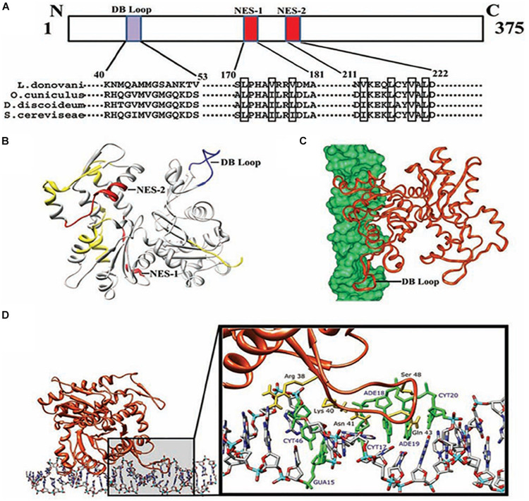
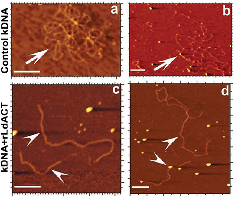
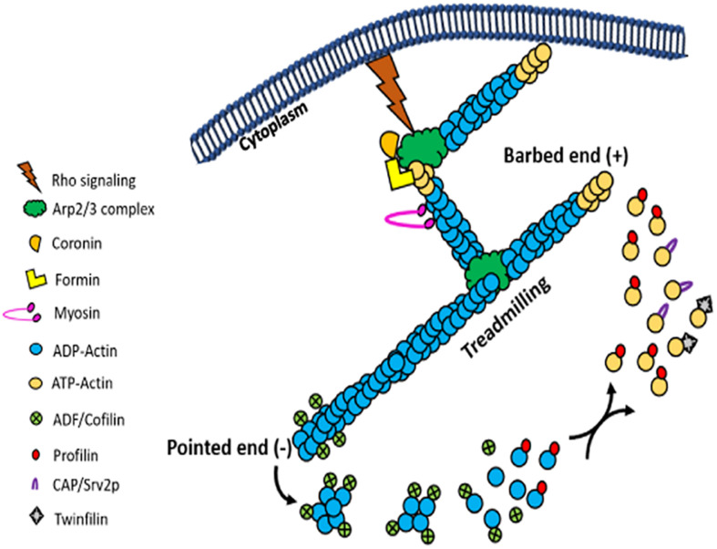
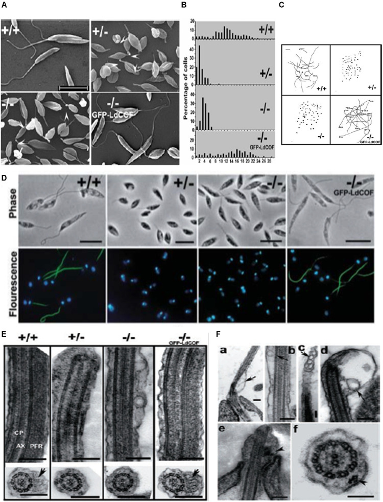
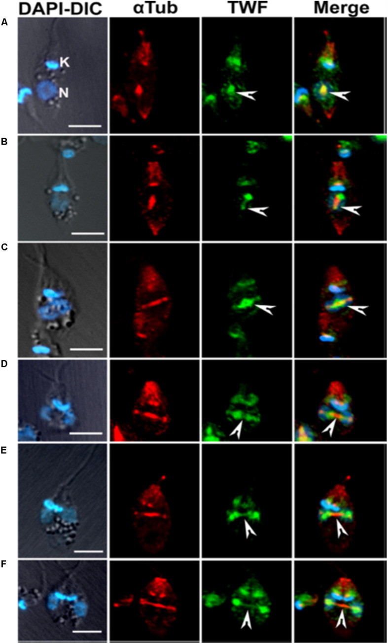
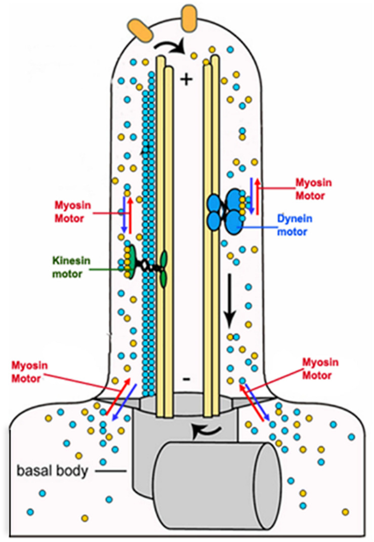
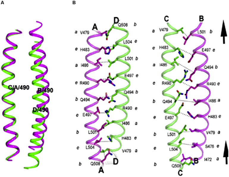
Actin is the major protein constituent of the cytoskeleton that performs wide range of cellular functions. It exists in monomeric and filamentous forms, dynamics of which is regulated by a large repertoire of actin binding proteins. However, not much was known about existence of these proteins in trypanosomatids, till the genome sequence data of three important organisms of this class, viz. Trypanosoma brucei, Trypanosoma cruzi and Leishmania major, became available. Here, we have reviewed most of the findings reported to date on the intracellular distribution, structure and functions of these proteins and based on them, we have hypothesized some of their functions. The major findings are as follows: (1) All the three organisms encode at least a set of ten actin binding proteins (profilin, twinfilin, ADF/cofilin, CAP/srv2, CAPz, coronin, two myosins, two formins) and one isoform of actin, except that T. cruzi encodes for three formins and several myosins along with four actins. (2) Actin 1 and a few actin binding proteins (ADF/cofilin, profilin, twinfilin, coronin and myosin13 in L. donovani; ADF/cofilin, profilin and myosin1 in T. brucei; profilin and myosin-F in T.cruzi) have been identified and characterized. (3) In all the three organisms, actin cytoskeleton has been shown to regulate endocytosis and intracellular trafficking. (4) Leishmania actin1 has been the most characterized protein among trypanosomatid actins. (5) This protein is localized to the cytoplasm as well as in the flagellum, nucleus and kinetoplast, and in vitro, it binds to DNA and displays scDNA relaxing and kDNA nicking activities. (6) The pure protein prefers to form bundles instead of thin filaments, and does not bind DNase1 or phalloidin. (7) Myosin13, myosin1 and myosin-F regulate endocytosis and intracellular trafficking, respectively, in Leishmania, T. brucei and T. cruzi. (8) Actin-dependent myosin13 motor is involved in dynamics and assembly of Leishmania flagellum. (9) Leishmania twinfilin localizes mostly to the nucleolus and coordinates karyokinesis by effecting splindle elongation and DNA synthesis. (10) Leishmania coronin binds and promotes actin filament formation and exists in tetrameric form rather than trimeric form, like other coronins. (11) Trypanosomatid profilins are essential for survival of all the three parasites.

Keywords: actin; actin binding proteins; functions; intracellular distribution; structure; tryanosomatids.


Figures

FIGURE 1
FIGURE 1
(A) Ribbon diagram of the actin molecule with space filling ATP (protein data bank [PDB]: 1ATN). N, amino terminus; C, carboxyl terminus. Numbers 1, 2, 3, and 4 label the four subdomains (re-printed from Pollard et al. (2016) with copyright permission from Elsevier Publishers). (B) Model of actin protofilaments derived from linear polymers along a single strand of F-actin.
FIGURE 2
FIGURE 2
Seven important phyla of subkingdom protozoa with schematic representations. Metamonada (intestinal flagellates, e.g., Giardia); Parabasalia (intestinal and related flagellates, e.g., Trichomonas); Percolozoa (flagellated amoebae, e.g., Naegleria); Euglenozoa (kinetoplastid flagellates, e.g., Trypanosoma, Leishmania); Amoebozoa (amoebae, e.g., Entamoeba); Sporozoa (sporozoans, e.g., Toxoplasma, Plasmodium) and Ciliophora (ciliates, e.g., Tetrahymena). Schematic images of the protozoan parasites.
FIGURE 3
FIGURE 3
An average molecular dynamics simulated homology model of LdAct showing colored stretches of diverged amino acid residues (aa 1–9 of subdomain 1, aa 40–53 of subdomain 2, aa 266–281 and aa 307–315 of subdomain 3, aa 194–200 and 229–240 of subdomain 4), brown ball and stick residues in the DNase-I binding loop are the diverged replacements in LdAct that are known to make strong interactions with DNase-I in the actin-DNase-I complex crystal structure, whereas green ball and stick residues are conserved amino acid residues that are known to make weak interactions with DNase-I (taken from Kapoor et al., 2008 with permission).
FIGURE 4
FIGURE 4
(A) Immunofluorescence micrograph of Leishmania promastigotes after treating them with 0. 5% NP-40 and staining with anti-LdAct antibodies and DAPI showing the presence of LdAct in the nucleus and kinetoplast and its association with nuclear DNA and kDNA (adapted from Sahasrabuddhe et al., 2004 with permission). (B) Electron micrographs of immunogold-labeled actin showing the presence (panel a) of LdAct in the nucleus (Nu), the kinetoplast (K), the flagellum (F), and the flagellar pocket (FP). In addition, the presence of LdAct on membranes of vacuoles (V) may also be noticed in panel (b), and its associations with kDNA network, nuclear membrane and subpellicular microtubules may clearly be seen in panels (c–e), respectively. The arrowheads in panel (e) mark the microtubules. Bar, 200 nm (Adapted from Sahasrabuddhe et al., 2004 with permission). (C) Chromatin Immuno-precipitation (ChIP) analysis using anti-LdAct antibodies showing the in vivo association of LdAct with chromatin (a) and kDNA network (b). Panels (a,b) are the agarose gels of PCR products after ChIP assay. Lanes are marked on the top with their respective antibodies used in the ChIP assay and arrows indicated the genes amplified after pull down. An irrelevant, non-DNA associating antibody, GRP78, was used as a negative control, whereas antibodies against DNA polβ, and UMSBP (universal minicircle sequence-binding protein), were used as positive controls for nuclear DNA and kDNA respectively. LdPfn, Leishmania profilin; NM12/17, specific minicircle primers (this was originally published in Nucleic Acids Research, Kapoor et al., 2010© Oxford University Press). (D,a) Negatively stained transmission electron micrograph of in vitro reconstituted rabbit muscle actin (RbAct) filaments in F-buffer (100 mM KCl, 2 mM MgCl2 and 2 mMATP; pH 8.0; 25°C) and (b) LdAct at 2 μM protein concentration, unlike RbAct, formed bundles rather thin filaments, under identical conditions. (c) LdAct forms very thin filaments at 0.2 μM G-LdAct concentration in F-buffer, pH7.0 at 25°C. RbAct under these conditions failed to form filaments (taken from Kapoor et al., 2008 with permission).
FIGURE 5
FIGURE 5
Computational docking of average simulated model of LdAct with DNA showing the interaction of the diverged DB-loop of LdAct with the major groove of DNA. (A) Sequence alignment of LdAct with other actins showing the presence of nuclear export signals (NES-1 and NES-2) in the LdAct aa sequence and the diverged DB-loop predicted to be involved in the DNA binding, by DP-Bind server. (B) Energy minimized average simulated model of LdAct showing positions of NES-1, NES-2 (red) and the diverged stretches of amino acid sequences (yellow) including the sequence that fall in DB loop (blue). (C) Docking of LdAct (orange) with DNA (green) using HADDOCK protocols. (D) Amino acid residues of the DB loop of LdAct (yellow) showing hydrogen bonding with the nucleotides (green) of DNA. DB, DNase I binding; NES, nuclear export signal (This was originally published in Nucleic Acids Research, Kapoor et al., 2010© Oxford University Press).
FIGURE 6
FIGURE 6
Atomic force micrographs of kDNA after its incubation in the presence and absence (control) of LdAct, showing decatenation of kDNA with LdAct. Panels (a,b) control kDNA, arrows indicate catenated kDNA. Panels (c,d) kDNA with rLdAct, arrowheads indicate decatenated nicked kDNA (scale bar: 500 nm) (This figure was originally published in Nucleic Acids Research, Kapoor et al., 2010© Oxford University Press).
FIGURE 7
FIGURE 7
Picture cartoon of actin treadmilling, showing that the rate of treadmilling is regulated by ADF/cofilins and profilin, which results in an increase and decrease in the size of actin filaments, respectively. It further shows that Arp2/3 complex nucleate new filaments by its binding with actin monomers and the side of actin filaments, while formins nucleate new filaments by binding actin monomers and through cooperation of profilin.
FIGURE 8
FIGURE 8
(A) Scanning electron micrographs, showing short and stumpy cell body with significantly shortened flagella of heterozygous (+/-) and homozygous (-/-) LdCof mutants, compared with wild type (+/+) cells. Episomal complementation of LdCof– /– cells with LdCof gene (-/- comp) restored the wild-type morphology and flagellar length. Bar, 10 μm. Arrowheads indicate the “blob-like” structures seen at the tip of the flagella of mutant cells (taken from Tammana et al., 2008 with permission). (B) Histogram, showing flagellar lengths of LdCof+/+, LdCof+/–, LdCof– /– and LdCof–/–comp cells (taken from Tammana et al., 2008 with permission). (C) Motility analysis of LdCof mutants by time lapse microscopy. Traces of paths of live, individual cells in the movies indicate that LdCof+/– and LdCof– /– cells are completely immotile. However, upon episomal complementation of LdCof– /– cells the motility is restored back to normal. Origin of the path is indicated by solid dots. Bar, 50 μm (taken from Tammana et al., 2008 with permission). (D) Immuno -flourescence micrographs, showing loss of paraflagellar rod proteins, PFR1 and PFR2, after staining the LdCof +/– and LdCof – /– mutants with mAb2E10 antibodies and their restoration after complimenting LdCof gene in the null mutants. Bar, 5 μm (taken form Tammana et al., 2008 with permission). (E) Transmission electron micrographs of thin sections of flagellum from chemically fixed whole cells showing the absence of PFR in LdCof +/– and LdCof – /– cells and its restoration upon episomal complementation. Longitudinal sections of the flagellum showing the axoneme (AX) with the central pair microtubules (CP) and PFR confined between the axoneme and the flagellar membrane in wild type cells and GFP–LdCof complemented mutants. Bar, 200 nm. Cross sections of the flagellum, showing the PFR in LdCof +/+ (marked by arrow) and GFP–LdCof-complemented mutant cells, and complete absence of this structure in the cross sections of LdCof+/– and LdCof– /– cells. Bar, 200 nm (taken from Tammana et al., 2008 with permission). (F) (a–d) Longitudinal sections of chemically fixed whole cells of LdCof– /– mutants, showing accumulation of membrane-bound vesicles at the base (a), along the length (b,c) and tip (d) of the flagellum. Arrows indicate the membrane-bound vesicles. Longitudinal section (e) and cross section (f) of chemically fixed whole cells of LdCof– /– mutant, showing IFT-like particles along the length of the flagellum. Arrowheads indicate IFT-like particles. Bar, for panels (a–e) 200 nm and for panel (f) 100 nm. IFT, intraflagellar transport (taken from Tammana et al., 2008 with permission).
FIGURE 9
FIGURE 9
Immunofluorescence micrographs (A–F) after staining the cells with anti-LdTwf antibodies, showing movement of twinfilin (Twf) from the nucleolus to origin of the mitotic spindle where it completely localized on the extending spindle microtubules and finally redistributed to the spindle poles. Arrow heads mark distribution patterns of TWF on the spindle, showing the presence of residual TWF on the spindle microtubules while the larger TWF bulk migrated to the poles in the later stages of karyokinesis. Mitotic spindle has been marked by anti α-tubulin (aTub) antibody. Bar, 5 mm (taken from Kumar et al., 2016 with permission).
FIGURE 10
FIGURE 10
Cartoon diagram showing involvement of the actin based LdMyo13 motor protein in assembly/disassembly of the paraflagellar road (PFR) and the flagellar membrane during remodeling of the Leishmania flagellum.
FIGURE 11
FIGURE 11
(A) Asymmetry observed in the LdCor coiled coil domain tetramer. Cartoon represents the pairs of dimers, highlighting the asymmetry. In left panel, Cα atoms of aa residues 475–507 of chain C were superposed to corresponding atoms of chain A using Superpose (Krissinel and Henrick, 2004) and the transformation applied to the BC dimer. The B and D helices of AD/BC dimers superpose with an RMSD of 3.4 Å. (B) Interactions at the BC dimer are different from that of AD dimer due to an upward shift in B helix by a heptad. Also, the distances across the interface are longer in the BC helical interface (Taken from Nayak et al., 2016 with permission).

References

    1. Adhiambo C., Forney J. D., Asai D. J., LeBowitz J. H. (2005). The two cytoplasmic dynein 2 isoforms in Leishmania mexicana perform separate functions. Mol. Biochem. Parasitol. 143 216–225. 10.1016/j.molbiopara.2005.04.017 - DOI - PubMed
    1. Ali H. Z., Harding C. R., Denny P. W. (2012). Endocytosis and sphingolipid scavenging in Leishmania mexicana amastigotes. Biochem. Res. Int. 2012:691363. 10.1155/2012/691363 - DOI - PMC - PubMed
    1. Alsford S., Turner D. J., Obado S. O., Sanchez-Flores A., Glover L., Berriman M., et al. (2011). High-throughput phenotyping using parallel sequencing of RNA interference targets in the African trypanosome. Genome Res. 21 915–924. 10.1101/gr.115089.110 - DOI - PMC - PubMed
    1. Ambaru B., Gopalsamy A., Tammana T. V. S., Subramanya H. S., Gupta C. M. (2020). Actin sequestering protein, profilin, regulates intracellular vesicle transport in Leishmania. Mol. Biochem. Parasitol. 238:111280. 10.1016/j.molbiopara.2020.111280 - DOI - PubMed
    1. Asano S., Mishima M., Nishida E. (2001). Coronin forms a stable dimer through its C-terminal coiled coil region: an implicated role in its localization to cell periphery. Genes Cells 6 225–235. 10.1046/j.1365-2443.2001.00416.x - DOI - PubMed