

Enzyme kinetics and inhibition parameters of human leukocyte glucosylceramidase
- PMID: 33163670
- PMCID: PMC7609449
- DOI: 10.1016/j.heliyon.2020.e05191
Enzyme kinetics and inhibition parameters of human leukocyte glucosylceramidase
Abstract
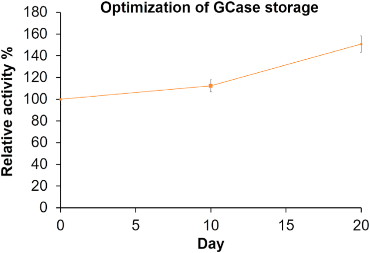
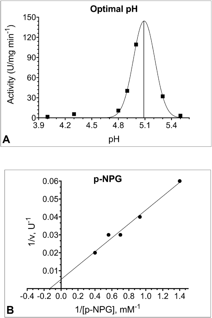
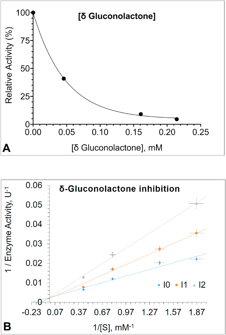
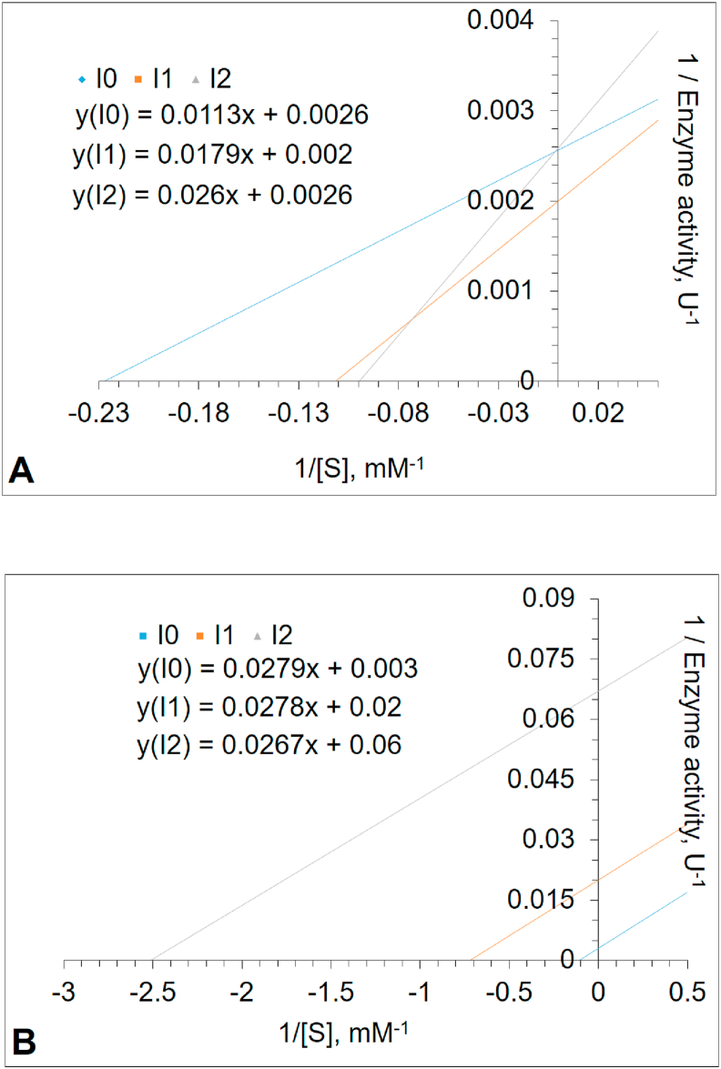
Glucosylceramidase (GCase) is a lysosomal enzyme that catalyzes the cleavage of β-glucosidic linkage of glucocerebroside (GC) into glucose and ceramide; thereby, plays an essential function in the degradation of complex lipids and the turnover of cellular membranes. The growing list of 460 mutations in the gene coding for it-glucosylceramidase beta acid 1 (GBA1)-is reported to abolish its catalytic activity and decrease its enzyme stability, associating it with severe health conditions such as Gaucher disease (GD), Parkinson Disease (PD) and Dementia with Lewy bodies (DLB). Although the three-dimensional structure of wild type glucosylceramidase is elucidated, little is known about its features in human cells. Moreover, alternative sources of GCase that prove to be effective in the treatment of diseases with enzyme treatment therapies, impose the need for a simple and cost-effective procedure to study the enzyme behavior. This work, for the first time, shows a well-established, yet simple, cost- and time-efficient protocol for the study of GCase enzyme in human leukocytes by the artificial substrate p-Nitrophenyl-β-D-glucopyranoside (PNPG). Characterization of the enzyme in human leukocytes for activation parameters (optimal pH, Km, and Vmax) and enzyme inhibition was done. The results indicate that the optimum pH of GCase enzyme with PNPG is 5.0. The Km and Vmax values are 12.6mM and 333 U/mg, respectively. Gluconolactone competitively inhibits GCase, with a Ki value of 0.023 mM and IC50 of 0.047 mM. Glucose inhibition is uncompetitive with a Ki of 1.94 mM and IC50 of 55.3 mM. This is the first report for the inhibitory effect of glucose, δ-gluconolactone on human leukocyte GCase activity.
Keywords: Biochemistry; Clinical research; Enzyme kinetics; Gaucher disease; Gluconolactone; Glucose; Glucosylceramidase; Glucosylceramide; Hematological system; Human leukocyte; Inhibition parameters; Molecular biology; PNPG; Proteins.
© 2020 The Authors.
Figures

Similar articles
-
Lysosomal functions and dysfunctions: Molecular and cellular mechanisms underlying Gaucher disease and its association with Parkinson disease.Adv Drug Deliv Rev. 2022 Aug;187:114402. doi: 10.1016/j.addr.2022.114402. Epub 2022 Jun 25. Adv Drug Deliv Rev. 2022. PMID: 35764179 Review.
-
Complement drives glucosylceramide accumulation and tissue inflammation in Gaucher disease.Nature. 2017 Mar 2;543(7643):108-112. doi: 10.1038/nature21368. Epub 2017 Feb 22. Nature. 2017. PMID: 28225753
-
Acid ceramidase inhibition ameliorates α-synuclein accumulation upon loss of GBA1 function.Hum Mol Genet. 2018 Jun 1;27(11):1972-1988. doi: 10.1093/hmg/ddy105. Hum Mol Genet. 2018. PMID: 29579237 Free PMC article.
-
Activation of β-Glucocerebrosidase Reduces Pathological α-Synuclein and Restores Lysosomal Function in Parkinson's Patient Midbrain Neurons.J Neurosci. 2016 Jul 20;36(29):7693-706. doi: 10.1523/JNEUROSCI.0628-16.2016. J Neurosci. 2016. PMID: 27445146 Free PMC article.
-
Targeting the GBA1 pathway to slow Parkinson disease: Insights into clinical aspects, pathogenic mechanisms and new therapeutic avenues.Pharmacol Ther. 2023 Jun;246:108419. doi: 10.1016/j.pharmthera.2023.108419. Epub 2023 Apr 19. Pharmacol Ther. 2023. PMID: 37080432 Review.
Cited by
-
Mapping Sphingolipid Metabolism Pathways during Phagosomal Maturation.ACS Chem Biol. 2021 Dec 17;16(12):2757-2765. doi: 10.1021/acschembio.1c00393. Epub 2021 Oct 14. ACS Chem Biol. 2021. PMID: 34647453 Free PMC article.
-
Patient centered guidelines for the laboratory diagnosis of Gaucher disease type 1.Orphanet J Rare Dis. 2022 Dec 21;17(1):442. doi: 10.1186/s13023-022-02573-6. Orphanet J Rare Dis. 2022. PMID: 36544230 Free PMC article.
-
LRRK2 kinase activity regulates Parkinson's disease-relevant lipids at the lysosome.Mol Neurodegener. 2025 Aug 6;20(1):89. doi: 10.1186/s13024-025-00880-7. Mol Neurodegener. 2025. PMID: 40770658 Free PMC article.
-
pH regulates potassium conductance and drives a constitutive proton current in human TMEM175.Sci Adv. 2022 Mar 25;8(12):eabm1568. doi: 10.1126/sciadv.abm1568. Epub 2022 Mar 25. Sci Adv. 2022. PMID: 35333573 Free PMC article.
References
-
- Smith L., Mullin S., Schapira A.H.V. Insights into the structural biology of Gaucher disease. Exp. Neurol. 2017;298:180–190. - PubMed
-
- Beutler E. Gaucher disease: new molecular approaches to diagnosis and treatment. Science. 1992;256:794–799. - PubMed
-
- Interplay between Glucocerebrosidase 1 and Glucocerebrosidase 2; Potential Implications for the Pathogenesis of Gaucher and Parkinson’s Diseases - UCL Discovery. https://discovery.ucl.ac.uk/id/eprint/1571208/
-
- Matern H., Boermans H., Lottspeich F., Matern S. Molecular cloning and expression of human bile acid beta-glucosidase. J. Biol. Chem. 2001 - PubMed
LinkOut - more resources
Full Text Sources
Miscellaneous
