. 2020 Nov 2;6(11):e05363.
doi: 10.1016/j.heliyon.2020.e05363. eCollection 2020 Nov.

The role of estrogen receptors in rat Sertoli cells at different stages of development

Affiliations

The role of estrogen receptors in rat Sertoli cells at different stages of development

Carla Macheroni et al. Heliyon. .

Abstract

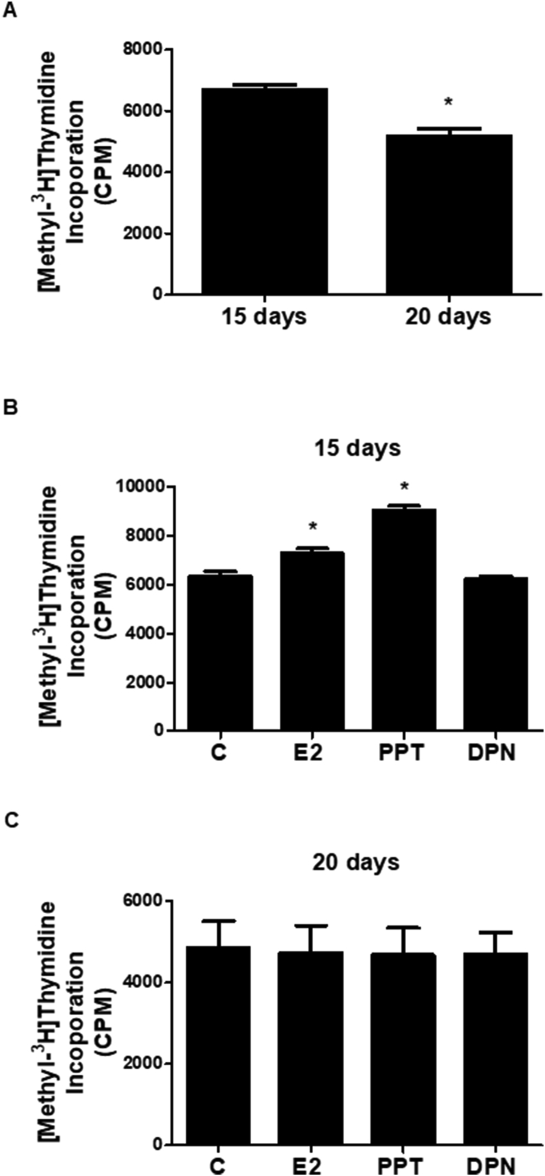
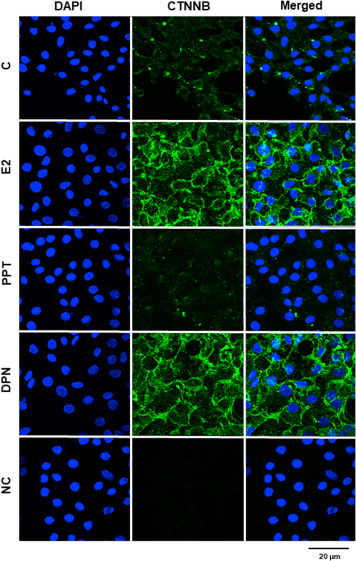
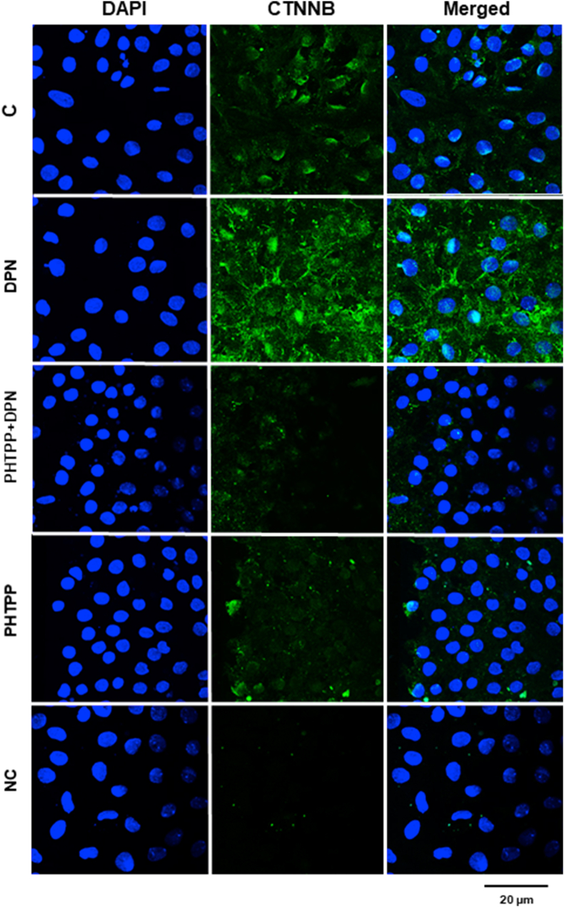
The aim of the study was to investigate the effects of estrogen receptors (ESR1 and ESR2) on the expression of the proteins involved with proliferation (CCND1) and differentiation (CDKN1B and CTNNB) of Sertoli cells from rat in different stages of development. ESR1-selective agonist PPT, but not ESR2-selective agonist DPN, increased CCND1 expression in Sertoli cells from 5- and 15-day old rats. PPT did not have any effect on CCND1 expression in Sertoli cells from 20- and 30-day-old rats. DPN, but not PPT, increased CDKN1B expression in Sertoli cells from 15-, 20-, 30-day-old rats. DPN did not have any effect on Sertoli cells from 5-day-old rats. 17β-estradiol (E2) and PPT enhanced the [Methyl-3H] thymidine incorporation in Sertoli cells from 15-day-old rats, whereas the treatment did not have any effect in 20-day-old rats. E2 and DPN, but not PPT, increased non-phosphorylated CTNNB expression in Sertoli cells from 20-day-old rats. This upregulation was blocked by ESR2-selective antagonist PHTPP. The activation of ESR1 and ESR2, respectively, plays a role in the proliferation and differentiation of Sertoli cells in a critical period of testicular development. Furthermore, in Sertoli cells from 20-day-old rats, upregulation of non-phosphorylated CTNNB by E2/ESR2, via c-SRC/ERK1/2 and PI3K/AKT, may play a role in the interaction between Sertoli cells and/or in cell-germ cell adhesion and/or in the stabilization and accumulation of CTNNB in the cytosol. CTNNB could be translocated to the nucleus and modulate the transcriptional activity of specific target genes. The present study reinforces the important role of estrogen in normal testis development.

Keywords: 17β-estradiol; Cell biology; Cell culture; Cell differentiation; Estrogen receptors; Reproductive hormone; Reproductive medicine; Sertoli cells; Steroid hormones; Systems biology.


Figures

Figure 1
Figure 1
Effects of ESR1-selective agonist PPT and ESR2-selective agonist DPN on the expression of CCND1 in the Sertoli cells from5-, 15-, 20- and 30-day-old rats. A. Sertoli cells from 5-, 15-, 20- and 30-day-old rats were incubated in the absence (C, control) and presence of PPT (10 nM) for 24 h. The relative positions of CCND1 (top panel) and ACT (bottom panel) proteins are shown at the right. The data shown are representative of four independent experiments (top and bottom panels). See, full image in Supplemental Fig S1. Bars represent the densitometric analysis of four independent experiments. Results were normalized to ACT expression in each sample and plotted (mean ± SEM) in relation to control, C (=1). ∗Significantly different from control (P < 0.05, Student t-test, n = 4). B. Sertoli cells from 5-day-old rats were incubated in the absence (C, control) and presence of PPT (10 nM) or DPN (10 nM) for 24 h. The data shown are representative of two independent experiments. See, full image in Supplemental Fig S1. C. Sertoli cells from 5-, 15-, 20- and 30-day-old rats were incubated in the absence of agonists (C, control, basal levels). Bars represent the densitometric analysis of four independent experiments. Results were normalized to ACT expression in each sample and plotted (mean ± SEM). # Significantly different from 20- or 30-day-od rats (P < 0.05, Newman-Keuls test).
Figure 2
Figure 2
Effects of 17β-estradiol (E2), ESR1-selective agonist PPT and ESR2-selective agonist DPN on the incorporation of [Methyl-3H thymidine in the Sertoli cells from 15- and 20-day-old rats. Sertoli cells from 15- and 20-day-old rats were initially incubated with 2 μCi/ml [Methyl-3H]thymidine for 6 h. After this incubation, A. cells were incubated in the absence of the agonists for 24 h (basal [Methyl-3H] thymidine incorporation). B and C. cells were incubated in the absence (C, control, basal [Methyl-3H] thymidine incorporation) and presence of E2 (0.1 nM), PPT (10 nM) or DPN (10 nM) for 24 h. Bound radioactivity was determined and the results plotted (mean ± SEM, n = 3 independent experiments). ∗Significantly different from control (P < 0.05, Student t-test or Newman-Keuls test).
Figure 3
Figure 3
Effects of ESR1-selective agonist PPT and ESR2-selective agonist DPN on the expression of CDKN1B in the Sertoli cells from 5-, 15-, 20- and 30-day-old rats. A. Sertoli cells from 30-, 20- and 15-day-old rats were incubated in the absence (C, control) and presence of DPN (10 nM) for 24 h. The relative positions of CDKN1B (top panel) and ACT (bottom panel) proteins are shown at the right. The data shown are representative of four independent experiments (top and bottom panels). See, full image in Supplemental Fig S3. Bars represent the densitometric analysis of four independent experiments. Results were normalized to ACT expression in each sample and plotted (mean ± SEM) in relation to control (C = 1). ∗Significantly different from control (P < 0.05, Student t-test, n = 4). B. Sertoli cells from 30- and 5-day-old rats were incubated in the absence (C, control) and presence of PPT (10 nM) or DPN (10 nM) for 24 h. The data shown are representative of two independent experiments. See, full image in Supplemental Fig S3. C. Sertoli cells from 5-, 15-, 20- and 30-day-old rats were incubated in the absence of agonists (C, control). Bars represent the densitometric analysis of four independent experiments. Results were normalized to ACT expression in each sample and plotted (mean ± SEM). # Significantly different from 20- or 30-day-od rats (P < 0.05, Newman-Keuls test).
Figure 4
Figure 4
Effects of 17β-estradiol (E2), ESR1-selective agonist PPT and ESR2-selective agonist DPN on the expression of non-phosphorylated CTNNB in the Sertoli cells from 20-day-old rats. Sertoli cells from 20-day-old rats were incubated in the absence (C, control) and presence of E2 (0.1 nM), PPT (10 nM) or DPN (10 nM) for 24 h. Immunostaining for CTNNB (green) was detected using an antibody specific for non-phosphorylated CTNNB and Alexa Fluor 488-labeled secondary antibody. Nuclei were stained with DAPI (blue). Negative control (NC) was performed using normal rabbit serum at the same dilution of antibody. Scale bar as indicated. The data shown are representative of three independent experiments.
Figure 5
Figure 5
Effects of ESR2-selective antagonist PHTPP on the expression of non-phosphorylated CTNNB induced by DPN in the Sertoli cells from 20-day-old rats. Sertoli cells from 20-day-old rats were incubated in the absence (C, control) and presence of DPN (10 nM) for 24 h. The cells were also untreated or pretreated with ESR2-selective antagonist PHTPP (10 nM) for 30 min. Afterward, the cells were stimulated with DPN (10 nM) 24 h. Immunostaining for CTNNB (green) was detected using an antibody specific for non-phosphorylated CTNNB and Alexa Fluor 488-labeled secondary antibody. Nuclei were stained with DAPI (blue). Negative control (NC) was performed using normal rabbit serum at the same dilution of antibody. Scale bar as indicated. The data shown are representative of three independent experiments.
Figure 6
Figure 6
Effects of proteins kinases inhibitors on the expression of non-phosphorylated CTNNB induced by DPN in the Sertoli cells from 20-day-old rats. Sertoli cells from 20-day-old rats were incubated in the absence (C, control) and presence of DPN (10 nM) for 24 h. The cells were also untreated or pretreated with PP2, an inhibitor of the SRC family of protein tyrosine kinases; U0126, an inhibitor of MEK1/2; Wortmannin, an inhibitor of PI3K and MK-2206, an inhibitor of AKT for 30 min. Afterward, the cells were stimulated with DPN (10 nM) 24h. Immunostaining for CTNNB (green) was detected using an antibody specific for non-phosphorylated CTNNB and Alexa Fluor 488-labeled secondary antibody. Nuclei were stained with DAPI (blue). Negative control was performed using normal rabbit serum at the same dilution of antibody. Scale bar as indicated. The data shown are representative of three independent experiments. Note: Control, C, and DPN are the same results of the Figure 5. All treatments were performed on the same day (Figures 5 and 6).
Fig. S1
Fig. S1
Effects of ESR1-selective agonist PPT and ESR2-selective agonist DPN on the expression of CCND1 in the Sertoli cells from 5-, 15-, 20- and 30-day-old rats. A. Sertoli cells from 5-, 15-, 20- and 30-day-old rats were incubated in the absence (C, control) and presence of PPT (10 nM) for 24 h. The relative positions of CCND1 (top panel) and ACT (bottom panel) proteins are shown at the right. The data shown are representative of four independent experiments (top and bottom panels). B. Sertoli cells from 5-day-old rats were incubated in the absence (C, control) and presence of PPT (10 nM) or DPN (10 nM) for 24 h. The data shown are representative of two independent experiments.
Fig. S3
Fig. S3
Effects of ESR1-selective agonist PPT and ESR2-selective agonist DPN on the expression of CDKN1B in the Sertoli cells from 5-, 15-, 20- and 30-day-old rats. A. Sertoli cells from 30-, 20- and 15-day-old rats were incubated in the absence (C, control) and presence of DPN (10 nM) for 24 h. The relative positions of CDKN1B (top panel) and ACT (bottom panel) proteins are shown at the right. The data shown are representative of four independent experiments (top and bottom panels). B. Sertoli cells from 30- and 5-day-old rats were incubated in the absence (C, control) and presence of PPT (10 nM) or DPN (10 nM) for 24 h. The data shown are representative of two independent experiments.
Fig. S4
Fig. S4
Effects of 17β-estradiol (E2) and ESR2-selective agonist DPN on the phosphorylation of ERK1/2 in the Sertoli cells from 20-day-old rats. Cells were incubated in the absence (C, control) and presence of E2 (0.1 nM) (A) or DPN (10 nM) (B) for different times. The relative positions of phosphorylated ERK1/2 (top panel) and total ERK1/2 (bottom panel) proteins are shown at the right. The data shown are representative of four independent experiments. Bars represent the densitometric analysis of four independent experiments. Results were normalized to total ERK1/2 expression in each sample and plotted (mean ± SEM) in relation to control (C = 1). Open bars ERK1 and closed bars ERK2. ∗Significantly different from control (P < 0.05, Student t-test, n = 4).
Fig. S5
Fig. S5
Effects of 17β-estradiol (E2) and ESR2-selective agonist DPN on the phosphorylation of ERK1/2 in the Sertoli cells from 20-day-old rats. Cells were incubated in the absence (C, control) and presence of E2 (0.1 nM) (A) or DPN (10 nM) (B) for different times. The relative positions of phosphorylated ERK1/2 (top panel) and total ERK1/2 (bottom panel) proteins are shown at the right. The data shown are representative of four independent experiments. Bars represent the densitometric analysis of four independent experiments. Results were normalized to total AKT expression in each sample and plotted (mean ± SEM) in relation to control (C = 1). ∗Significantly different from control (P < 0.05, Student t-test, n = 4).

Similar articles

Cited by

References

    1. Antal M.C., Krust A., Chambon P., Mark M. Sterility and absence of histopathological defects in nonreproductive organs of a mouse ER-null mutant. Proc. Natl. Acad. Sci. U.S.A. 2008;105:2433–2438. - PMC - PubMed
    1. Auharek S.A., Avelar G.F., Lara N.L., Sharpe R.M., França L.R. Sertoli cell numbers and spermatogenic efficiency are increased in inducible nitric oxide synthase mutant mice. Int. J. Androl. 2011;34:e621. - PubMed
    1. Bergmann M., Dierichs R. Postnatal formation of the blood-testis barrier in the rat with special reference to the initiation of meiosis. Anat. Embryol. 1983;168:269–275. - PubMed
    1. Beumer T.L., Kiyokawa H., Roepers-Gajadien H.L., van den Bos L.A., Lock T.M., Gademan I.S., Rutgers D.H., Koff A., de Rooij D.G. Regulatory role of CDKN1B in the mouse and human testis. Endocrinology. 1999;140:1834–1840. - PubMed
    1. Buzzard J.J., Wreford N.G., Morrison J.R. Thyroid hormone, retinoic acid, and testosterone suppress proliferation and induce markers of differentiation in cultured rat Sertoli cells. Endocrinology. 2003;144:3722–3731. - PubMed