Review
. 2020 Jul 21;5(11):1870-1893.
doi: 10.1016/j.ekir.2020.07.013. eCollection 2020 Nov.

Understanding Mesangial Pathobiology in AL-Amyloidosis and Monoclonal Ig Light Chain Deposition Disease

Affiliations
Review

Understanding Mesangial Pathobiology in AL-Amyloidosis and Monoclonal Ig Light Chain Deposition Disease

Guillermo A Herrera et al. Kidney Int Rep. .

Abstract

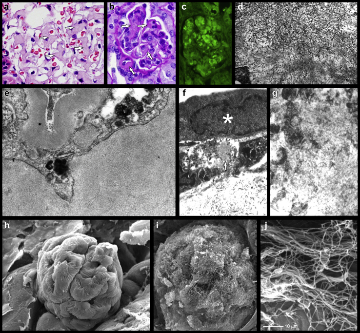
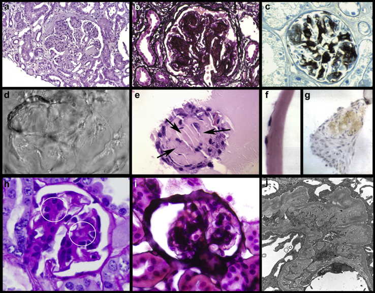
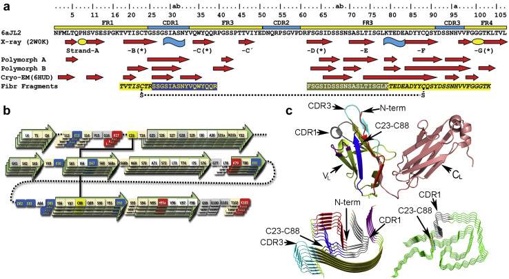
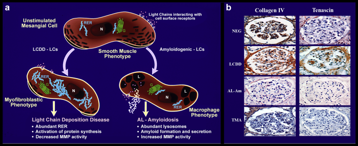
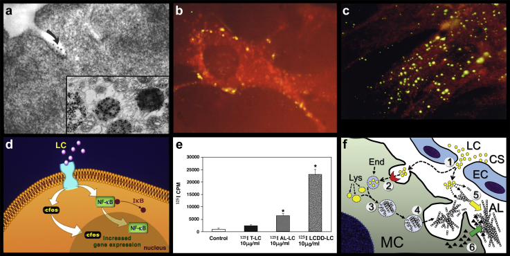
Patients with plasma cell dyscrasias produce free abnormal monoclonal Ig light chains that circulate in the blood stream. Some of them, termed glomerulopathic light chains, interact with the mesangial cells and trigger, in a manner dependent of their structural and physicochemical properties, a sequence of pathological events that results in either light chain-derived (AL) amyloidosis (AL-Am) or light chain deposition disease (LCDD). The mesangial cells play a key role in the pathogenesis of both diseases. The interaction with the pathogenic light chain elicits specific cellular processes, which include apoptosis, phenotype transformation, and secretion of extracellular matrix components and metalloproteinases. Monoclonal light chains associated with AL-Am but not those producing LCDD are avidly endocytosed by mesangial cells and delivered to the mature lysosomal compartment where amyloid fibrils are formed. Light chains from patients with LCDD exert their pathogenic signaling effect at the cell surface of mesangial cells. These events are generic mesangial responses to a variety of adverse stimuli, and they are similar to those characterizing other more frequent glomerulopathies responsible for many cases of end-stage renal disease. The pathophysiologic events that have been elucidated allow to propose future therapeutic approaches aimed at preventing, stopping, ameliorating, or reversing the adverse effects resulting from the interactions between glomerulopathic light chains and mesangium.

Keywords: AL-amyloidosis; kidney; kidney repair; light chain deposition disease; mesangium; monoclonal Ig deposition disease; monoclonal light chains; stem cells.


Figures

Figure 1
Figure 1
Light chain–derived amyloidosis (AL-Am). Comparison of findings in experimental platforms and renal biopsy of patient with AL-Am. (a) Hematoxylin and eosin staining in renal biopsy from patient with AL-Am. Note the expanded mesangial areas with eosinophilic, amorphous material replacing normal mesangial matrix (arrows). (b–d) Samples of renal tissue obtained from rats in vivo perfused with an amyloidogenic λ light chains (LCs) through penile vein. (b) Periodic acid–Schiff (PAS) stain showing expanded mesangial areas with similar eosinophilic, amorphous material in some mesangial areas (arrows), as shown in (a). Original magnification ×750. (c) Thioflavin T staining showing fluorescence in areas with amyloid deposition. Original magnification ×750. (d) Transmission electron microscopy (TEM) showing randomly disposed, nonbranching 7- to 13-nm fibrils replacing normal mesangial matrix. Original magnification ×18,500. (e) TEM micrograph showing transformed mesangial cell (MC) with macrophage phenotype and surrounding amyloid fibrils in a renal biopsy of a patient with AL-Am. Sample stained with uranyl and lead citrate. Original magnification ×32,500. (f) TEM micrograph showing MC grown in Matrigel with amyloidogenic LC for 72 hours. Formation of amyloid (arrow) by surrounding transformed MC (with macrophage phenotype, and normal MC [∗]) on top with smooth muscle phenotype not participating in the process of amyloid formation. (g) Magnified area shown with the arrow in (f). Sample stained with uranyl and lead citrate stain. Original magnification is ×7500 and ×18,500 in (f) and (g), respectively. (h–j) Scanning electron microscopy (SEM) image of renal samples taken from rat in vivo perfused with an amyloidogenic λ LCs through penile vein. (h) Normal-appearing rat glomerulus. (i) Fibrillary material in rat glomerulus with advanced amyloid deposition. (h,i) Original magnification ×700. (j) High-magnification (×22,500) SEM micrograph showing details of amyloid fibrils. (d–g) TEM samples stained with uranyl acetate and lead citrate. (e) Reprinted from Teng J, Turbat-Herrera EA, Herrera GA. Extrusion of amyloid fibrils to the extracellular space in experimental mesangial AL-amyloidosis: transmission and scanning electron microscopy studies and correlation with renal biopsy observations. Ultrastruct Pathol. 2014;38:104–115, with permission from Taylor & Francis Ltd., http://www.tandfonline.com. (f,g) Reprinted with permission from Tagouri YM, Sanders PW, Picken MM, et al. In vitro AL-amyloid formation by rat and human mesangial cells. Lab Invest. 1996;74:290–302. Copyright © 1996, Springer Nature. (h–j) Reprinted with permission from Teng J, Turbat-Herrera EA, Herrera GA. An animal model of glomerular light-chain-associated amyloidogenesis depicts the crucial role of lysosomes. Kidney Int. 2014;86:738–746.
Figure 2
Figure 2
Comparison of findings in experimental cellular platform and renal biopsy of patient with light chain deposition disease (LCDD). (a–c) Renal biopsy of a patient with LCDD. (a) Hematoxylin and eosin staining showing mesangial nodularity. Similarity with nodules shown in the experimental platforms is striking. Original magnification ×350. (b) Silver methenamine stain highlighting increased matrix in the mesangial areas. Original magnification ×500. (c) Immunohistochemical stain for tenascin showing abundant tenascin deposition in expanded mesangial areas. Analysis performed using Avidin Biotin Complex method method and 3,3′-diaminobenzidine as chromogen. Original magnification ×500. (d–g) Mesangial cells (MCs) grown on Matrigel incubated with monoclonal light chains (LCs) purified from urine of a patient with κ LCDD. (d) Phase-contrast microscopy showing a 3-dimensional view of accumulated material creating a nodule. Original magnification ×300. (e) Light microscopy. Hematoxylin and eosin staining showing nodule with eosinophilic staining proteinaceous material in center, which is similar to the mesangial nodules in renal biopsy (a). Arrows pointing to extracellular matrix in center and mesangial nodules. MCs incubated with LCDD-LC purified from urine of a patient for 72 hours. Original magnification ×850. (f) Control MCs incubated with tubulopathic LC from the urine of a patient with myeloma cast nephropathy. MCs growing on Matrigel as a single layer. Note difference from (e) and (f). (g) MCs incubated in Matrigel and LCDD-LC for 72 hours. Immunohistochemical stain for tenascin. Brown staining in the mesangial nodule indicating abundant tenascin deposition. Avidin-biotin technique, diaminobenzidine as marker. Original magnification ×700. (h–j) In vivo rat model of LCDD. (h) Periodic acid–Schiff (PAS) stain showing expanded mesangial with increased PAS-positive extracellular matrix (circles). (i) Silver methenamine stain showing the silver-positive expanded mesangial matrix. (h,i) Original magnification ×500. (j) TEM micrograph showing increase mesangial matrix and scattered powdery LC deposits. Uranyl acetate and lead citrate stain. Original magnification ×1500. (c) Reprinted with permission from Turbat-Herrera EA, Isaac J, Sanders PW, et al. Integrated expression of glomerular extracellular matrix proteins and beta 1 integrins in monoclonal light chain-related renal diseases. Mod Pathol. 1997;10:485–495. Copyright © 1997, Springer Nature. (e) Reprinted with permission from Teng J, Zhang PL, Russell WJ, et al. Insights into mechanisms responsible for mesangial alterations associated with fibrogenic glomerulopathic light chains. Nephron Physiol. 2003;94:28–38. Copyright © 2003 Karger Publishers, Basel, Switzerland. (g) From Keeling J, Herrera GA. An in vitro model of light chain deposition disease. Kidney Int. 2009;75:634–645. (h,j) Reprinted with permission from Herrera GA, Turbat-Herrera EA, Teng J. Animal models of light chain deposition disease provide a better understanding of nodular glomerulosclerosis. Nephron. 2016;132:119–136. Copyright © 2016 Karger Publishers, Basel, Switzerland.
Figure 3
Figure 3
(a) Comparison of the secondary structure arrangement of the λ6 VL protein 6aJL2 in native state determined by X-ray crystallography (PDB 2W0K) with that of the polymorphs A and B of the amyloid-like fibrils of the λ6 VL protein 6aJL2-R25G (determined by solid-state nuclear magnetic resonance [NMR] analysis24) and the amyloid fibrils of a cardiotoxic λ6 light chain (LC) (determined by cryogenic electron microscopy, PDB 6HUD). The sequence of 3 fibrillogenic fragments (Fibr fragments) of protein 6aJL2 identified by limited proteolysis with trypsin is also displayed. Note that one of the fibrillogenic fragments is composed of 2 protein segments covalently linked by the conserved intradomain disulfide bond between cysteines 23 and 88 (Cys23-Cys88), represented as a dotted line. β-strands and α-helices are represented as red arrows and blue curved ribbons, respectively. β-strand in native 6aJL2 protein are indicated according to the standard identification (strand-A to -G). ∗The edge strands of the VL β-sandwich. Residue numbering and the complementarity determining (CDRs) and framework (FRs) regions are according to Al-Lazikani et al. The green ovals represent the structural motifs “sheet-switch” and “β-bulge” that interrupt β-strands A and G, respectively, playing an anti-aggregation role. Note that most of the regions adopting β-strand conformation the amyloid fibrils overlap with the fibrillogenic fragments. (b) Schematic representation of 6aJL2-G25G fibril organization of polymorph A, according to solid-state NMR analysis. Residues showing in-register parallel orientation are represented by several layers. Protein segments in β-strand are represented as arrows. The disulfide bond Cys23-Cys88 is displayed as a line. Color code: red denotes positively charged (basic) residues, blue denotes negatively charged (acid) residues, beige denotes polar noncharged residues, white denotes hydrophobic residues, yellow denotes cysteine residues, and gray denotes glycine residues. (c) Structure of a native full-length λ6 LC (top) and 2 LC-derived (AL) amyloid fibrils, one λ6 (bottom-left) and the other λ1 (bottom-right). The structure of the λ6 LC was determined by X-ray crystallography as part of a hepcidin-Fab complex (PDB 3H0T). The variable (VL) and constant (CL) domains are indicated. The structure of both AL fibrils was determined by cryogenic electron microscopy (λ6 AL PDB 6HUD, λ1 AL PDB 6IC3). Note that both AL fibrils are assembled by the stacking of the LC monomers, one on top of each other (only 5 monomers are represented). Homologous regions of the VL of the full-length λ6 LC and the monomers of the λ6 AL fibril are colored the same. The N-terminal, CDR1, CDR3, and the disulfide bond between Cys23-Cys88 are indicated in the structures. For comparative purposes, the protein segment corresponding to the CDR1 in the native LC is colored the same in the AL structures.
Figure 4
Figure 4
Impact of glomerulopathic long chains (GLCs) in protein expression in renal glomeruli. (a) Schematic representation of phenotypic transformation that occurs in mesangial cells (MCs) after interaction with GLCs (but not tubulopathic LCs [TLCs]). G, Golgi complex; L, lysosome; M, myofilaments; N, nucleus; RER, rough endoplasmic reticulum. (b) Immunohistochemical stain for collagen IV and tenascin in renal biopsies from patients with light chain deposition disease (LCDD), light chain–derived amyloidosis (AL-Am), thin glomerular basement membrane disease (NEG), and thrombotic microangiopathy (TMA). NEG and TMA biopsies were included as negative and positive controls, respectively. Collagen IV is by far the most prominent extracellular matrix protein in the normal glomeruli with essentially no tenascin. In LCDD, tenascin replaces collagen IV in the expanded (nodular) mesangial areas. No tenascin remains in AL-Am and in TMA, less than normal collagen IV is present. Analysis performed using biotin avidin complex method and 3,3'-diaminobenzidine as chromogen. (a) Reprinted with permission from Keeling J, Teng J, Herrera GA. AL-amyloidosis and light-chain deposition disease light chains induce divergent phenotypic transformations of human mesangial cells. Lab Invest. 2004;84:1322–1338. Copyright © 2004, Springer Nature. (b) Reprinted with permission from Keeling J, Herrera GA. Matrix metalloproteinases and mesangial remodeling in light chain-related glomerular damage. Kidney Int. 2005;68:1590–1603.
Figure 5
Figure 5
Mechanism of amyloidogenesis in mesangial cells (MCs) incubated with amyloidogenic light chains (LCs). (a) Initial interaction of the LC with surface caveolae in MCs where receptor resides, after 30 seconds post incubation. The image shows transmission electron microscopy analysis with immunogold labeling (10-nm gold particles) for λ LCs in MCs incubated with an amyloidogenic λ LC. The insert shows the same LC after 5 hours post incubation in mature lysosomes. Original magnification in (a) and in insert is ×35,000 and ×17,500, respectively. (b) Direct fluorescence of MC co-incubated with Texas red-labeled (red) amyloidogenic LC and fluorescein (green)-labeled light chain deposition disease (LCDD) LC. Both LCDD and amyloidogenic LCs compete for the same receptor on the surface of MCs (colocalization showing yellow staining). Note that amyloidogenic LCs are avidly internalized. Original magnification ×500. (c) Internalization of fluorescein isothiocyanate-labeled amyloidogenic LCs into MCs detected by direct fluorescence. Original magnification ×500. (d) Schematic representation of interactions of glomerulopathic LCs (GLCs) with MCs and activation of c-fos and nuclear factor (NF)–κB to activate downward cellular pathways. (e) Comparison of various types of 125I-labeled LCs binding to MCs at 30 minutes of incubation. Note prominent interaction of LCDD LCs with surface of MCs and lesser but significant of amyloidogenic LCs (statistically significant when compared with tubulopathic LCs). The amyloidogenic LCs are avidly internalized. (f) Diagrammatic representation of LC-derived (AL) amyloid formation by MCs: (1) The unstable and misfolding-prone monoclonal LC (yellow circles) is attracted from the glomerular capillary to the MC. (2) The LC internalizes into the MCs by a receptor-mediated mechanism. (3) The early endosome (End) containing the misfolded LC fuses with the lysosomes (Lys), transforming into a late lysosome, where the self-assembly of the LC into amyloid fibrils occur. (4) The fibrils formed inside the lysosomes are extruded from the MC, accumulating in the extracellular space. (5) Soluble monomers of the monoclonal LCs aggregate into the preformed fibrils, which seed the aggregation reaction. Fibrils accumulate in the extracellular space. (6) Matrix metalloproteinases and other proteases secreted by the MCs proteolyze the AL, removing the protease-sensitive LC constant domain (CL), as well as other components of the extracellular matrix. This results in substitution of extracellular matrix by AL amyloid fibrils. Experimental data support steps (1–4). In steps (5) and (6), a possible mechanism of AL amyloid deposition in the glomeruli mediated by MCs is proposed. CS, capillary space; EC, endothelial cells. (a) From Herrera GA, Russell WJ, Isaac J, et al. Glomerulopathic light chain-mesangial cell interactions modulate in vitro extracellular matrix remodeling and reproduce mesangiopathic findings documented in vivo. Ultrastruct Pathol. 1999;23:107–126, with permission from Taylor & Francis Ltd., http://www.tandfonline.com. (b,c,e) Reprinted with permission from Teng J, Russell WJ, Gu X, et al. Different types of glomerulopathic light chains interact with mesangial cells using a common receptor but exhibit different intracellular trafficking patterns. Lab Invest. 2004;84:440–451. Copyright © 2004, Springer Nature. (d) Reprinted with permission from Herrera GA, Turbat-Herrera EA, Teng J. Understanding mesangial damage and repair: insights from an experimental model of immunoglobulin light chain-associated mesangiopathy. J Cell Biol Cell Metab. 2014;1:003. Copyright © 2014 Guillermo A Herrera, et al.
Figure 6
Figure 6
Interactions of mesenchymal stem cells (MSCs) with mesangial cells (MCs) incubated with amyloidogenic light chains (LCs) in the process of amyloid formation. (a) Hematoxylin and eosin staining of mouse glomerulus with amyloid deposits (asterisk) and MSCs (arrows) surrounding deposits (original magnification ×500). (b) MCs growing on Matrigel incubated with AL-LC and 48 hours later MSCs added. Note the MSCs (arrows) surrounding the amyloid deposits (asterisks). The insert shows green-labeled MSCs, allowing identification of them in the experimental media. (c) MSC in capillary space (original magnification ×7000) and (d) identifying mesangial area with damage and embracing area that needs repair (circles and insert) (original magnification in [d] and insert is ×7000 and ×14,000, respectively). (e) Incorporation of MSCs into the damaged mesangial area (original magnification ×15,500) and (f) eventual morphologic transformation into MCs with attachment plaques and myofilaments (circles) (original magnification ×20,500). Reprinted with permission from Herrera GA, Teng J, Liu X, et al. Mesenchymal stem cells in mesangial repair in a model of immunoglobulin light-chain mediated mesangial injury. J Stem Cell Res Ther. 2014;4:13. © 2014 Guillermo A. Herrera, et al.
Figure 7
Figure 7
Process of mesangial repair in vitro illustrating early and late phases of the process and the value of “cocktail” administration. First column (a,e,i): mesangial cells (MCs) in culture with no light chains (LCs). These images portray normal MCs in culture. Note clean background and cells with intracytoplasmic filaments and attachment plaques. Second column (b,f,j): Transformed MCs (macrophage phenotype) incubated with amyloidogenic LCs forming amyloid. Note the presence of amyloid deposits formed by transformed (macrophage-like) MCs. Third column (c,g,k): MCs incubated with amyloidogenic LCs and mesenchymal stem cells (SCs) without “cocktail.” Note the presence of transformed mesenchymal SCs with numerous lysosomes cleaning the damaged mesangial area. Fourth column (d,h,l): MCs incubated with amyloidogenic LCs, mesenchymal SCs, and “cocktail.” The mesenchymal SCs have transformed to MCs with peripheral intracytoplasmic myofilaments and attachment plaques (late repair process). This shows that the repair process is far more efficient (and faster) when the “cocktail” is used. (a–d) Toluidine blue staining. (e–l) Transmission electron microscopy with uranyl and lead citrate staining. Original magnification (a–d) ×500, (e–h) ×1000, (i, k) ×2000, (g) ×2500, and (l) ×12,500. (a–l) From Herrera GA, Teng J, Zeng C, et al. Phenotypic plasticity of mesenchymal stem cells is crucial for mesangial repair in a model of immunoglobulin light chain-associated mesangial damage. Ultrastruct Pathol. 2018;42:262–288, with permission from Taylor & Francis Ltd., http://www.tandfonline.com.

Similar articles

Cited by

References

    1. Martin T., Huff C.A. Multiple myeloma: current advances and future directions. Clin Lymphoma Myeloma Leuk. 2019;19:255–263. - PubMed
    1. Yadav P., Cook M., Cockwell P. Current trends of renal impairment in multiple myeloma. Kidney Dis (Basel) 2016;1:241–257. - PMC - PubMed
    1. Sanders P.W., Herrera G.A. Monoclonal immunoglobulin light chain-related renal diseases. Semin Nephrol. 1993;13:324–341. - PubMed
    1. Leung N., Bridoux F., Batuman V. The evaluation of monoclonal gammopathy of renal significance: a consensus report of the International Kidney and Monoclonal Gammopathy Research Group. Nat Rev Nephrol. 2019;15:45–59. - PMC - PubMed
    1. Leung N., Bridoux F., Hutchison C.A. Monoclonal gammopathy of renal significance:when MGUS is no longer undetermined or insignificant. Blood. 2012;120:4292–4295. - PubMed

LinkOut - more resources