

Dermoscopic rainbow pattern: A strong clue to malignancy or just a light show?
- PMID: 33163886
- PMCID: PMC7603844
- DOI: 10.14744/nci.2020.32656
Dermoscopic rainbow pattern: A strong clue to malignancy or just a light show?
Abstract
Objective: Rainbow pattern is a dermoscopic finding composed of multiple colors simulating a rainbow. It is known as a characteristic feature of Kaposi's sarcoma. Here, we reported different non-Kaposi's sarcoma conditions with a rainbow pattern aiming to discuss the diagnostic significance of the finding.
Methods: In this multicenter study, dermoscopic images of the non-Kaposi's sarcoma lesions having a histopathological diagnosis were reviewed for the presence of a rainbow pattern. Dermoscopic examination was performed by a polarized handheld dermoscope with x10 magnification.
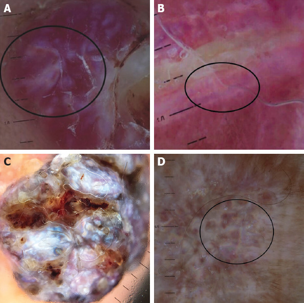
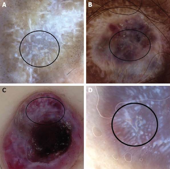
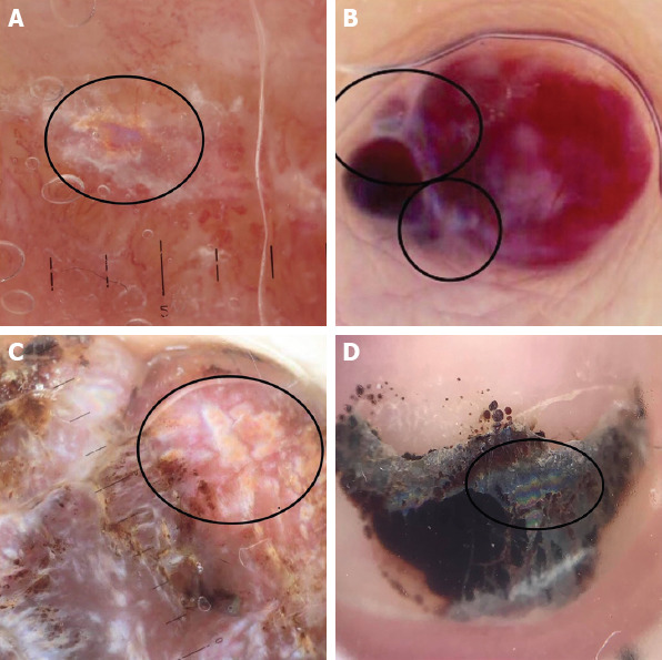
Results: A total of 840 lesions were reviewed and 21 (2%) non-Kaposi sarcoma lesions having dermoscopic rainbow pattern were detected. These lesions were as follows; pyogenic granuloma (n=4, 19%), hypertrophic scar (n=4, 19%), basal cell carcinoma (n=2, 10%), dermatofibroma (n=2, 10%), angiokeratoma (n=2, 10%), blue nevus (n=1, 5%), granuloma annulare (n=1, 5%), strawberry angioma (n=1, 5%), epidermal cyst (n=1, 5%), malignant melanoma (n=1, 5%), dissecting cellulitis (n=1, 5%) and subungual hematoma (n=1, 5%). The most common localization was limb (n=14, 67%) followed by face (n=3, 14%).
Conclusion: We suggest that the rainbow pattern is a complex and quite unspecific optic phenomenon which can be seen both in vascular and non-vascular lesions. Its diagnostic significance should be considered in the context of the other structural dermoscopic finding. To the best of our knowledge, to our knowledge, this is the most comprehensive study focusing on rainbow pattern in non-Kaposi's sarcoma lesions. Here, we also reported rainbow pattern in dissecting cellulitis, granuloma annulare and subungual hematoma which has not been shown to have rainbow pattern previously.
Keywords: Dermoscopy; Kaposi’s sarcoma; optic phenomenon; rainbow pattern.
Copyright: © 2020 by Istanbul Northern Anatolian Association of Public Hospitals.
Conflict of interest statement
Conflict of Interest: No conflict of interest was declared by the authors.
Figures
Similar articles
-
"Chasing Rainbows" Beyond Kaposi Sarcoma's Dermoscopy: A Mini-Review.Dermatopathology (Basel). 2024 Nov 25;11(4):333-341. doi: 10.3390/dermatopathology11040035. Dermatopathology (Basel). 2024. PMID: 39727617 Free PMC article. Review.
-
Dermoscopy of Kaposi's sarcoma: areas exhibiting the multicoloured 'rainbow pattern'.J Eur Acad Dermatol Venereol. 2009 Oct;23(10):1128-32. doi: 10.1111/j.1468-3083.2009.03239.x. Epub 2009 Apr 10. J Eur Acad Dermatol Venereol. 2009. PMID: 19438977
-
Rainbow pattern in Kaposi's sarcoma under polarized dermoscopy: a dermoscopic pathological study.Br J Dermatol. 2009 Apr;160(4):801-9. doi: 10.1111/j.1365-2133.2008.08940.x. Epub 2008 Dec 1. Br J Dermatol. 2009. PMID: 19067686
-
The Dermoscopic Rainbow Pattern - A Review of the Literature.Acta Dermatovenerol Croat. 2019 Jun;27(2):111-115. Acta Dermatovenerol Croat. 2019. PMID: 31351506 Review.
-
Pyogenic granuloma and nodular Kaposi’s sarcoma: dermoscopic clues for the differential diagnosis.Turk J Med Sci. 2019 Oct 24;49(5):1471-1478. doi: 10.3906/sag-1902-60. Turk J Med Sci. 2019. PMID: 31651116 Free PMC article.
Cited by
-
"Chasing Rainbows" Beyond Kaposi Sarcoma's Dermoscopy: A Mini-Review.Dermatopathology (Basel). 2024 Nov 25;11(4):333-341. doi: 10.3390/dermatopathology11040035. Dermatopathology (Basel). 2024. PMID: 39727617 Free PMC article. Review.
-
Iridescent Changes Observed During Dynamic Cross-polarized Dermoscopy.Dermatol Pract Concept. 2022 Jul 1;12(3):e2022119. doi: 10.5826/dpc.1203a119. eCollection 2022 Jul. Dermatol Pract Concept. 2022. PMID: 36159112 Free PMC article. No abstract available.
-
A spectrum of diseases with the dermatoscopic rainbow pattern.JAAD Case Rep. 2022 Jan 21;21:144-147. doi: 10.1016/j.jdcr.2022.01.001. eCollection 2022 Mar. JAAD Case Rep. 2022. PMID: 35242968 Free PMC article. No abstract available.
-
Non-Invasive Diagnostic Imaging in Kaposi Sarcoma Evaluation.Diagnostics (Basel). 2025 Jun 30;15(13):1665. doi: 10.3390/diagnostics15131665. Diagnostics (Basel). 2025. PMID: 40647663 Free PMC article.
References
-
- Hu SC, Ke CL, Lee CH, Wu CS, Chen GS, Cheng ST. Dermoscopy of Kaposi's sarcoma:areas exhibiting the multicoloured 'rainbow pattern'. J Eur Acad Dermatol Venereol. 2009;23:1128–132. - PubMed
-
- Cheng ST, Ke CL, Lee CH, Wu CS, Chen GS, Hu SC. Rainbow pattern in Kaposi's sarcoma under polarized dermoscopy: a dermoscopic pathological study. Br J Dermatol. 2009;160:801–9. - PubMed
-
- Vázquez-López F, García-García B, Rajadhyaksha M, Marghoob AA. Dermoscopic rainbow pattern in non-Kaposi sarcoma lesions. Br J Dermatol. 2009;161:474–5. - PubMed
LinkOut - more resources
Full Text Sources