. 2020 Nov 4;3(5):1791-1810.
doi: 10.1016/j.matt.2020.10.012. Epub 2020 Oct 29.

On-Mask Chemical Modulation of Respiratory Droplets

Affiliations

On-Mask Chemical Modulation of Respiratory Droplets

Haiyue Huang et al. Matter. .

Abstract

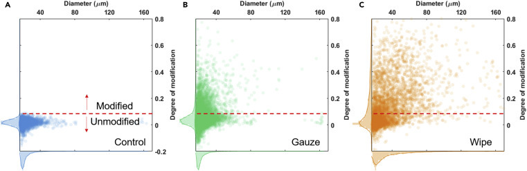
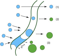
Transmission of infectious respiratory diseases starts from pathogen-laden respiratory droplets released during coughing, sneezing, or speaking. Here we report an on-mask chemical modulation strategy, whereby droplets escaping a masking layer are chemically contaminated with antipathogen molecules (e.g., mineral acids or copper salts) preloaded on polyaniline-coated fabrics. A colorimetric method based on the color change of polyaniline and a fluorometric method utilizing fluorescence quenching microscopy are developed for visualizing the degree of modification of the escaped droplets by H+ and Cu2+, respectively. It is found that even fabrics with low fiber-packing densities (e.g., 19%) can readily modify 49% of the escaped droplets by number, which accounts for about 82% by volume. The chemical modulation strategy could offer additional public health benefits to the use of face covering to make the sources less infectious, helping to strengthen the response to the current pandemic or future outbreaks of infectious respiratory diseases.

Keywords: MAP4: Demonstrate.


Conflict of interest statement

Northwestern University has filed a patent application including discoveries made in this work.

Figures

None
Graphical abstract
Figure 1
Figure 1
Three Necessary Steps for the Transmission of an Infectious Disease A source that can release the pathogens, possible transmission pathways along which the pathogens remain structurally intact and infectious before eventually reaching other hosts, and susceptible persons that the pathogens can successfully infect to duplicate themselves and start a new cycle. Mitigation strategies must address one or more of these steps. Isolating the source or making it less infectious should be most effective, as it cuts down the source of the spread chain and greatly reduces the burden at the later stages along the transmission pathways.
Figure 2
Figure 2
On-Mask Chemical Modulation of Respiratory Droplets: Hypothesis and Approach (A) Exhalation pushes warmer and wetter air outward, along with respiratory droplets through the mask to the environment. In contrast, inhalation draws colder and drier air from the environment, but without droplets. A chemical modulation layer can be inserted to leach antipathogen agents into the outgoing droplets. To avoid inhaling antipathogen agents (e.g., chemicals or particles), their release mechanism should be activated by the droplets. (B) In the experimental design, droplets of model respiratory fluids were pneumatically pulsed to simulate those released during coughing and sneezing with comparable range of size distribution, number density, and velocity. The droplets were directed toward a modulation layer (e.g., a nonwoven fabric loaded with chemical modifiers). A detector film was placed at the downstream to collect the escaped droplets, so that their degrees of chemical modification could be visualized directly.
Figure 3
Figure 3
Preparation of the Chemical Modulation Layer (A) A roll of nonwoven fabric was coated with polyaniline by in situ polymerization of aniline. The fabric was then washed with deionized water and dried in air. The polyaniline coating can be doped with molecular antipathogen agents such as mineral acids or Cu ions by soaking in H3PO4 or CuSO4 solutions, respectively. (B–G) Optical microscopy images showing the gauze fibers (B) before and (C) after polyaniline growth. The insets are photos of the gauze fabrics (2 cm × 2 cm). (D) SEM image of coated gauze fibers. Corresponding images of a lint-free wipe are shown in (E), (F), and (G).
Figure 4
Figure 4
Preparation of the Colorimetric Detector Film (A) A layer of polyaniline was grown on one side of a smooth plastic film floating on the surface of the polymerization bath. (B–D) (B) The as-grown green film (5 cm × 5 cm) turned (C) blue after dedoping, which yielded (D) a highly uniform blue field of view under an optical microscope. (E) AFM image showing a torn edge of the polyaniline layer. (F) Height profiles taken from (E) show that the coating thickness is around 100 nm.
Figure 5
Figure 5
Colorimetric Visualization of Modified Droplets (A–C) (A) Unmodified, nearly neutral droplets leave drying stains made of polymer and salts that are highly visible under (B) the reflection mode of imaging, but not under (C) the transmission mode. (D) In contrast, since modified droplets contain acid and can dope polyaniline, they leave green stains that are visible under both modes of imaging. (E–H) (E and F) and (G and H) are representative images of stains of droplets passing through a gauze cloth and a lint-free wipe, respectively. Note that not all the droplets observed under the reflection mode (E and G) can be seen under the transmission mode, due to insufficient level of acid modification. All scale bars represent 200 μm.
Figure 6
Figure 6
Quantitative Analysis of Colorimetric Microscopy Images (A) Since both unmodified and modified droplets are visible in the reflection mode of image, it was used to create marks to identify the positions of all droplets in (D) the corresponding transmission mode of image, in which only modified droplets are visible. (B) The reflection image underwent a few steps of processing to yield a noise-reduced, binarized image to prepare for recognition using Hough circle transformation. (C) Eventually the droplets were approximated as circular marks, which were used to identify the locations and sizes of the droplets in the transmission image. (D and E) (E) The green channel of (D) was extracted to calculate pixel intensity values. (F–H) (F) By overlapping the marking patterns (C) and (E), the pixel intensity inside each droplet, as illustrated by the yellow circles in (H), can be measured, together with the pixel intensity of local background around each droplet (red circle). Pixel intensities inside and outside a droplet were used to calculate its degree of modification based on Equation 1 (see also Supplemental Information). Such analysis produced the calculated degree of modification of all droplets and their corresponding radii based on optical microscopy images from (A) and (D), which are plotted in (G). Each dot represents one droplet.
Figure 7
Figure 7
Calibration of the Degree of Modification Droplets from stock solutions of six known pH values (1.3, 1.6, 1.9, 2.1, 2.3, and 2.5) were sprayed onto the colorimetric screen, and (A) the resulting images taken under transmission mode were used to calibrate (B) how the color changes (i.e., greener dot means higher degree of modification) correlate with the pH values of the droplets. Microscopy observation in (A) shows that for a droplet to produce a distinguishable green dot on the blue polyaniline film, its pH value needs to be ≤2.3. All images in (A) are 200 μm × 200 μm.
Figure 8
Figure 8
Effect of Droplet Modulation by the Gauze and Wipe The histograms show the distributions of the size and the corresponding degree of modification of droplets, after they passed through (A) a blank gauze, (B) a gauze, and (C) a lint-free wipe coated with acid-doped polyaniline, respectively. Each dot represents one droplet on the detector screen, and each histogram consists of over 4,000 droplets. The red dashed lines denote the threshold of modification chosen to calculate modulation efficiency, which corresponds to the peak value of pH 2.3 curve in Figure 7B.
Figure 9
Figure 9
Possible Ways for Droplets to Escape the Modulation Layer Schematic drawings showing that (1) droplets can escape the layer unmodified due to lack of any or significant interaction with the fibers, the fraction of which is largely determined by the packing density of the fabric; (2) droplets colliding with the fibers have higher chances of being modified; and (3) droplets can get caught on the fibers and grow larger through coalescence and/or condensation of water vapor, which leads to longer retention time (i.e., higher degree of modification), before they break apart via the air flow and escape.
Figure 10
Figure 10
Modulation of Droplets by Cu Ions and Fluorometric Visualization (A) Droplets passing through polyaniline-coated fabrics doped with CuSO4 can dissolve Cu ions. A Nc/PMMA film spin-coated on a coverglass was used as a fluorometric detector to capture the droplets, which was then imaged by FQM. The Cu-modified droplets can quench the fluorescence of their underlying Nc dye and thus appear as dark spots under FQM. (B) Fluorescence image of a control sample of Cu-free droplets on the detector film, showing drying marks of droplets, which did not quench the fluorescence. (C) Fluorescence image of another control sample of Cu-containing droplets on the detector film. The stains quenched fluorescence strongly and appeared as dark spots. (D) Transmission mode of image of a sample collected after they passed a Cu-loaded gauze, which clearly shows the drying marks of the droplets, but cannot differentiate which droplets were modified with Cu. (E) Under fluorescence mode, Cu-modified droplets are clearly visible as dark spots. The yellow circles highlight modified droplets and the red circles mark the unmodified ones. A total of 18 out of 58 droplets were modified. All scale bars represent 50 μm.

Similar articles

Cited by

References

    1. Gates B. Responding to Covid-19—a once-in-a-century pandemic? N. Engl. J. Med. 2020;382:1677–1679. - PubMed
    1. Huang H., Fan C., Li M., Nie H.-L., Wang F.-B., Wang H., Wang R., Xia J., Zheng X., Zuo X. COVID-19: a call for physical scientists and engineers. ACS Nano. 2020;14:3747–3754. - PubMed
    1. Flint J., Racaniello V.R., Rall G.F., Skalka A.M. John Wiley & Sons; 2015. Principles of Virology.
    1. Siegel J.D., Rhinehart E., Jackson M., Chiarello L., Practices H.C.I.C. 2007 guideline for isolation precautions: preventing transmission of infectious agents in health care settings. Am. J. Infect. Control. 2007;35:S65–S164. - PMC - PubMed
    1. World Health Organization Transmission of SARS-CoV-2: Implications for Infection Prevention Precautions. https://www.who.int/publications/i/item/modes-of-transmission-of-virus-c...

LinkOut - more resources