. 2020 Nov 17;1(8):100142.
doi: 10.1016/j.xcrm.2020.100142. Epub 2020 Oct 29.

Pharmacological Inhibition of Acid Sphingomyelinase Prevents Uptake of SARS-CoV-2 by Epithelial Cells

Affiliations

Pharmacological Inhibition of Acid Sphingomyelinase Prevents Uptake of SARS-CoV-2 by Epithelial Cells

Alexander Carpinteiro et al. Cell Rep Med. .

Abstract

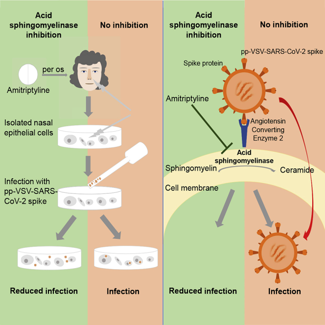
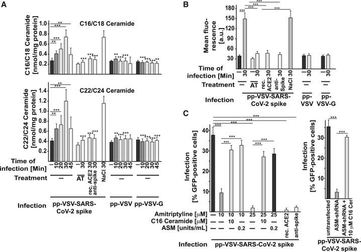
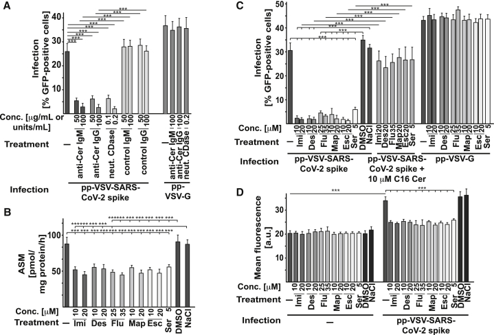
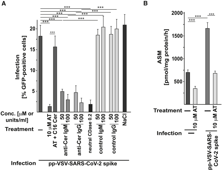
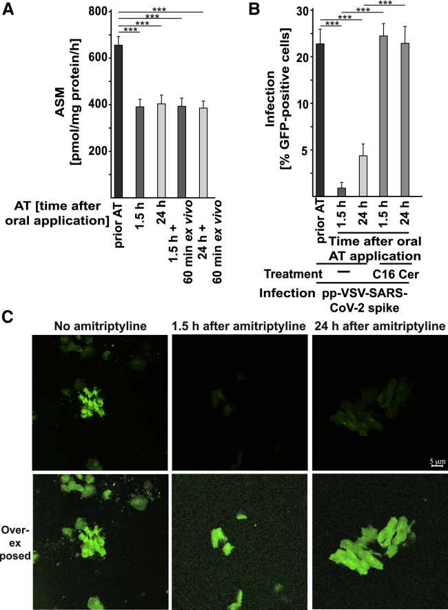
The acid sphingomyelinase/ceramide system plays an important role in bacterial and viral infections. Here, we report that either pharmacological inhibition of acid sphingomyelinase with amitriptyline, imipramine, fluoxetine, sertraline, escitalopram, or maprotiline or genetic downregulation of the enzyme prevents infection of cultured cells or freshy isolated human nasal epithelial cells with severe acute respiratory syndrome coronavirus 2 (SARS-CoV-2) or vesicular stomatitis virus (VSV) pseudoviral particles (pp-VSV) presenting SARS-CoV-2 spike protein (pp-VSV-SARS-CoV-2 spike), a bona fide system mimicking SARS-CoV-2 infection. Infection activates acid sphingomyelinase and triggers a release of ceramide on the cell surface. Neutralization or consumption of surface ceramide reduces infection with pp-VSV-SARS-CoV-2 spike. Treating volunteers with a low dose of amitriptyline prevents infection of freshly isolated nasal epithelial cells with pp-VSV-SARS-CoV-2 spike. The data justify clinical studies investigating whether amitriptyline, a safe drug used clinically for almost 60 years, or other antidepressants that functionally block acid sphingomyelinase prevent SARS-CoV-2 infection.

Keywords: SARS-CoV-2; acid sphingomyelinase; amitriptyline; antidepressants; ceramide; escitalopram; fluoxetine; human nasal epithelial cells; infection.


Conflict of interest statement

The authors declare no competing financial interests.

Figures

None
Graphical abstract
Figure 1
Figure 1
Amitriptyline Prevents Infection with pp-VSV-SARS-CoV-2 Spike (A) Vero cells were infected for 24 h with vesicular stomatitis virus (VSV) pseudoviral particles presenting severe acute respiratory syndrome coronavirus 2 spike protein (pp-VSV-SARS-CoV-2 spike) or with VSV particles that express the glycoprotein of VSV (pp-VSV-G). Cells were pretreated with amitriptyline (AT) and infected with pp-VSV-SARS-CoV-2 in the presence or absence of 5, 10, 20, or 25 μM AT. The solvent was 0.9% NaCl. AT treatment was initiated 4 h before infection of the pseudoviral particles. Expression of enhanced green fluorescent protein (EGFP) was quantified after 24 h by counting positive cells in 2,000 cells per sample in randomly chosen microscopic fields. Shown are representative fluorescence microscopy results or the means ± SD of the percentage of infected cells from 8 independent experiments. ∗∗∗p < 0.001 compared to infection without AT treatment (0); +++p < 0.001 compared between infection with pp-VSV-SARS-CoV-2 spike and pp-VSV-G; ANOVA, followed by post hoc Student’s t tests. (B) Surface expression of angiotensin-converting enzyme 2 (ACE2) was determined by flow cytometry 24 h after infection. Cells were stained with fluorescein isothiocyanate (FITC)-coupled anti-ACE2 antibodies. Shown are the means of the fluorescence ± SD, in random units, from 6 independent experiments. ∗∗∗p < 0.001 compared to not infected; +++p < 0.001 compared between infection with pp-VSV-SARS-CoV-2 spike and pp-VSV-G; ANOVA, followed by post hoc Student’s t tests. (C and D) AT did not exhibit cytotoxicity after treatment for 24 h. Cells were exposed for 24 h to 10 μM or 25 μM AT, and toxicity was measured by FITC-Annexin V staining (C) and terminal deoxynucleotidyl transferase dUTP nick end labeling (TUNEL) assay (D). FITC-Annexin V staining and TUNEL assays were analyzed by flow cytometry; staining of cellular actin was analyzed by confocal microscopy. Permeabilized cells served as positive controls for the FITC-Annexin V staining. Permeabilized and DNase-treated cells served as positive controls for TUNEL assays. Shown are representative results from 4 independent studies or the means ± SD of the fluorescence in the flow cytometry studies (n = 4); ∗∗∗p < 0.001; ANOVA, followed by post hoc Student’s t tests. Results are presented in arbitrary units (a.u.).
Figure 2
Figure 2
AT Prevents Infection with SARS-CoV-2 Human Caco-2 cells were pretreated with 10 or 25 μM AT for 4 h or left untreated and were infected with SARS-CoV-2 with a multiplicity of infection (MOI) of 0.001 in the presence or absence of AT. The titer of progeny in the culture supernatants was determined 24 h post infection (p.i.) by plaque assay. Viral transcripts were also detected by quantitative real-time RT-PCR with two separate primer sets in the lysates of Caco-2 cells treated as above but infected with a MOI of 0.1 for 24 h. Shown are the means ± SD of 3 experiments; ∗p < 0.05, ∗∗p < 0.01, ∗∗∗p < 0.001 compared to mock or infected without AT; Dunnets (for plaque-forming units [PFUs]) or Sidak’s (for PCR) multiple comparisons test.
Figure 3
Figure 3
VSV-SARS-CoV-2 Spike Particles Activate and Use the Acid Sphingomyelinase for Infection (A) Genetic downregulation of acid sphingomyelinase (ASM) with short hairpin RNA (shRNA) prevented infection of Caco-2 cells with pp-VSV-SARS-CoV-2 spike but had no impact on infection with particles that express the pp-VSV-G. Downregulation of neutral sphingomyelinase 2 (NSM2) did not affect the infection of Caco-2 cells with pp-VSV-SARS-CoV-2 spike. Control shRNA did not change the infection of Caco-2 cells by pp-VSV-SARS-CoV-2 spike. Cells were infected with pp-VSV-SARS-CoV-2 spike or pp-VSV-G, and cells positive for EGFP were counted in 2,000 cells per sample. Downregulation of ASM or NSM was confirmed by measuring the activity of the enzymes (right panels). (B) Vero cells were infected with pp-VSV-SARS-CoV-2 spike, pp-VSV, or pp-VSV-G for the indicated times or were left uninfected. Samples were pre-incubated with AT or with the solvent (0.9% NaCl) or were left untreated. In addition, recombinant (rec.) ACE2 protein or neutralizing anti-spike antibodies was added prior to and maintained during the infection. Cells were lysed in 250 mM sodium acetate (pH 5.0) and 0.2% NP40. ASM activity was determined by measuring the consumption of [14C]sphingomyelin. Shown are the means ± SD of the percentage of infected cells or the activity of the ASM or NSM from 6 independent experiments. ∗p < 0.05, ∗∗p < 0.01, ∗∗∗p < 0.001; ANOVA, followed by post hoc Student’s t tests.
Figure 4
Figure 4
VSV-SARS-CoV-2 Spike Particles Induce Ceramide Formation, Facilitating Infection (A) To determine ceramide levels, we organically extracted the samples and quantified ceramide levels by using the ceramide kinase method. We quantified C16 (C16-Cer)/C18 ceramide and C22/C24 ceramide. If indicated, cells were preincubated with 5, 10, 20, or 25 μM AT for 4 h before the addition of pp-VSV-SARS-CoV-2 spike. The rec. ACE2 protein or neutralizing anti-spike antibodies were added as above. In addition, cells were infected with pp-VSV or pp-VSV-G. Displayed are the means ± SD of the ceramide concentrations from each 5 independent experiments. ∗∗p < 0.01, ∗∗∗p < 0.001; ANOVA, followed by post hoc Student’s t tests, as indicated or compared to the corresponding value without inhibitor. (B) Flow cytometry reveals the formation of ceramide in the outer leaflet of the cell membrane. Cells were treated with 25 μM AT, each 2 μg rec. ACE2 protein or neutralizing anti-spike antibodies and infected with pp-VSV-SARS-CoV-2 spike. Controls were infected with pp-VSV or pp-VSV-G. Shown are the mean fluorescence values (in a.u.) ± SD of 6 independent flow cytometry studies; ∗∗∗p < 0.001; ANOVA, followed by post hoc Student’s t tests. (C) Reconstitution of ceramide in Vero cells (left panel) that had been treated with 10 or 25 μM AT or in Caco-2 cells (right panel) transfected with shRNA targeting ASM by the addition of C16-Cer (10 μM) or ASM (0.2 U/mL) during the infection with pp-VSV-SARS-CoV-2 spike restores viral infection of the cells. The rec. ACE2 protein or neutralizing anti-spike antibodies prevented the infection. Displayed are the means ± SD of the percentage of infected cells from 6 independent experiments. ∗∗∗p < 0.001; ANOVA, followed by post hoc Student’s t tests. See also Figure S3.
Figure 5
Figure 5
Neutralization of Surface Ceramide or Treatment with Other Antidepressants Prevents Infection with pp-VSV-SARS-CoV-2 Spike (A) Vero cells were infected with pp-VSV-SARS-CoV-2 spike in the presence or absence of 50 or 100 μg/mL anti-ceramide (anti-Cer) IgM antibodies, clone S58-9, or a monoclonal anti-Cer IgG, or of 0.1 or 0.2 units/mL of neutral ceramidase (neut. CDase) to either neutralize or consume surface ceramide. Infection was measured by the expression of EGFP in the cells. Control IgM or IgG exerted no effect. Anti-Cer antibodies or neut. CDase were without effect on infection with pp-VSV-G. Shown are means ± SD from 5 independent experiments. ∗∗∗p < 0.001; ANOVA, followed by post hoc Student’s t tests. Conc., concentration. (B–D) Incubation of Vero cells with imipramine (Imi), desipramine (Des), fluoxetine (Flu), sertraline (Ser), escitalopram (Esc), or maprotiline (Map) inhibits ASM (B) and prevents the infection of Vero cells with pp-VSV-SARS-CoV-2 spike but not with pp-VSV-G measured as uptake (C) and upregulation of ACE2 expression as measurement for infection (D). Reconstitution of ceramide in treated cells with 10 μM C16-Cer restored infection of the cells with pp-VSV-SARS-CoV-2 spike (C); see also Figure S3. The solvents of the drugs (DMSO or 0.9% NaCl) did not affect ASM activity or viral infection (B–D). Shown are the means ± SD of 6 independent experiments. ∗∗∗p < 0.001; ANOVA, followed by post hoc Student’s t tests.
Figure 6
Figure 6
Infection of Freshly Isolated Human Nasal Epithelial Cells Is Prevented by Ex Vivo Treatment with AT, Anti-Cer Antibodies, or neut. CDase (A) Freshly isolated human nasal epithelial cells were infected with pp-VSV-SARS-CoV-2 spike for 60 min. Anti-Cer antibodies (IgM or IgG) or neut. CDase were co-applied with the virus. AT (10 μM) was applied for 60 min before the infection and was also present during the infection. All of these treatments blocked the infection of freshly isolated human nasal epithelial cells with pp-VSV-SARS-CoV-2 spike. Control IgM, IgG, or 0.9% NaCl (solvent for AT) exerted no effect. Ceramide was reconstituted in AT-treated nasal epithelial cells with 10 μM C16-Cer; this reconstitution restored the infection of the cells with pp-VSV-SARS-CoV-2 spike. (B) AT reduced ASM activity in nasal epithelial cells upon infection. Shown are the means ± SD of 6 (A) or 5 (B) independent experiments. ∗∗∗p < 0.001; ANOVA, followed by post hoc Student’s t tests.
Figure 7
Figure 7
In Vivo Administration of AT Prevents Infection of Nasal Epithelial Cells with pp-VSV-SARS-CoV-2 Spike Ex Vivo Volunteers were orally treated with 0.5 mg/kg AT. Nasal epithelial cells were isolated from volunteers before and 1.5 h and 24 h after administration of AT. (A) Activity of ASM was determined by measurement of the consumption of [14C]sphingomyelin in aliquots of freshly isolated nasal epithelial cells or after an additional 1-h culture ex vivo. (B) After isolation, nasal epithelial cells were infected for 1 h, with pp-VSV-SARS-CoV-2 spike and washed. The expression of EGFP was determined after 24 h in at least 500 epithelial cells per sample in randomly chosen microscopic fields. Ceramide in nasal epithelial cells isolated from volunteers who had taken AT was reconstituted with 10 μM C16-Cer. This reconstitution restored the infection of the cells with pp-VSV-SARS-CoV-2 spike. The percentage of infected nasal epithelial cells is displayed. (C) Shown is a typical result from 6 independent infection studies with nasal epithelial cells from volunteers before and after oral administration of AT. The bottom panel shows an over-exposed microphotograph to visualize all cells. Shown are the means ± SD from 6 volunteers. ∗∗∗p < 0.001; ANOVA, followed by post hoc Student’s t tests.

References

    1. Zhou P., Yang X.L., Wang X.G., Hu B., Zhang L., Zhang W., Si H.R., Zhu Y., Li B., Huang C.L. A pneumonia outbreak associated with a new coronavirus of probable bat origin. Nature. 2020;579:270–273. - PMC - PubMed
    1. Yang X., Yu Y., Xu J., Shu H., Xia J., Liu H., Wu Y., Zhang L., Yu Z., Fang M. Clinical course and outcomes of critically ill patients with SARS-CoV-2 pneumonia in Wuhan, China: a single-centered, retrospective, observational study. Lancet Respir. Med. 2020;8:475–481. - PMC - PubMed
    1. World Health Organization Coronavirus disease (COVID-19): similarities and differences with influenza. https://www.who.int/emergencies/diseases/novel-coronavirus-2019/question...
    1. John Hopkins University Mortality analyses. https://coronavirus.jhu.edu/data/mortality
    1. Hoffmann M., Kleine-Weber H., Schroeder S., Krüger N., Herrler T., Erichsen S., Schiergens T.S., Herrler G., Wu N.H., Nitsche A. SARS-CoV-2 cell entry depends on ACE2 and TMPRSS2 and is blocked by a clinically proven protease inhibitor. Cell. 2020;181:271–280.e8. - PMC - PubMed

Publication types

MeSH terms