. 2020 Nov 18;12(11):275-289.
doi: 10.1093/intbio/zyaa022.

Endothelial cell apicobasal polarity coordinates distinct responses to luminally versus abluminally delivered TNF-α in a microvascular mimetic

Affiliations

Endothelial cell apicobasal polarity coordinates distinct responses to luminally versus abluminally delivered TNF-α in a microvascular mimetic

Alec T Salminen et al. Integr Biol (Camb). .

Abstract

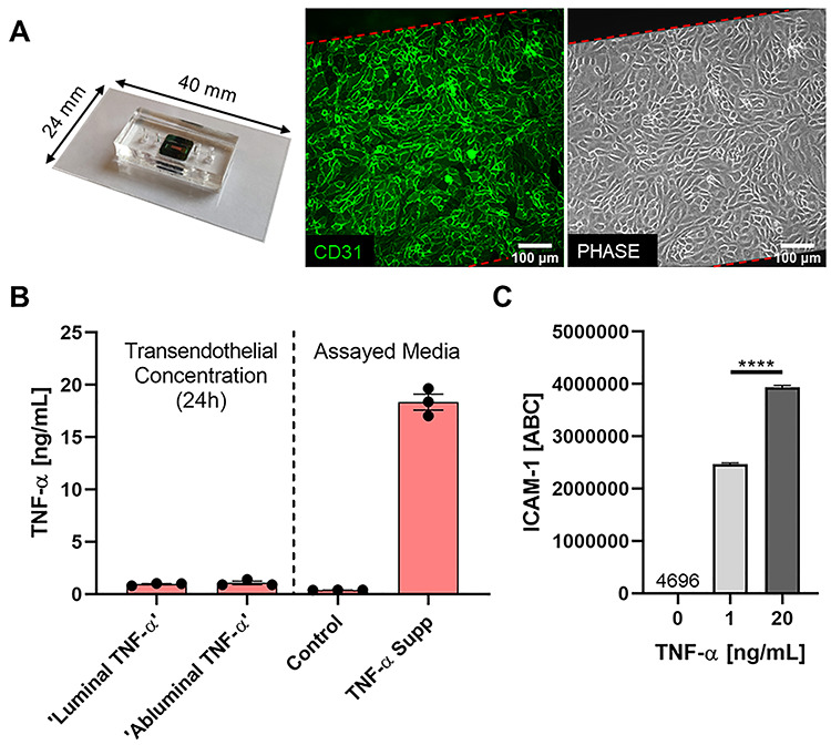
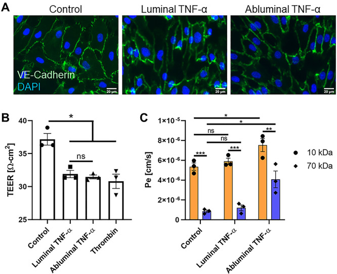
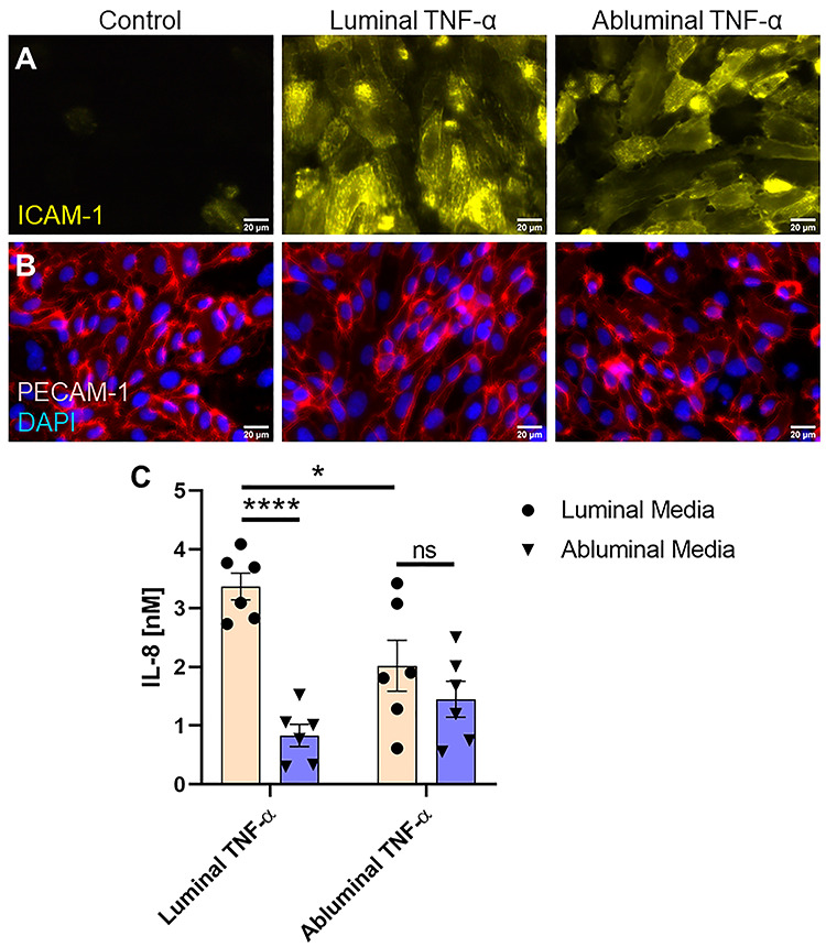
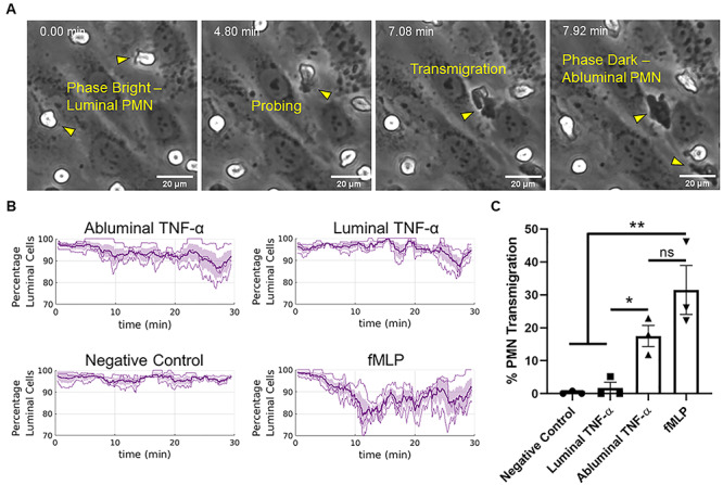
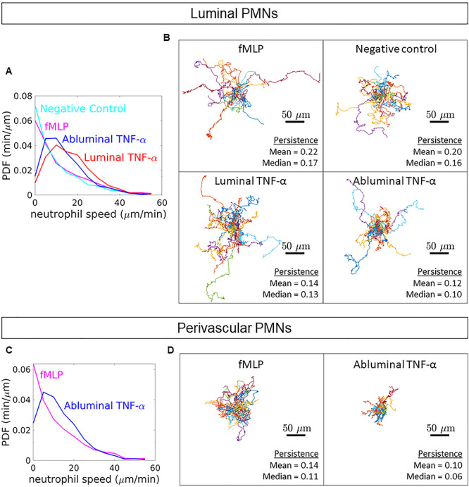
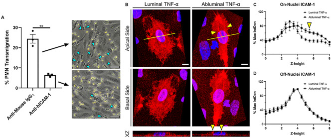
Endothelial cells (ECs) are an active component of the immune system and interact directly with inflammatory cytokines. While ECs are known to be polarized cells, the potential role of apicobasal polarity in response to inflammatory mediators has been scarcely studied. Acute inflammation is vital in maintaining healthy tissue in response to infection; however, chronic inflammation can lead to the production of systemic inflammatory cytokines and deregulated leukocyte trafficking, even in the absence of a local infection. Elevated levels of cytokines in circulation underlie the pathogenesis of sepsis, the leading cause of intensive care death. Because ECs constitute a key barrier between circulation (luminal interface) and tissue (abluminal interface), we hypothesize that ECs respond differentially to inflammatory challenge originating in the tissue versus circulation as in local and systemic inflammation, respectively. To begin this investigation, we stimulated ECs abluminally and luminally with the inflammatory cytokine tumor necrosis factor alpha (TNF-α) to mimic a key feature of local and systemic inflammation, respectively, in a microvascular mimetic (μSiM-MVM). Polarized IL-8 secretion and polymorphonuclear neutrophil (PMN) transmigration were quantified to characterize the EC response to luminal versus abluminal TNF-α. We observed that ECs uniformly secrete IL-8 in response to abluminal TNF-α and is followed by PMN transmigration. The response to abluminal treatment was coupled with the formation of ICAM-1-rich membrane ruffles on the apical surface of ECs. In contrast, luminally stimulated ECs secreted five times more IL-8 into the luminal compartment than the abluminal compartment and sequestered PMNs on the apical EC surface. Our results identify clear differences in the response of ECs to TNF-α originating from the abluminal versus luminal side of a monolayer for the first time and may provide novel insight into future inflammatory disease intervention strategies.

Keywords: apicobasal polarity; endothelial cells; inflammation; neutrophils; sepsis; tissue-chip.


Figures

Figure 1
Figure 1
Modeling luminal versus abluminal inflammation in a microvascular mimetic. (A) Microfluidic, silicon membrane-enabled microvascular mimetics (μSiM-MVMs) were assembled and sterilized, and HUVECs were seeded at high density (40 000 cells/cm2). Epifluorescence (green: CD31, PECAM-1) and phase microscopy were utilized to characterize HUVECs in the μSiM-MVM unit and ensure confluency. (B) TNF-α transendothelial flux was quantified for assay development. TNF-α supplemented media (TNF-α Supp) and control media were added to their respective channels (e.g. ‘Luminal TNF-α’ group was loaded with TNF-α supplemented media luminally and control media abluminally) and devices were incubated for 24 h. Trans-compartment media was collected and TNF-α concentration was determined by ELISA. Control and TNF-α media (right) were assayed in parallel to ensure that proper TNF-α concentrations were achieved at hour 0 and build confidence in ELISA results. In either case, ~1-ng/ml TNF-α was observed in the trans-compartment following 24-h incubation, thus confluent HUVECs function as a restrictive barrier to TNF-α and cells are primarily exposed in a polarized manor (20:0–1 ng/ml) for the 24-h experiment. (C) Flow cytometry analysis of HUVEC ICAM-1 expression following 24-h single-side stimulation with 0 (control)-, 1- or 20-ng/ml TNF-α. Statistically different levels of inflammation-induced ICAM-1 expression, as measured by ABC, ensure HUVECs differentially respond to 1- versus 20-ng/ml TNF-α. ABCs were statistically compared by ANOVA with Tukey’s multiple comparisons tests, ****P < 0.0001.
Figure 2
Figure 2
VE-cadherin and vascular permeability analysis following luminal or abluminal TNF-α delivery. (A) VE-cadherin immunofluorescence staining on HUVECs upon luminal or abluminal TNF-α stimulation revealed potential, yet equivocal changes in intensity and morphology across treatment groups. Scale bars = 20 μm, n = 3 independent experiments and devices, representative images shown. (B) TEER measurements were taken in traditional Transwell™ permeable support systems 24 h post-HUVEC treatment to determine the functional impact of polarized TNF-α stimulation of HUVEC barrier function. TEER revealed a statistically significant increase in EC permeability following cytokine stimulation (when compared with negative control), independent of EC apicobasal polarity. N = 3 independent experiments, error bars represent standard error of mean, means were statistically compared by ANOVA with Tukey’s multiple comparisons test, *P < 0.05 and ns = no significance. (C) Small-molecule [10 (orange) and 70 (blue) kDa FITC-dextran] permeability measurements performed in the μSiM-MVM showed a significant increase in HUVEC permeability in response to abluminal TNF-α with respect to control, which was not observed following luminal treatment. N = 3 independent experiments and devices, error bars represent standard error of mean, means were statistically compared by ANOVA with Tukey’s multiple comparisons test, *P < 0.05 and ns = no significance.
Figure 3
Figure 3
EC expression of insoluble and soluble proteins in response to luminal or abluminal TNF-α. (A) Immunofluorescence staining of EC ICAM-1 revealed upregulation of the surface adhesion protein, with no visible difference between luminal and abluminal stimulation groups. Scale bars = 20 μm, n = 3 independent experiments and devices, representative images shown. (B) PECAM-1 staining showed no obvious differences between treatment groups and controls all together. Scale bars = 20 μm, n = 3 independent experiments and devices, representative images shown. (C) Soluble IL-8 content in CCM was quantified by ELISA. Luminal TNF-α exposure induced a gradient of soluble IL-8 favoring luminal presentation. Abluminal TNF-α exposure, however, failed to induce a statistically significant difference in IL-8 secretion directed toward the luminal versus abluminal compartments, suggesting uniform secretion of the chemoattractant. N = 6 independent experiments and devices across treatment groups, error bars represent standard error of mean, means were statistically compared by ANOVA with Tukey’s multiple comparisons test, *P < 0.05, ****P < 0.0001 and ns = no significance.
Figure 4
Figure 4
Abluminal TNF-α drives PMN transmigration. (A) PMNs transition from phase bright (luminal) to phase dark (perivascular/abluminal) as they traverse the endothelium within the μSiM-MVM, allowing for automated tracking of PMN transmigration without exogenous dyes. (B) Automated tracking was used to quantify the ratios of luminal PMNs to total PMNs (percentage luminal cells) following luminal or abluminal TNF-α treatment of ECs (20 ng/ml for 24 h). Additionally, fMLP was perfused in the abluminal compartment of independent devices as a positive control for transmigration. N = 3 independent experiments and devices, dark lines represent time-dependent mean, shaded regions represent standard error of mean. (C) Percent PMN transmigration was also quantified by hand as a means of validating the automated tracking and drawing statistically robust conclusions from the data set. Percent transmigration was defined as the ratio of the number of cells in the first frame of the time lapse movie to the number of cells that transmigrate over the course of 30 min. Both automated and hand tracking revealed a robust transmigration response to abluminal TNF-α EC stimulation. N = 3 independent experiments and devices, means were statistically compared with fMLP (positive control) by ANOVA with Tukey’s multiple comparisons test, treatments were compared with unpaired t-tests, *P < 0.05, **P < 0.005 and ns = no significance.
Figure 5
Figure 5
PMN transmigration potential following exogenous IL-8 addition. (A) μSiM-MVMs were loaded luminally with PMNs as well as luminally and abluminally with recombinant human IL-8 at the concentrations determined by ELISA: Luminal (L) TNF-α Mock = 3.4-nM IL-8 luminally and 0.8-nM IL-8 abluminally, Abluminal (A) TNF-α Mock = 2-nM IL-8 luminally and 1.4-nM IL-8 abluminally, Positive Gradient (PG) = 0-nM IL-8 luminally and 2-nM IL-8 abluminally. Phase images were collected at time of PMN loading (first column) and following 30 min of incubation (second column). Neither mock treatment resulted in PMN transmigration. PMN transmigration was observed, however, in the positive gradient group. Representative images shown. N = 3 independent experiments and devices. (B) Experiments were repeated in devices exposed to abluminal TNF-α [20 ng/ml, 24 h]. PMN transmigration was again not observed in the luminal TNF-α mock group but was recovered in the abluminal TNF-α mock group. Scale bars = 20 μm, n = 3, representative images shown. Yellow arrows emphasize transmigration events. Quantification was performed by hand by dividing the number of phase white PMNs in the first image to the number of phase dark PMNs in the final image (listed as mean ± SEM on respective images). (C) Mean percent PMN transmigration was statistically compared by two-way ANOVA with Sidak’s multiple comparisons test, *P < 0.05 and ns = no significance.
Figure 6
Figure 6
PMN dynamics following directional cytokine stimulation. (A) PDFs of luminal PMN speeds were calculated from hand tracks. Mean luminal PMN speeds were calculated from instantaneous velocities as follows: negative control = 10.87 ± 2.44 μm/min, fMLP = 13.30 ± 3.39 μm/min, luminal TNF-α = 17.37 ± 2.13 μm/min and abluminal TNF-α = 14.88 ± 1.92 μm/min. (B) Additionally, PMN persistence, defined as the ratio of displacement to total pathlength, was quantified. Spider plots depict hand tracks stemming from a universal origin. (C) Analyses were repeated for PMNs crawling in the perivascular space (between the HUVECs and the nanoporous membrane). Mean perivascular PMN speeds were calculated from instantaneous velocities as follows: fMLP = 13.14 ± 3.18 μm/min and abluminal TNF-α = 14.51 ± 2.16 μm/min. (D) Persistence of perivascular PMNs was determined as well; spider plots shown. For all conditions: n = 30, three independent experiments and devices, 10 PMNs per device. A comprehensive statistical summary based on population means can be found in the online supplement (Supplementary Figs S3 and S4).
Figure 7
Figure 7
ICAM-1 reorganization on membrane ruffles following abluminal stimulation of ECs. (A) Percent PMN transmigration across abluminally stimulated ECs following ICAM-1 (Anti-hICAM-1) or isotype control (Anti-Mouse-IgG1) antibody blocking. Scale bar = 50 μm, n = 3, representative images shown. Mean percent transmigration was statistically compared by unpaired t-test, **P < 0.005. (B) Representative 3D reconstruction of confocal stacks depicting apical (top row, view from apical side of the cell) and basal (bottom row, inverted image showing view from basal side of cell) ICAM-1 expression following polarized cytokine (TNF-α [20 ng/ml]; 24 h) stimulation. Yellow arrows emphasize ICAM-1 rich ruffles present above EC nuclei following abluminal TNF-α treatment. N = 3 independent experiments and devices, 2 cells per device, representative images shown. XZ projection was collected at the mid-nucleus cross-section (yellow line) to show ruffles extension in the Z plane. Scale bars = 10 μm. ICAM-1 label intensity was quantified as follows: (C) ICAM-1 label integrated density at each z-height in the nuclear region as marked by DAPI and (D) ICAM-1 label integrated density at each z-height on the periphery of the cell. Integrated densities were normalized to respective image maximum values. Plots represent mean ± SEM values at each z-stack height (0.49-μm increments, 8-μm total height). Zero micrometer represents nanoporous membrane focus. For both on- and off-nuclei measurements, ICAM-1 intensity following luminal or abluminal TNF-α exposure was statistically compared at each respective z-height by two-way ANOVA with Sidak’s multiple comparisons test, *P < 0.05.

Similar articles

Cited by

References

    1. Hotchkiss RS, Moldawer LL, Opal SM et al. Sepsis and septic shock. Nat Rev Dis Primers 2016;2:16045. - PMC - PubMed
    1. Zeni F, Freeman B, Natanson C. Anti-inflammatory therapies to treat sepsis and septic shock: a reassessment. Crit Care Med 1997;25:1095–100. - PubMed
    1. Goldblum SE, Hennig B, Jay M et al. Tumor necrosis factor alpha-induced pulmonary vascular endothelial injury. Infect Immun 1989;57:1218–26. - PMC - PubMed
    1. Puhlmann M, Weinreich DM, Farma JM et al. Interleukin-1beta induced vascular permeability is dependent on induction of endothelial tissue factor (TF) activity. J Transl Med 2005;3:37. - PMC - PubMed
    1. Zheng X, Zhang W, Hu X. Different concentrations of lipopolysaccharide regulate barrier function through the PI3K/Akt signalling pathway in human pulmonary microvascular endothelial cells. Sci Rep 2018;8:9963. - PMC - PubMed

Publication types

MeSH terms