Comparative Study
. 2021 Jan 1;320(1):H108-H116.
doi: 10.1152/ajpheart.00639.2020. Epub 2020 Nov 8.

Progression of infarct-mediated arrhythmogenesis in a rodent model of heart failure

Affiliations
Comparative Study

Progression of infarct-mediated arrhythmogenesis in a rodent model of heart failure

Ikeotunye Royal Chinyere et al. Am J Physiol Heart Circ Physiol. .

Abstract

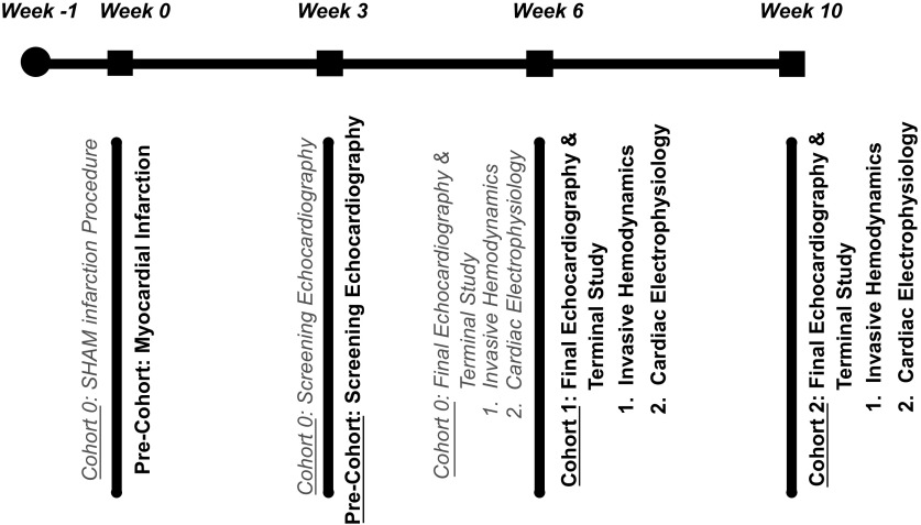
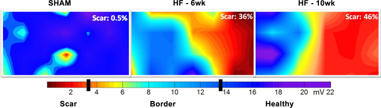
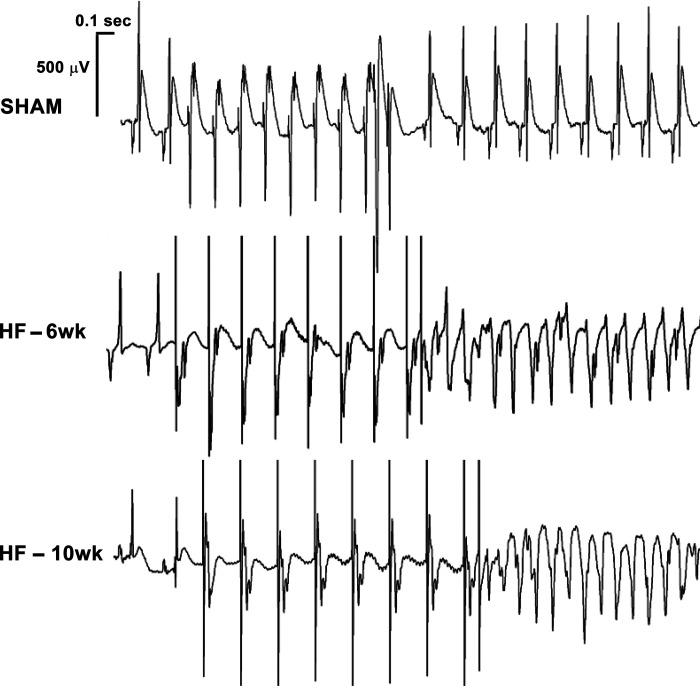
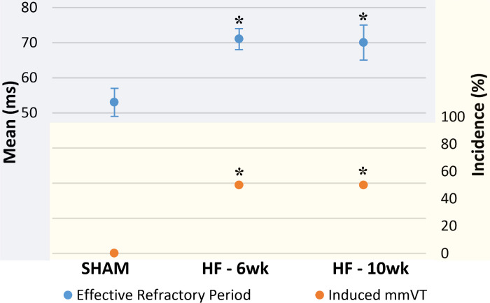
Heart failure (HF) post-myocardial infarction (MI) presents with increased vulnerability to monomorphic ventricular tachycardia (mmVT). To appropriately evaluate new therapies for infarct-mediated reentrant arrhythmia in the preclinical setting, chronologic characterization of the preclinical animal model pathophysiology is critical. This study aimed to evaluate the rigor and reproducibility of mmVT incidence in a rodent model of HF. We hypothesize a progressive increase in the incidence of mmVT as the duration of HF increases. Adult male Sprague-Dawley rats underwent permanent left coronary artery ligation or SHAM surgery and were maintained for either 6 or 10 wk. At end point, SHAM and HF rats underwent echocardiographic and invasive hemodynamic evaluation. Finally, rats underwent electrophysiologic (EP) assessment to assess susceptibility to mmVT and define ventricular effective refractory period (ERP). In 6-wk HF rats (n = 20), left ventricular (LV) ejection fraction (EF) decreased (P < 0.05) and LV end-diastolic pressure (EDP) increased (P < 0.05) compared with SHAM (n = 10). Ten-week HF (n = 12) revealed maintenance of LVEF and LVEDP (P > 0.05), (P > 0.05). Electrophysiology studies revealed an increase in incidence of mmVT between SHAM and 6-wk HF (P = 0.0016) and ERP prolongation (P = 0.0186). The incidence of mmVT and ventricular ERP did not differ between 6- and 10-wk HF (P = 1.0000), (P = 0.9831). Findings from this rodent model of HF suggest that once the ischemia-mediated infarct stabilizes, proarrhythmic deterioration ceases. Within the 6- and 10-wk period post-MI, no echocardiographic, invasive hemodynamic, or electrophysiologic changes were observed, suggesting stable HF. This is the necessary context for the evaluation of experimental therapies in rodent HF.NEW & NOTEWORTHY Rodent model of ischemic cardiomyopathy exhibits a plateau of inducible monomorphic ventricular tachycardia incidence between 6 and 10 wk postinfarction.

Keywords: adverse remodeling; ischemia; monophasic action potential; rigor and reproducibility; ventricular tachycardia.


Figures

Figure 1.
Figure 1.
Study timeline. A graphical display of the study design. All rats were acclimated upon arrival for 1 wk (week 1) until myocardial infarction (MI; sample size = 62). SHAM rats (cohort 0; gray text) (sample size = 10) underwent a left thoracotomy without left coronary artery ligation (week 0) to serve as appropriate surgical controls. Left ventricular ejection fraction below 40% was confirmed via screening echocardiography (week 3) for surviving infarcted precohort rats (sample size = 32) before random assignment to either cohort 1 (6-wk HF; sample size = 20) or cohort 2 (10-week HF; sample size = 12). Cohort 1 was survived for 6 wk post-MI (week 6), and cohort 2 was survived for 10 wk post-MI (week 10). At each respective end point, rats underwent a final echocardiographic study before returning to the surgical table for their terminal study. The terminal study began with invasive hemodynamic assessment and was completed after the cardiac electrophysiology study. Overall survival post-MI was 60%, and 5 rats were excluded for ejection fractions above 40%.
Figure 2.
Figure 2.
Epicardial monophasic action potentials are of excellent quality. Three epicardial monophasic action potential tracings from a single 10-wk heart failure (HF) rat. The tracings highlight the substantial difference in monophasic action potential amplitude in millivolts (y-axis) between healthy myocardium, border tissue, and scar tissue, likely due to the decreased number of viable cardiomyocytes. A difference can also be observed in the monophasic action potential duration to 90% repolarization (x-axis). Differences in the waveform slope, particularly in the 2nd and 3rd phase, are thought to be due to impaired potassium efflux associated with ischemic remodeling.
Figure 3.
Figure 3.
Two-dimensional electroanatomic colormaps reveal comparable substrate. Epicardial monophasic action potential amplitude colormaps from 3 randomly selected rats, each representing its respective group namely SHAM, 6-wk heart failure (HF), or 10-wk HF. The colormaps reveal a normal epicardium in SHAM, with a single area of red highlighting the sensitivity of monophasic action potential amplitude (MAPA) in characterizing the epicardium. The 6-wk HF colormap shows gross MAPA defects on the right, which corresponds to the left coronary artery myocardial territory. The HF-10-wk map exhibits maintenance of the electrical infarct with a widening of the border region but improvement in MAPA millivoltage in the scar. Although scar burden is different between these 2 randomly selected HF rats representing the 6-wk end point and the 10-wk end point, the qualities of the substrates are similar and the hemodynamic and echocardiographic group averages support the notion that the degree of myocardial infarction-mediated HF was comparable between the 2 groups.
Figure 4.
Figure 4.
Programmed electrical stimulation-induced ventricular tachycardia in heart failure (HF). Surface electrocardiogram (lead II) tracings from individual rats representing each group. Each tracing begins with 2 cardiac cycles of intrinsic electrical activity, followed by the programmed electrical stimulation (PES) drivetrain of 8 S1 stimulations and 1 premature S2 stimulation. The resulting rhythm is two premature ventricular contractions followed by a brief delay and then spontaneous return to normal sinus rhythm for the SHAM rat, monomorphic ventricular tachycardia (mmVT) for the HF-6-wk rat, and a comparable mmVT for the HF-10wk rat. Scale bars are provided before the SHAM tracing. For SHAM and HF 6-wk tracings, the P wave observed during intrinsic electrical activity can be observed during PES.
Figure 5.
Figure 5.
Plateau of both ventricular tachycardia and effective refractory period. A graph summarizing the electrophysiologic results for incidence of inducible monomorphic ventricular tachycardia (mmVT) and ventricular effective refractory period (ERP; means ±SE. Changes between SHAM (n = 15) and 6-wk heart failure (HF) (n = 20) include an increase in inducible mmVT and a prolongation of the ventricular ERP. These changes do not continue between 6 wk and 10 wk of HF (n = 10). *P < 0.01 vs. SHAM (ERP: ANOVA-Tukey; mmVT: Fisher’s exact).

Similar articles

Cited by

References

    1. Benjamin EJ, Muntner P, Alonso A, Bittencourt MS, Callaway CW, Carson AP , et al. Heart Disease and Stroke Statistics-2019 updated: a report from the American Heart Association. Circulation 139: e56–e528, 2019. [Erratum in Circulation 141: e33, 2020]. - PubMed
    1. Basuray A, French B, Ky B, Vorovic E, Olt C, Sweitzer NK, Cappola TP, Fang JC. Heart failure with recovered ejection fraction: clinical description, biomarkers, and outcomes. Circulation 129: 2380–2387, 2014. doi:10.1161/CIRCULATIONAHA.113.006855. - DOI - PMC - PubMed
    1. Cutler MJ, Rosenbaum DS, Dunlap ME. Structural and electrical remodeling as therapeutic targets in heart failure. J Electrocardiol 40: S1–S7, 2007. - PubMed
    1. Ebinger MW, Krishnan S, Schuger CD. Mechanisms of ventricular arrhythmias in heart failure. Curr Heart Fail Rep 2: 111–117, 2005. doi:10.1007/s11897-005-0018-y. - DOI - PubMed
    1. Roden DM, Abraham RL. Refining repolarization reserve. Heart Rhythm 8: 1756–1757, 2011. doi:10.1016/j.hrthm.2011.06.024. - DOI - PMC - PubMed

Publication types