. 2021 Mar:229:100-114.
doi: 10.1016/j.trsl.2020.10.005. Epub 2020 Oct 22.

Improved chemosensitivity following mucolytic therapy in patient-derived models of mucinous appendix cancer

Affiliations

Improved chemosensitivity following mucolytic therapy in patient-derived models of mucinous appendix cancer

Ashok K Dilly et al. Transl Res. 2021 Mar.

Abstract

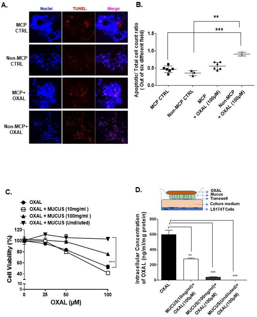
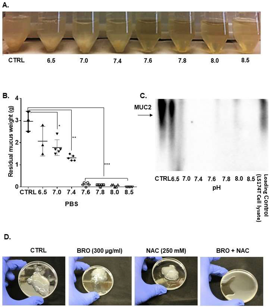
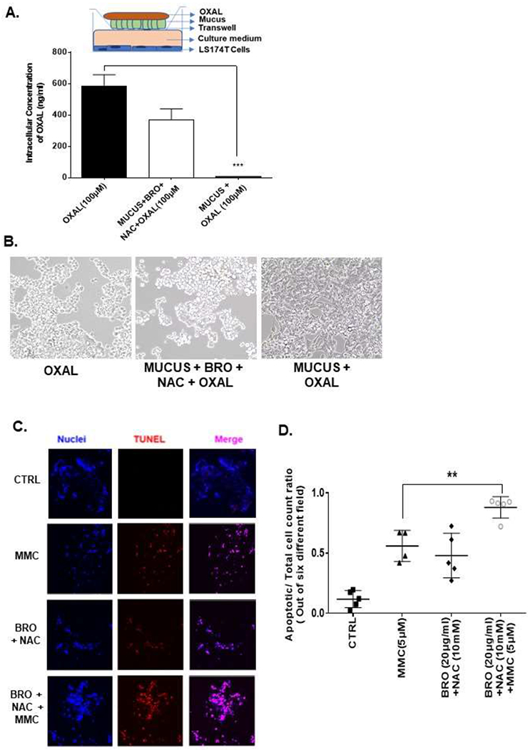
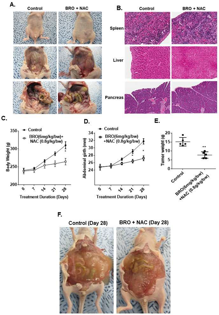
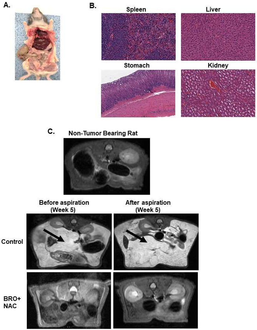
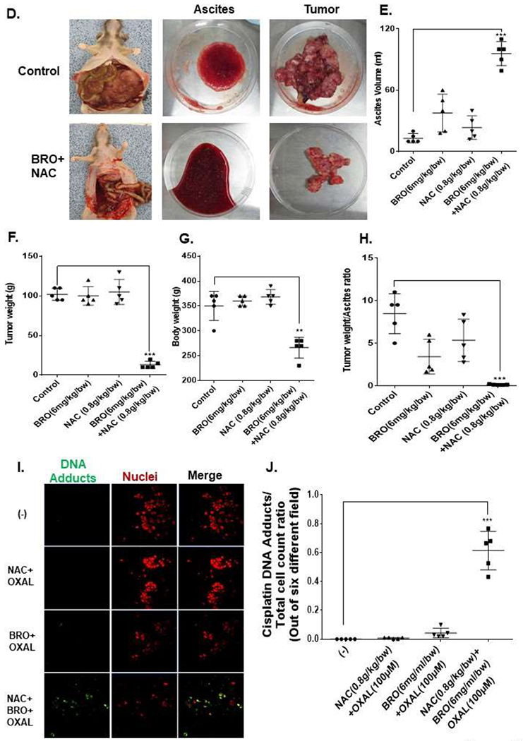
Abundant intraperitoneal (IP) accumulation of extracellular mucus in patients with appendiceal mucinous carcinoma peritonei (MCP) causes compressive organ dysfunction and prevents delivery of chemotherapeutic drugs to cancer cells. We hypothesized that reducing extracellular mucus would decrease tumor-related symptoms and improve chemotherapeutic effect in patient-derived models of MCP. Mucolysis was achieved using a combination of bromelain (BRO) and N-acetylcysteine (NAC). Ex vivo experiments of mucolysis and chemotherapeutic drug delivery/effect were conducted with MCP and non-MCP tissue explants. In vivo experiments were performed in mouse and rat patient-derived xenograft (PDX) models of early and late (advanced) MCP. MCP tumor explants were less chemosensitive than non-MCP explants. Chronic IP administration of BRO + NAC in a mouse PDX model of early MCP and a rat PDX model of late (advanced) MCP converted solid mucinous tumors into mucinous ascites (mucolysis) that could be drained via a percutaneous catheter (rat model only), significantly reduced solid mucinous tumor growth and improved the efficacy of chemotherapeutic drugs. Combination of BRO + NAC efficiently lyses extracellular mucus in clinically relevant models of MCP. Conversion of solid mucinous tumors into mucinous ascites decreases tumor bulk and allows for minimally invasive drainage of liquified tumors. Lysis of extracellular mucus removes the protective mucinous coating surrounding cancer cells and improves chemotherapeutic drug delivery/efficacy in cancer cells. Our data provide a preclinical rationale for the clinical evaluation of BRO + NAC as a therapeutic strategy for MCP.


Conflict of interest statement

Conflict of Interest: All authors have read the journals policy on disclosure of potential conflicts, and have no competing interests to declare. All authors have read the journal’s authorship agreement, and approved the final version of the manuscript.

Figures

Figure 1.
Figure 1.. Extracellular mucus is cytoprotective for cancer cells and imparts chemoresistance.
TUNEL assay was performed on MCP and non-MCP tumor tissues, with or without OXAL (100μM) treatment, for 24h ex vivo (A) and TUNEL-positive cell were quantified; images were randomly taken of 6 different fields and analyzed using Image J software to quantify apoptosis (B). LS174T cells were coated with undiluted or diluted mucus (diluted to 10 mg/ml or 100 mg/ml in PBS), treated with OXAL (0-100μM) for 24h, following which cell viability was analyzed by MTS assay (C). In a tranwell assay, delivery of OXAL (upper chamber) to LS174T cells (lower chamber) through undiluted or diluted mucus separating the chambers was determined by quantifying intracellular platinum using AAS; the insert is a pictorial representation of the transwell setup (D). Error bars represent standard deviation (SD) from triplicate experiments (**P< .01, ***P< .001).
Figure 2.
Figure 2.. Extracellular mucus can be lysed with a combination of BRO and NAC.
Extracellular mucus (1g) from MCP patients were treated with BRO (300 μg/ml) + NAC (250 mM) in 10 ml PBS solution or PBS alone (CTRL) at 37°C for 24h at varying pH (pH 6.5-8.5) (A). At 24h, the mucus solution from (A) was filtered and the residual mucus gel was weighed (B). Protein was extracted from the filtered solution and western blot assay for MUC2 protein was performed (C). Complete mucolysis of 1g of mucus was achieved when treated with BRO (300 μg/ml) + NAC (250mM) in 10 ml PBS (pH 8.0) at 37°C for 2h, while minimal mucolysis was achieved by BRO alone or NAC alone under the same condtions (D). Error bars represent standard deviation (SD) from triplicate experiments (**P< .01, ***P< .001).
Figure 3.
Figure 3.. Mucolytic therapy enhances drug delivery to cancer cells and improves chemosensitivity.
In a tranwell assay undiluted mucus, or mucus lysed with BRO (300 μg/ml) + NAC (250mM) in 10 ml PBS solution at 37°C for 2h, was coated onto the insert, OXAL (100μM) was added to the upper chamber for 24h, following which intracellular platinum in LS174T cells in the lower chamber was measured by AAS; insert is a pictorial representation of the transwell setup (A). Apoptotic cells following treatment in (A) were analyzed by phase-contrast microscopy (B). MCP tumor explants were treated with MMC (5 μM), BRO (20 μg/ml) + NAC (10 mM) or combination of MMC + BRO + NAC for 24h and apoptosis was measured by TUNEL assay (C). Confocal images were randomly taken and analyzed using Image J software to quantify the average intensity of TUNEL positivity (D). Error bars represent standard deviation (SD) from triplicate experiments (***P< .001).
Figure 4.
Figure 4.. Mucolytic therapy decreases mucinous tumor growth in a mouse PDX model of early MCP.
Non-tumor bearing nude mice were administered 200 μl of BRO (6 mg/kg bw) + NAC (0.8 g/kg bw) solution (in PBS, pH 8.0) or PBS (control) IP every other day for 2 weeks; gross evaluation (A) and pathological microscopic evaluation of abdominal organs (with H&E staining) (B) for toxicity is shown. Early MCP model of IP mucolytic (versus control) therapy, at same dose/frequency as in toxicity studies, demonstrating serial changes in gross body weight (C) and abdominal girth (D); and abdominal tumor content weight following sacrifice at day 28 (E). Late (advanced) MCP model of IP mucolytic (versus control) therapy showing gross pictures of advanced tumor burden unresponsive to mucolytic therapy at day 28 (F). Error bars represent standard deviation (SD) from triplicate experiments (*P< .05, **P< .01).
Figure 5.
Figure 5.. Mucolytic therapy decreases mucinous tumor growth in a rat PDX model of late (advanced) MCP.
Non-tumor bearing nude rats were administered 5 ml of BRO (6 mg/kg bw) + NAC (0.8 g/kg bw) solution (in PBS, pH 8.0) or PBS (control) IP every other day for 2 weeks; gross evaluation (A) and pathological microscopic evaluation of abdominal organs (with H&E staining) (B) for toxicity is shown. Late (advanced) MCP model of IP mucolytic (versus control) therapy at same dose/frequency as in toxicity studies; pre-and post ascites drainage MRIs at week 5 (MRI of non-tumor bearing rat is also shown) (C); gross pictures of mucinous ascites drained and residual abdominal mucinous tumor at sacrifice (week 5) (D); volume of muinous ascites (E), residual mucinous tumor weight (F), gross body weight (G), and ratio of ascites volume to tumor weight (H). Residual mucinous tumor fragments following IP mucolytic (or control) therapy, treated with OXAL (100 μM) for 24h ex vivo, and subjected to IF assay to quantify DNA adducts (I, J). Error bars represent standard deviation (SD) from triplicate experiments (**P< .01, ***P< .001).

References

    1. Hugen N, Brown G, Glynne-Jones R, de Wilt JH, Nagtegaal ID. Advances in the care of patients with mucinous colorectal cancer. Nat Rev Clin Oncol. 2016;13(6):361–9. - PubMed
    1. Khan M, Loree JM, Advani SM, Ning J, Li W, Pereira AAL, et al. Prognostic Implications of Mucinous Differentiation in Metastatic Colorectal Carcinoma Can Be Explained by Distinct Molecular and Clinicopathologic Characteristics. Clin Colorectal Cancer. 2018;17(4):e699–e709. - PMC - PubMed
    1. Luo C, Cen S, Ding G, Wu W. Mucinous colorectal adenocarcinoma: clinical pathology and treatment options. Cancer Commun (Lond). 2019;39(1):13. - PMC - PubMed
    1. O’Connell JT, Hacker CM, Barsky SH. MUC2 is a molecular marker for pseudomyxoma peritonei. Mod Pathol. 2002;15(9):958–72. - PubMed
    1. O’Connell JT, Tomlinson JS, Roberts AA, McGonigle KF, Barsky SH. Pseudomyxoma peritonei is a disease of MUC2-expressing goblet cells. Am J Pathol. 2002;161(2):551–64. - PMC - PubMed

Publication types

MeSH terms

LinkOut - more resources