. 2021 Feb 1;78(2):149-156.
doi: 10.1001/jamaneurol.2020.4201.

Associations of Plasma Phospho-Tau217 Levels With Tau Positron Emission Tomography in Early Alzheimer Disease

Affiliations

Associations of Plasma Phospho-Tau217 Levels With Tau Positron Emission Tomography in Early Alzheimer Disease

Shorena Janelidze et al. JAMA Neurol. .

Abstract

Importance: There is an urgent need for inexpensive and minimally invasive blood biomarkers for Alzheimer disease (AD) that could be used to detect early disease changes.

Objective: To assess how early in the course of AD plasma levels of tau phosphorylated at threonine 217 (P-tau217) start to change compared with levels of established cerebrospinal fluid (CSF) and positron emission tomography (PET) biomarkers of AD pathology.

Design, setting, and participants: This cohort study included cognitively healthy control individuals (n = 225) and participants with subjective cognitive decline (n = 89) or mild cognitive impairment (n = 176) from the BioFINDER-2 study. Participants were enrolled at 2 different hospitals in Sweden from January 2017 to October 2019. All study participants underwent plasma P-tau217 assessments and tau- and amyloid-β (Aβ)-PET imaging. A subcohort of 111 participants had 2 or 3 tau-PET scans.

Main outcomes and measures: Changes in plasma P-tau217 levels in preclinical and prodromal AD compared with changes in CSF P-tau217 and PET measures.

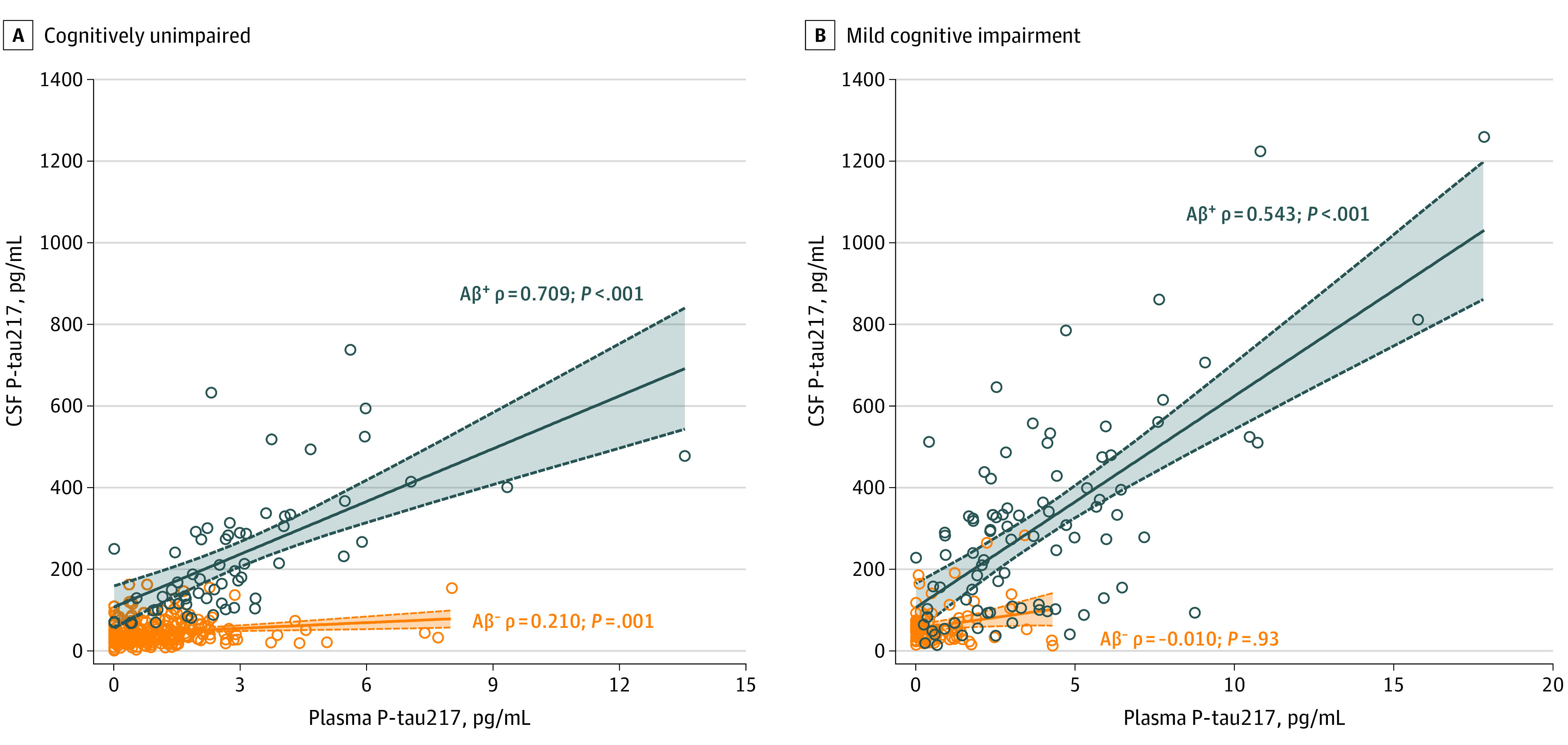
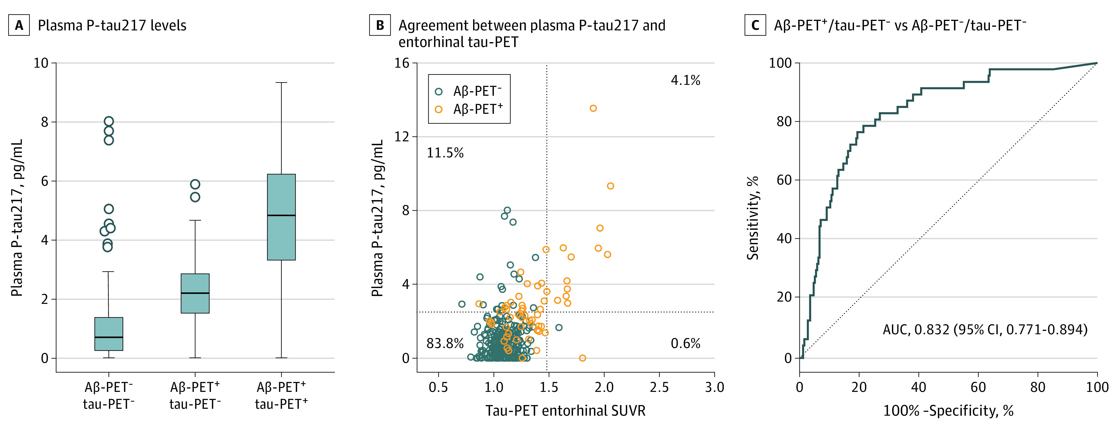
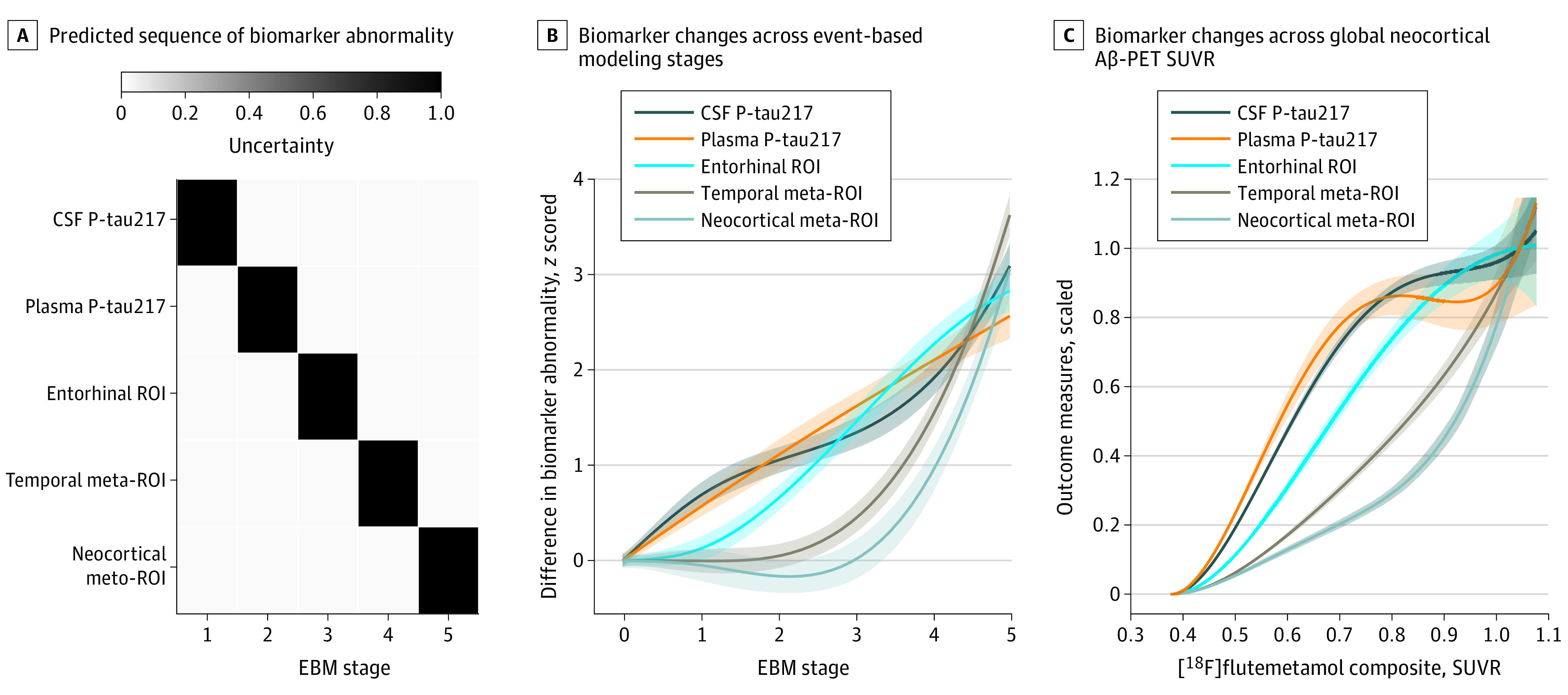
Results: Of 490 participants, 251 were women (51.2%) and the mean (SD) age was 65.9 (13.1) years. Plasma P-tau217 levels were increased in cognitively unimpaired participants with abnormal Aβ-PET but normal tau-PET in the entorhinal cortex (Aβ-PET+/ tau-PET- group vs Aβ-PET-/ tau-PET- group: median, 2.2 pg/mL [interquartile range (IQR), 1.5-2.9 pg/mL] vs 0.7 pg/mL [IQR, 0.3-1.4 pg/mL]). Most cognitively unimpaired participants who were discordant for plasma P-tau217 and tau-PET were positive for plasma P-tau217 and negative for tau-PET (P-tau217+/tau-PET-: 36 [94.7%]; P-tau217-/tau-PET+: 2 [5.3%]). Event-based modeling of cross-sectional data predicted that in cognitively unimpaired participants and in those with mild cognitive impairment, both plasma and CSF P-tau217 would change before the tau-PET signal in the entorhinal cortex, followed by more widespread cortical tau-PET changes. When testing the association with global Aβ load in nonlinear spline models, both plasma and CSF P-tau217 were increased at lower Aβ-PET values compared with tau-PET measures. Among participants with normal baseline tau-PET, the rates of longitudinal increase in tau-PET in the entorhinal cortex were higher in those with abnormal plasma P-tau217 at baseline (median standardized uptake value ratio, 0.029 [IQR, -0.006 to 0.041] vs -0.001 [IQR, -0.021 to 0.020]; Mann-Whitney U, P = .02).

Conclusions and relevance: In this cohort study, plasma P-tau217 levels were increased during the early preclinical stages of AD when insoluble tau aggregates were not yet detectable by tau-PET. Plasma P-tau217 may hold promise as a biomarker for early AD brain pathology.


Conflict of interest statement

Conflict of Interest Disclosures: Dr Berron reported being a cofounder of neotiv GmbH outside the submitted work. Mr Proctor reported being an employee and stockholder of Eli Lilly and Company. Dr Dage reported being an employee and stockholder of Eli Lilly and Company. Dr Hansson reported receiving grants from the Swedish Research Council, the Knut and Alice Wallenberg Foundation, Roche, and Biogen; receiving financial support from GE Healthcare outside the submitted work; receiving research support from Roche, Pfizer, GE Healthcare, Biogen, Eli Lilly and Company, and AVID Radiopharmaceuticals; and receiving consultancy and speaker fees (paid to the institution) from Biogen and Roche. No other disclosures were reported.

Figures

Figure 1.
Figure 1.. Association Between Plasma and Cerebrospinal Fluid (CSF) Tau Phosphorylated at Threonine 217 (P-tau 217)
A and B, Association between plasma and CSF P-tau217 in cognitively unimpaired participants (amyloid-β positron emission tomography negative [Aβ-PET], n = 252; Aβ-PET+, n = 61) (A) and participants with mild cognitive impairment (Aβ-PET, n = 83; Aβ-PET+, n = 89) (B).
Figure 2.
Figure 2.. Plasma Tau Phosphorylated at Threonine 217 (P-tau217) and Tau–Positron Emission Tomography (PET) Positivity in the Entorhinal Cortex of Cognitively Unimpaired Participants
A, Plasma P-tau217 levels in participants with amyloid-β (Aβ)–PET/ tau-PET (n = 252), participants with Aβ-PET+/ tau-PET(n = 47), and participants with Aβ-PET+/ tau-PET+ (n = 14) (P < .001 for Aβ-PET+/ tau-PET– vs Aβ-PET–/tau-PET– and Aβ-PET+/tau-PET+ vs Aβ-PET–/ tau-PET–). P values are from univariate general linear models adjusted for age and sex as described in the Methods section. Boxes indicate 25th to 75th percentiles, center lines indicate median, whiskers extend to the upper and lower adjacent values or the most extreme points within 1.5 × interquartile range of the 25th and 75th percentiles, and dots indicate outliers. B, Agreement between plasma P-tau217 and entorhinal tau-PET. The dotted lines represent cutoffs for plasma P-tau217 (2.5 pg/mL) and entorhinal tau-PET (standardized uptake value ratio [SUVR], 1.48). C, Receiver operating characteristic curve analysis for differentiating participants with Aβ-PET+/ tau-PET (n = 47) from those with Aβ-PET/ tau-PET (n = 252). Cutoffs for plasma P-tau217, entorhinal tau-PET, and Aβ-PET (SUVR, 0.53) were determined as described in the Methods section. AUC indicates area under the curve.
Figure 3.
Figure 3.. Order of Change in Plasma Tau Phosphorylated at Threonine 217 (P-tau217), Cerebrospinal Fluid (CSF) P-tau217, and Tau–Positron Emission Tomography (PET) Abnormality
Order of change in plasma P-tau217, CSF P-tau217, and tau-PET abnormality in cognitively unimpaired participants and those with mild cognitive impairment (n = 484). A, Predicted sequence of biomarker abnormality from event-based modeling (EBM). Gray scale coding indicates uncertainty. B, Visualization of biomarker changes across EBM stages using nonlinear spline models. Uncertainties represent 95% CIs from the model-estimated variance-covariance matrix. C, Summary of biomarker changes in relation to global neocortical amyloid β (Aβ)–PET. All biomarkers are on a common scale ranging from 0 (baseline levels) to 1 (the mean levels in the top 10 percentiles). ROI, region of interest; and SUVR, standardized uptake value ratio.
Figure 4.
Figure 4.. Mediated Effect of Plasma Tau Phosphorylated at Threonine 217 (P-tau217) on the Association of Amyloid-β (Aβ) Positron Emission Tomography (PET) With Tau-PET
Mediated effect of plasma P-tau217 on the association of Aβ-PET with tau-PET in cognitively unimpaired participants and in those with mild cognitive impairment (n = 484). A, The direct association (c) of Aβ-PET with tau-PET. B, The mediated effect of plasma P-tau217 is designated c-c′. The remaining association of Aβ-PET with tau-PET after adjusting for plasma P-tau217 is designated c′. The direct association of Aβ-PET with plasma P-tau217 is a, and the association of plasma P-tau217 with tau-PET is b (c-c′ = 0.357; 95% CI, 0.281-0.435; 76.4%). This indicates that plasma P-tau217 mediated 76.4% of the effect of Aβ-PET on tau-PET.

Comment in

References

    1. Scheltens P, Blennow K, Breteler MM, et al. . Alzheimer’s disease. Lancet. 2016;388(10043):505-517. doi:10.1016/S0140-6736(15)01124-1 - DOI - PubMed
    1. Beach TG, Monsell SE, Phillips LE, Kukull W. Accuracy of the clinical diagnosis of Alzheimer disease at National Institute on Aging Alzheimer Disease Centers, 2005-2010. J Neuropathol Exp Neurol. 2012;71(4):266-273. doi:10.1097/NEN.0b013e31824b211b - DOI - PMC - PubMed
    1. Jack CR Jr, Bennett DA, Blennow K, et al. ; NIA-AA research framework: toward a biological definition of Alzheimer’s disease. Alzheimers Dement. 2018;14(4):535-562. doi:10.1016/j.jalz.2018.02.018 - DOI - PMC - PubMed
    1. Mielke MM, Hagen CE, Xu J, et al. . Plasma phospho-tau181 increases with Alzheimer’s disease clinical severity and is associated with tau- and amyloid-positron emission tomography. Alzheimers Dement. 2018;14(8):989-997. doi:10.1016/j.jalz.2018.02.013 - DOI - PMC - PubMed
    1. Tatebe H, Kasai T, Ohmichi T, et al. . Quantification of plasma phosphorylated tau to use as a biomarker for brain Alzheimer pathology: pilot case-control studies including patients with Alzheimer’s disease and Down syndrome. Mol Neurodegener. 2017;12(1):63. doi:10.1186/s13024-017-0206-8 - DOI - PMC - PubMed

Publication types