

Laparoscopic approach to ureteroinguinal hernia
- PMID: 33166812
- PMCID: PMC7653005
- DOI: 10.1016/j.ijscr.2020.10.127
Laparoscopic approach to ureteroinguinal hernia
Abstract
Introduction: Herniation of the ureter into the inguinal canal is a rare occurrence. There have been reports of inadvertent injury to the ureter during routine inguinal hernia repair. After an extensive search of the literature, we believe that this is the first case to be managed via laparoscopic Trans Abdominal Pre-Peritoneal Repair and would like to highlight the technical details of the laparoscopic procedure and is presented in line with SCARE 2018 Guidelines [1].
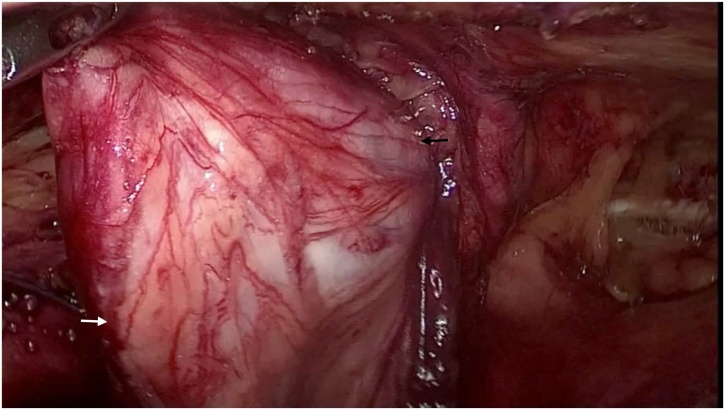
Presentation of case: A 60-year-old male presented with left inguinal hernia. He also complained of an increase in frequency of micturition, with an occasional radiating pain from loin to the groin. Imaging revealed the left ureter coursing into the left inguinal canal, descending into the scrotum, and looping back to enter the bladder with mild hydroureteronephrosis. Patient underwent a laparoscopic repair of the inguinal hernia with reduction of ureter under ureteroscope guidance and stent placement.
Discussion: The presence of ureter buried in a large amount of fat can be mistaken for a lipoma of the cord or extraperitoneal fat and injured with blind clamping and division. Presence of fat without an obvious sac should alert the surgeon to the possibility of ureter being a content.
Conclusion: Laparoscopy is safe, technically feasible, offers good visualization of all hernial orifices, demonstrates complete reduction of ureter from inguinal canal under vision, allows manipulation of ureter under the vision for ureteroscopy and stenting, making sure there are no loops or kinking and allows placement of mesh in the preperitoneal space.
Keywords: Hydro-ureteronephrosis; Laparoscopic approach; Uretero-inguinal hernia.
Copyright © 2020 The Authors. Published by Elsevier Ltd.. All rights reserved.
Figures


References
-
- Agha R.A., Borrelli M.R., Farwana R., Koshy K., Fowler A., Orgill D.P., For the SCARE Group The SCARE 2018 statement: updating consensus surgical CAse REport (SCARE) guidelines. Int. J. Surg. 2018;60:132–136. - PubMed
Publication types
LinkOut - more resources
Full Text Sources
Research Materials
