Review
. 2021 Feb:68:98-106.
doi: 10.1016/j.coi.2020.09.006. Epub 2020 Nov 6.

Tuning tuft cells: new ligands and effector functions reveal tissue-specific function

Affiliations
Review

Tuning tuft cells: new ligands and effector functions reveal tissue-specific function

Tyler E Billipp et al. Curr Opin Immunol. 2021 Feb.

Abstract

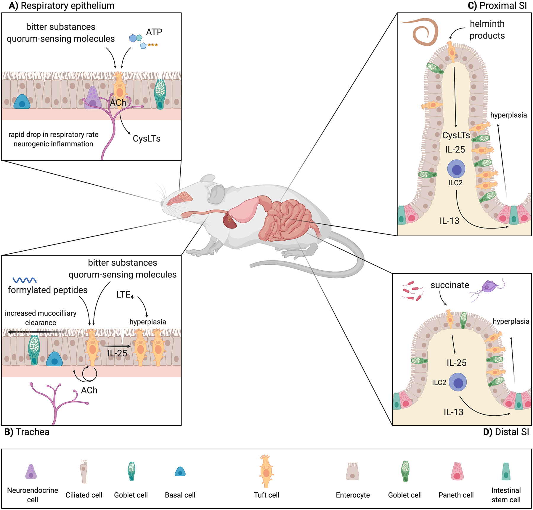
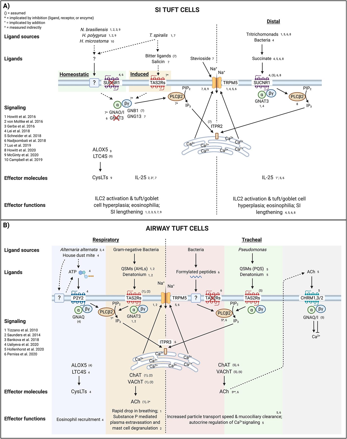
Tuft cells are rare chemosensory epithelial cells that monitor their environment and relay messages to the surrounding tissue via secretion of neuromodulatory and immunomodulatory molecules. In the small intestine tuft cells detect helminth infection, protist colonization, and bacterial dysbiosis, and initiate a type 2 immune response characterized by tissue remodeling. In the airways, tuft cells sense bacteria, allergens, and noxious stimuli and drive evasive behavior, neuroinflammation, and anti-bacterial responses. Here we summarize the most recent tuft cell research and discuss how these findings have provided insight into tuft cell diversity. Built around a core program of chemosensing, tuft cell receptors and effector functions are tuned to the unique environmental exposure and physiology of their surrounding tissue.


Conflict of interest statement

Declaration of Interests

The authors declare no competing interests.

Figures

Figure 1:
Figure 1:. Tuft cell ligands and effector functions are tissue-specific.
A) In the respiratory epithelium, tuft cells sense irritants, bacterial dysbiosis, and tissue homeostasis through bitter substances, quorum-sensing molecules, and ATP, respectively. These tufts cells are innervated, making ACh an effective effector molecule with which tuft cells can modulate breathing rate as well as neurogenic inflammation. Additionally, respiratory tuft cells can alter tissue immunity through ACh and CysLTs. B) Tracheal tuft cells sense bitter ligands and bacterial secreted formylated peptides. In the trachea, tuft cells are not as innervated but still use ACh to modulate tissue physiology, in this case through autocrine and paracrine signaling to epithelial cells to regulate mucociliary clearance. There is some evidence of tuft cell hyperplasia in the trachea downstream of LTE4 and IL-25, but the mechanism is not fully elucidated. C) The ligands and receptors by which tuft cells in the SI sense luminal helminths are still unknown. Nevertheless, SI tuft cells are critical for the initiation of rapid type 2 immune responses to luminal helminths. These helminths primarily reside in the proximal SI, but the type 2 immune response and resulting tuft and goblet cell hyperplasia is seen throughout the SI. D) In the distal SI, tuft cells sense Tritrichomonas protists and bacterial dysbiosis though the metabolite succinate. Tuft cells in this portion of the SI seem to be particularly poised to sense succinate, as they express higher levels of Sucnr1 compared to tuft cells in the proximal SI. Accordingly, tuft cell hyperplasia following succinate treatment is most prominent in the distal SI, although still occurs throughout the SI. Abbreviations: Acetylcholine (ACh); Choline acetyltransferase (ChAT); Cysteinyl leukotrienes (CysLTs); Leukotriene E4 (LTE4); Group 2 innate lymphoid cell (ILC2); Small intestine (SI); Succinate receptor (SUCNR1). Created with BioRender.com.
Figure 2:
Figure 2:. Airway and small intestinal tuft cell signaling
A) Small intestinal tuft cells sense the presence of helminth worms via an unidentified receptor and ligand. Tuft cell-derived IL-25 and CysLTs activate lamina propria ILC2s to drive epithelial remodeling, eosinophilia, and worm clearance. The calcium-gated cation channel TRPM5, downstream of canonical taste transduction in taste cells, is also required for helminth sensing, though Gnat3−/− mice are not affected. Additionally, while some helminths can produce succinate as a metabolic byproduct, Sucnr1−/− mice have no defect in tuft hyperplasia in response to helminth infection. Following initial activation and IL-13 signaling, tuft cells express TAS2Rs, which may sense “bitter” substances found in T. spiralis excretory-secretory products, and result in additional release of IL-25. G-alpha O/I subunits, as well as the G-gamma 13 subunit and PLCB2, have also been implicated in T. spiralis sensing and IL-25 release. Potentiating TRPM5 with stevioside is sufficient to cause release of IL-25. Specifically in the distal SI, tuft cells monitor the metabolite succinate, which can be produced by Tritrichomonas protists as well as dysbiotic bacteria. Unlike helminth sensing, GNAT3 is required for the succinate response, as are PLCB2 and TRPM5, indicating a canonical taste transduction pathway. B) Airway tuft cells have been studied most thoroughly in the respiratory and tracheal epithelia. Respiratory tuft cells (and olfactory tuft cells) use the receptor P2Y2 to sense ATP released from cells via an unknown mechanism following treatment with Alternaria alternata or house dust mite. ATP signaling drives increased intracellular Ca2+ and release of CysLTs that recruit eosinophils and amplify type 2 inflammation. It remains unclear if tracheal tuft cells produce CysLTs. Respiratory tuft cells also express numerous TAS2Rs, which bind denatonium and the “bitter” QSMs produced by certain Gram-negative bacteria. Signaling activates the canonical taste transduction pathway, resulting in release of ACh that signals on peptidergic neurons to rapidly induce a drop in breathing rate and neurogenic inflammation. In the trachea, where tuft cells are less extensively innervated, sensing of bacterial formylated peptides, QSMs, or denatonium activate PLCB2, Ca2+ flux, and TRPM5-dependent ACh release. Via CHRMs, ACh signals in both a paracrine fashion on neighboring ciliated cells to increase ciliary beat frequency and in an autocrine fashion on tuft cells to enhance or suppress the magnitude of Ca2+ flux. While TAS2Rs are assumed to be required for sensing QSMs and denatonium, they are not involved in formylated peptide sensing; the actual receptor remains unidentified. Dashed arrows indicate unconfirmed pathways. Abbreviations: Acetylcholine (ACh); Acyl-homoserine lactones (AHL); Arachidonate 5-lipoxygenase (ALOX5); Bitter taste receptor family (TAS2Rs); Choline acetyltransferase (ChAT); Cysteinyl leukotrienes (CysLTs); G Subunit Alpha Q/I/O (GNAQ/I/O); G Protein Subunit Alpha Transducin 3 (GNAT3); G Subunit Beta 1 (GNB1); G Subunit Gamma 13 (GNG13); Inositol 1,4,5-Trisphosphate Receptor Type 3 (ITPR); Leukotriene C4 synthase (LTC4S); Muscarinic ACh receptors (CHRMs); Phospholipase C Beta 2 (PLCB2); Pseudomonas quinolone signal (PQS); P2Y Purinoceptor 2 (P2Y2); Quorum-sensing molecules (QSMs); Small intestine (SI); Succinate receptor (SUCNR1); Transient Receptor Potential Cation Channel Subfamily M Member 5 (TRPM5); Vesicular acetylcholine transporter (VAChT). Created with BioRender.com.

References

    1. Järvi O, Keyriläinen O: On the cellular structures of the epithelial invasions in the gladular stomach of mice caused by intramural application of 20-methylcholantren. Acta Pathol Microbiol Scand 1956, 38:72–73. - PubMed
    1. Rhodin J, Dalhamn T: Electron microscopy of the tracheal ciliated mucosa in rat. Zeitschrift für Zellforsch und Mikroskopische Anat 1956, 44:345–412. - PubMed
    1. Tizzano M, Gulbransen BD, Vandenbeuch A, Clapp TR, Herman JP, Sibhatu HM, Churchill MEA, Silver WL, Kinnamon SC, Finger TE: Nasal chemosensory cells use bitter taste signaling to detect irritants and bacterial signals. Proc Natl Acad Sci U S A 2010, 107:3210–3215. - PMC - PubMed
    1. Krasteva G, Canning BJ, Hartmann P, Veres TZ, Papadakis T, Mühlfeld C, Schliecker K, Tallini YN, Braun A, Hackstein H, et al.: Cholinergic chemosensory cells in the trachea regulate breathing. Proc Natl Acad Sci U S A 2011, 108:9478–9483. - PMC - PubMed
    1. Saunders CJ, Christensen M, Finger TE, Tizzano M: Cholinergic neurotransmission links solitary chemosensory cells to nasal inflammation. Proc Natl Acad Sci U S A 2014, 111:6075–6080. - PMC - PubMed

Publication types

LinkOut - more resources