. 2020 Nov 5;21(21):8287.
doi: 10.3390/ijms21218287.

AHR Signaling Interacting with Nutritional Factors Regulating the Expression of Markers in Vascular Inflammation and Atherogenesis

Affiliations

AHR Signaling Interacting with Nutritional Factors Regulating the Expression of Markers in Vascular Inflammation and Atherogenesis

Carla Dahlem et al. Int J Mol Sci. .

Abstract

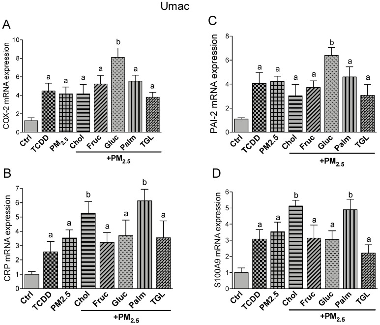
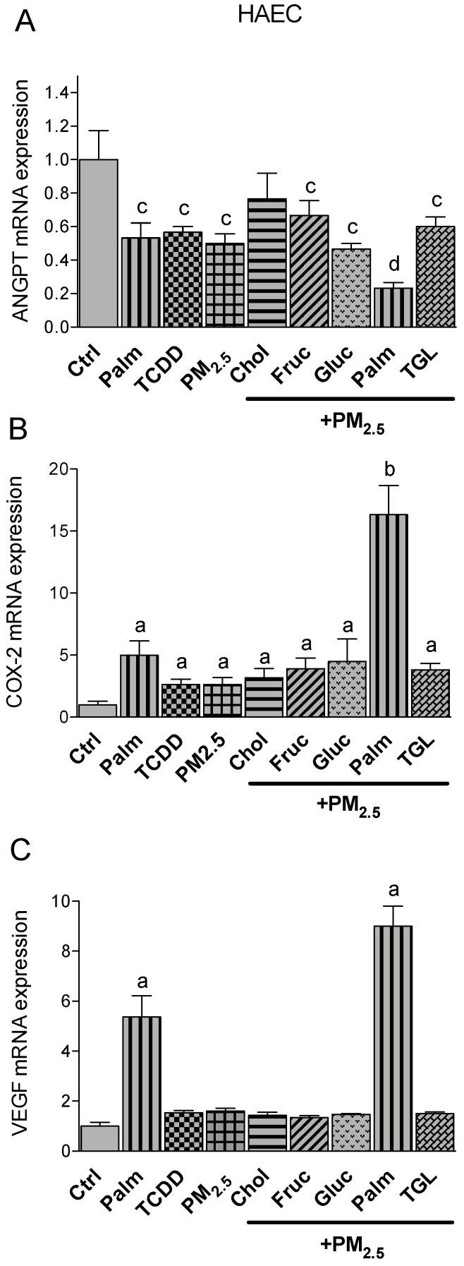
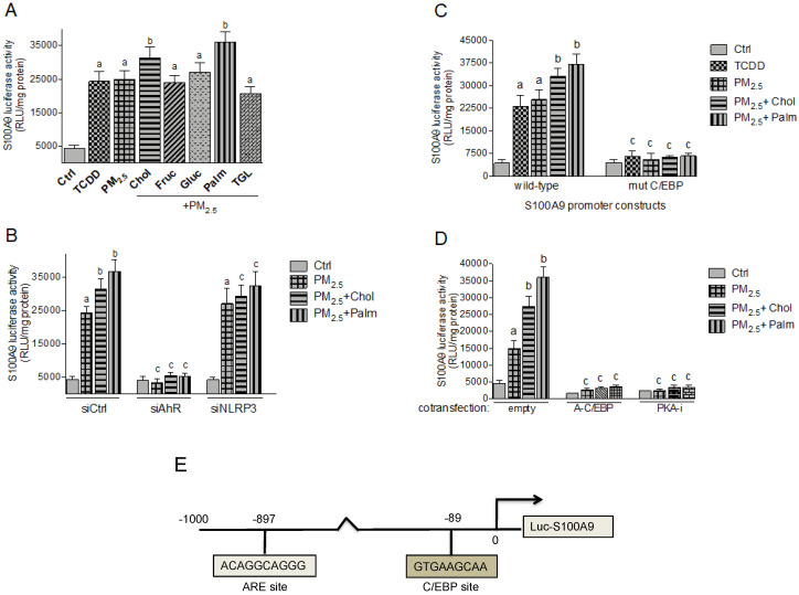
There is strong evidence that exposure to fine particulate matter (PM2.5) and a high-fat diet (HFD) increase the risk of mortality from atherosclerotic cardiovascular diseases. Recent studies indicate that PM2.5 generated by combustion activates the Aryl Hydrocarbon Receptor (AHR) and inflammatory cytokines contributing to PM2.5-mediated atherogenesis. Here we investigate the effects of components of a HFD on PM-mediated activation of AHR in macrophages. Cells were treated with components of a HFD and AHR-activating PM and the expression of biomarkers of vascular inflammation was analyzed. The results show that glucose and triglyceride increase AHR-activity and PM2.5-mediated induction of cytochrome P450 (CYP)1A1 mRNA in macrophages. Cholesterol, fructose, and palmitic acid increased the PM- and AHR-mediated induction of proinflammatory cytokines in macrophages. Treatment with palmitic acid significantly increased the expression of inflammatory cytokines and markers of vascular injury in human aortic endothelial cells (HAEC) after treatment with PM2.5. The PM2.5-mediated activation of the atherogenic markers C-reactive protein (CRP) and S100A9, a damage-associated molecular pattern molecule, was found to be AHR-dependent and involved protein kinase A (PKA) and a CCAAT/enhancer-binding protein (C/EBP) binding element. This study identified nutritional factors interacting with AHR signaling and contributing to PM2.5-induced markers of atherogenesis and future cardiovascular risk.

Keywords: AHR; PM; TCDD; atherosclerosis; cytokines; inflammation; macrophages; obesity.


Conflict of interest statement

The authors declare no conflict of interest.

Figures

Figure 1
Figure 1
Effect of nutritional compounds on tetrachlorodibenzo-p-dioxin (TCDD)- and particulate matter (PM2.5)-induced cytochrome P450 (CYP)1A1 expression and Aryl Hydrocarbon Receptor (AHR) activity. (A) U937-derived macrophages (Umac) and (B) human aortic endothelial cells (HAEC) were treated with cholesterol (Chol, 10 μg/mL), fructose (Fruc, 25 mM), glucose (Gluc, 25 mM), palmitic acid (Palm, 5 μM), triglyceride (TGL, 10 μg/mL), TCDD (1 nM), and PM2.5 (10 μg/mL) for 24 h. Cells were treated with 3-methoxy-4-nitroflavone (MNF) (5 μM) to block activation of AHR by TCDD and PM2.5. The effect of nutritional factors on CYP1A1 was tested in presence of PM2.5. The mRNA expression of CYP1A1 and the housekeeping ß-actin was analyzed by qPCR. Control cells were treated with the corresponding vehicle. (C) Effect of nutritional compounds on PM2.5-induced dioxin responsive element (DRE) luciferase activity. Umac were transiently transfected with a DRE luciferase reporter construct for 24 h and then treated with TCDD or PM2.5 in presence or absence of MNF for 16 h. The effect of nutritional factors on DRE activity was tested in presence of PM2.5; a significantly higher than Ctrl; b significantly higher than cells treated with PM2.5 only; c significantly lower than cells treated with TCDD or PM2.5 only; p < 0.05; (D) PM2.5-induced AHR binding activity. Bone marrow-derived macrophages (BMM) from AHR+/+ and AHR−/− mice were treated for 1 h with 10 μg/mL of PM2.5, cells were also treated with 1 nM TCDD as a positive control. Nuclear proteins were extracted and incubated with 32P-labeled oligonucleotides containing the consensus site of DRE and loaded on a native gel for gel-mobility shift assay (GMSA). A 100-fold excess of the unlabeled specific (cold) DRE oligonucleotides was added to confirm specificity.
Figure 2
Figure 2
Effect of nutritional compounds on PM2.5-induced cytokine expression in Umac. Cells were treated with 1 nM TCDD and 10 μg/mL PM2.5 for 24 h. The effect of nutritional factors cholesterol (Chol, 10 μg/mL), fructose (Fruc, 25 mM), glucose (Gluc, 25 mM), palmitic acid (Palm, 5 μM), and triglyceride (TGL, 10 μg/mL) was tested in presence of PM2.5 after 24 h treatment. Control cells were treated with the corresponding vehicle. The mRNA expression of (A) IL-1β; (B) IL-8, and (C) IL-33 was analyzed by qPCR. The expression was corrected against the housekeeping gene ß-actin. a significantly higher than Ctrl; b significantly higher than cells treated with PM2.5 only p < 0.05.
Figure 3
Figure 3
Effect of nutritional compounds on PM2.5-induced cytokine expression in HAEC. Cells were treated with 1 nM TCDD and 10 μg/mL PM2.5 for 24 h. The effect of nutritional factors cholesterol (Chol, 10 μg/mL), fructose (Fruc, 25 mM), glucose (Gluc, 25 mM), palmitic acid (Palm, 5 μM), and triglyceride (TGL, 10 μg/mL) was tested in presence of PM2.5 after 24 h of treatment. Control cells were treated with the corresponding vehicle. The mRNA expression of (A) IL-6 and (B) IL-8 was analyzed by qPCR. The expression was corrected against the housekeeping gene β-actin. a significantly higher than Ctrl; b significantly higher than cells treated with PM2.5 only p < 0.05.
Figure 4
Figure 4
Effect of nutritional compounds on PM2.5-induced expression of atherogenic markers in Umac; Cells were treated with 1 nM TCDD and 10 μg/mL PM2.5 for 24 h. The effect of nutritional factors cholesterol (Chol, 10 μg/mL), fructose (Fruc, 25 mM), glucose (Gluc, 25 mM), palmitic acid (Palm, 5 μM), and triglyceride (TGL, 10 μg/mL) was tested in presence of PM2.5 after 24 h treatment. Control cells were treated with the corresponding vehicle. The mRNA expression of (A) COX-2; (B) CRP; (C) PAI-2, and (D) S100A9 was analyzed by qPCR. The expression was corrected against the housekeeping gene β-actin. a significantly higher than Ctrl; b significantly higher than cells treated with PM2.5 only p < 0.05.
Figure 5
Figure 5
Effect of nutritional compounds on PM2.5-induced expression of atherogenic markers in HAEC; Cells were treated with 1 nM TCDD and 10 μg/mL PM2.5 for 24 h. The effect of nutritional factors cholesterol (Chol, 10 μg/mL), fructose (Fruc, 25 mM), glucose (Gluc, 25 mM), palmitic acid (Palm, 5 μM), and triglyceride (TGL, 10 μg/mL) was tested in presence of PM2.5 after 24 h treatment. Control cells were treated with the corresponding vehicle. The mRNA expression of (A) Angiopoetin (ANGPT), (B) COX-2, and (C) VEGF was analyzed by qPCR. The expression was corrected against the housekeeping gene ß-actin. a significantly higher than Ctrl; b significantly higher than cells treated with PM2.5 or Palm only; c significantly lower than Ctrl; d significantly lower than cells treated with Palm or PM2.5 only p < 0.05.
Figure 6
Figure 6
Effect of nutritional compounds on PM2.5-induced CRP promoter activity. (A) Umac were transiently transfected with a luciferase promoter construct containing 300 bp upstream of the regulatory sequence of the human CRP gene promoter. After 24 h cells were treated with 1 nM TCDD and 10 μg/mL PM2.5 in presence or absence of nutritional factors cholesterol (Chol, 10 μg/mL), fructose (Fruc, 25 mM), glucose (Gluc, 25 mM), palmitic acid (Palm, 5 μM), and triglyceride (TGL, 10 μg/mL) for 6 h. Control cells were treated with the corresponding vehicle only. (B) Umac were transiently transfected with the CRP wt luciferase promoter construct and co-transfected with a scrambled control siRNA, a AHR-specific or NLRP3-specific siRNA. After 24 h cells were treated with 10 μg/mL PM2.5 in the presence or absence of cholesterol (Chol, 10 μg/mL) or palmitic acid (Palm, 5 μM). (C) Umac were transiently transfected with the luciferase reporter constructs of the wt CRP promoter or a CRP construct containing a mutation in the NF-kB (mut NF-kB) or C/EBP binding site (mut C/EBP). After 24 h cells were treated with 1 nM TCDD or 10 μg/mL PM2.5 in the presence or absence of cholesterol (Chol, 10 μg/mL) or palmitic acid (Palm, 5 μM). (D) PM2.5-induced CRP promoter activity is C/EBP- and PKA-dependent. Cells were co-transfected with an empty, C/EBP-A dominant negative expression plasmid, or a PKA inhibitor (PKA-i) expression plasmid for 24 h and treated with 10 μg/mL PM2.5 in presence or absence of cholesterol (Chol, 10 μg/mL) or palmitic acid (Palm, 5 μM) for 6h. Relative luciferase activity units (RLU) are given as mean values of triplicates as a result of three independent experiments. a significantly higher than control (p < 0.05); b significantly higher than cells treated with PM2.5 only (p < 0.05); c significantly lower than cells transfected with ctrl siRNA, wild-type CRP, or an empty vector (p < 0.05). (E) Schematic illustration of the promoter construct of the human CRP gene containing 300 bp upstream of the transcriptional start site (indicated by an arrow) cloned into a luciferase (luc) reporter vector. Positions of the C/EBP, NF-κB, and STAT3 recognition sites are presented.
Figure 7
Figure 7
Effect of nutritional compounds on PM2.5-induced S100A9 promoter activity. (A) Umac were transiently transfected with a luciferase promoter construct containing 1000 bp upstream of the regulatory sequence of the human S100A9 gene promoter. After 24 h cells were treated with 1 nM TCDD and 10 μg/mL PM2.5 in the presence or absence of nutritional factors cholesterol (Chol, 10 μg/mL), fructose (Fruc, 25 mM), glucose (Gluc, 25 mM), palmitic acid (Palm, 5 μM), and triglyceride (TGL, 10 μg/mL) for 6 h. Control cells were treated with the corresponding vehicle. (B) Umac were transiently transfected with the S100A9 wt luciferase promoter construct and co-transfected with a scrambled control siRNA, a AHR-specific or NLRP3-specific siRNA. After 24 h cells were treated with 10 μg/mL PM2.5 in the presence or absence of cholesterol (Chol, 10 μg/mL) or palmitic acid (Palm, 5 μM). (C) Umac were transiently transfected with the luciferase reporter constructs of the wt S100A9 promoter or a construct containing a mutation in the C/EBP binding site. After 24 h cells were treated with 1 nM TCDD or 10 μg/mL PM2.5 in the presence or absence of cholesterol (Chol, 10 μg/mL) or palmitic acid (Palm, 5 μM). (D) Cells were co-transfected with an empty, C/EBP-A dominant negative expression plasmid, or a PKA inhibitor (PKA-i) expression plasmid for 24 h and treated with 10 μg/mL PM2.5 for 6h in the presence or absence of cholesterol (Chol, 10 μg/mL) or palmitic acid (Palm, 5 μM). Relative luciferase activity units (RLU) are given as mean values of triplicates as a result of three independent experiments. a significantly higher than control (p < 0.05); b significantly higher than cells treated with PM2.5 only (p < 0.05); c significantly lower than cells transfected with ctrl siRNA, wild-type S100A9, or an empty vector (p < 0.05). (E) Schematic illustration of promoter construct of the human S100A9 gene containing 1000 bp upstream of the transcriptional start site (indicated by an arrow) cloned into a luciferase (luc) reporter vector. Positions of the C/EBP recognition site and an antioxidant response element (ARE) are presented.
Figure 8
Figure 8
Effects of PM2.5 derived from traffic related air pollution (TRAP) and dietary factors of a high fat diet (HFD) on target cells of the cardiovascular system such as aortic endothelial cells and macrophages. AHR and NLRP3 mediate the activation of atherogenic markers and cardiovascular diseases induced by PM2.5 and HFD. The earliest visible lesion of atherosclerosis is the fatty streak as indicated by arrows. The fatty streak is due to an accumulation of lipid-laden foam cells in the intimal layer of the artery.

Similar articles

Cited by

References

    1. Denison M.S., Nagy S.R. Activation of the aryl hydrocarbon receptor by structurally diverse exogenous and endogenous chemicals. Annu. Rev. Pharmacol. Toxicol. 2003;43:309–334. doi: 10.1146/annurev.pharmtox.43.100901.135828. - DOI - PubMed
    1. Bohonowych J.E., Denison M.S. Persistent binding of ligands to the aryl hydrocarbon receptor. Toxicol. Sci. 2007;98:99–109. doi: 10.1093/toxsci/kfm085. - DOI - PMC - PubMed
    1. Rückerl R., Greven S., Ljungman P., Aalto P., Antoniades C., Bellander T., Berglind N., Chrysohoou C., Forastiere F. Air pollution and inflammation (interleukin-6, C-reactive protein, fibrinogen) in myocardial infarction survivors. Environ. Health Perspect. 2007;115:1072–1080. doi: 10.1289/ehp.10021. - DOI - PMC - PubMed
    1. Grunig G., Marsh L.M., Esmaeil N., Jackson K., Gordon T., Reibman J., Kwapiszewska G., Park S.H. Perspective: Ambient air pollution: Inflammatory response and effects on the lung’s vasculature. Pulm. Circ. 2014;4:25–35. doi: 10.1086/674902. - DOI - PMC - PubMed
    1. Libby P. Inflammation in atherosclerosis. Nature. 2002;420:868–874. doi: 10.1038/nature01323. - DOI - PubMed

MeSH terms