. 2020 Nov 5;21(21):8313.
doi: 10.3390/ijms21218313.

Histone H3K9 Demethylase JMJD2B Plays a Role in LXRα-Dependent Lipogenesis

Affiliations

Histone H3K9 Demethylase JMJD2B Plays a Role in LXRα-Dependent Lipogenesis

Ji-Hyun Kim et al. Int J Mol Sci. .

Abstract

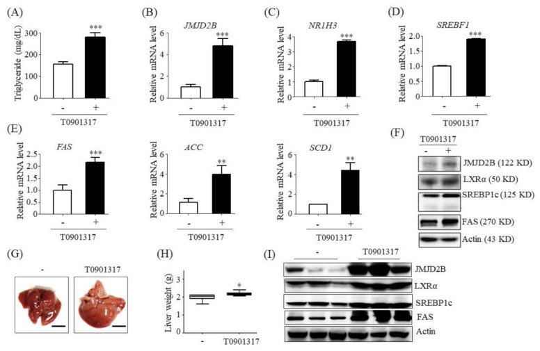
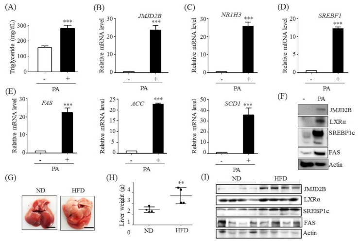
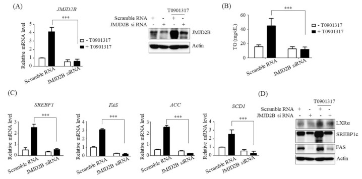
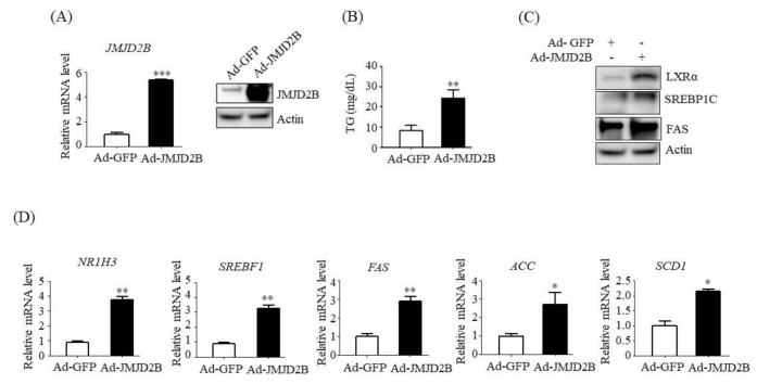
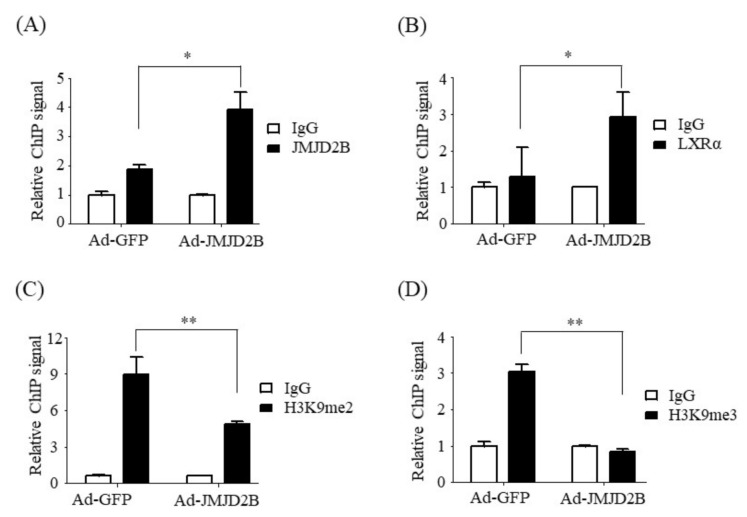
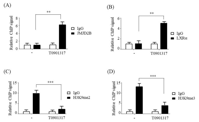
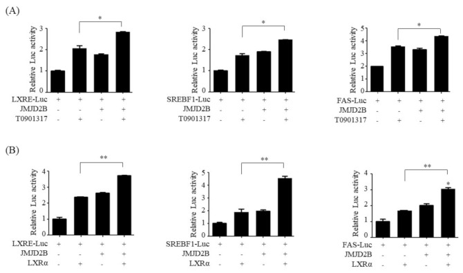
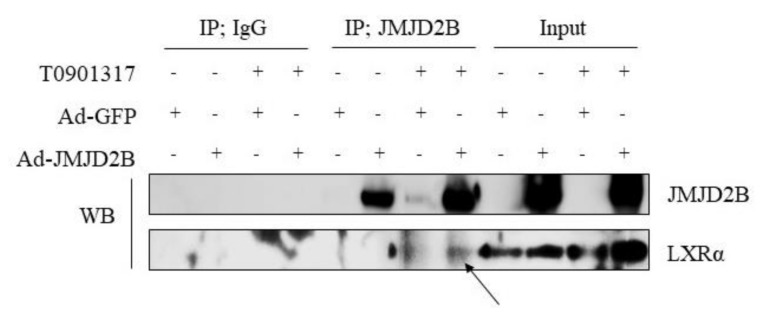
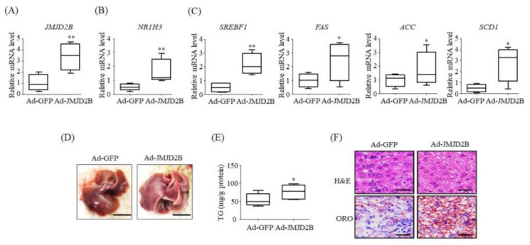
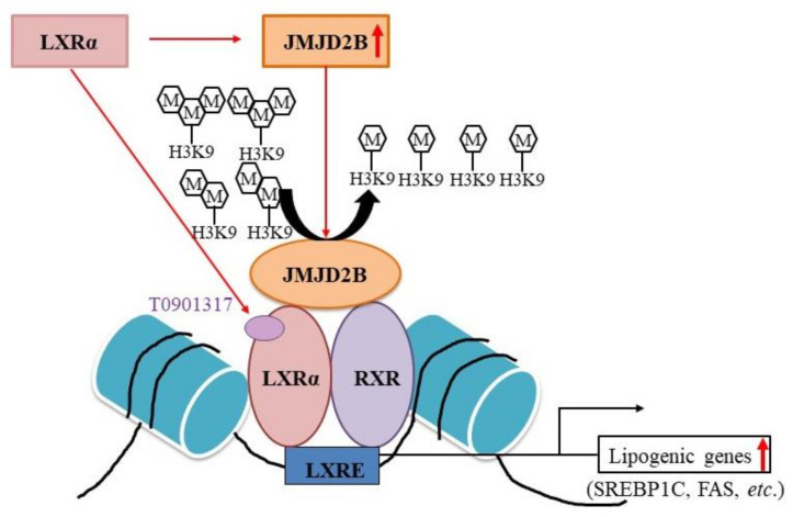
Ligand-activated liver X receptor α (LXRα) upregulates the expression of hepatic lipogenic genes, which leads to triglyceride (TG) accumulation, resulting in nonalcoholic fatty liver disease (NAFLD). Thus, LXRα regulation may provide a novel therapeutic target against NAFLD. However, histone methylation-mediated epigenetic regulation involved in LXRα-dependent lipogenesis is poorly understood. In this study, we investigated the functional role of the histone demethylase Jumonji domain-containing protein 2B (JMJD2B) in LXRα-dependent lipogenesis. JMJD2B expression level was upregulated in HepG2 cells treated with LXRα agonist T0901317 or palmitate and the liver of mice administered with T0901317 or fed a high-fat diet. Knockdown of JMJD2B using siRNA abrogated T0901317-induced LXRα-dependent lipogenic gene expression and lowered intracellular TG accumulation. Conversely, overexpression of JMJD2B in HepG2 cells upregulated the expression of LXRα-dependent lipogenic genes, in line with increased intracellular TG levels. JMJD2B overexpression or T0901317 treatment induced the recruitment of JMJD2B and LXRα to LXR response elements (LXRE) in the promoter region of LXRα-target gene and reduced the enrichment of H3K9me2 and H3K9me3 in the vicinity of the LXRE. Furthermore, JMJD2B enhanced T0901317 or LXRα-induced transcriptional activities of reporters containing LXRE. A co-immunoprecipitation assay revealed that JMJD2B interacted with activated LXRα. Moreover, overexpression of JMJD2B in mice resulted in upregulation of hepatic LXRα-dependent lipogenic genes, consistent with development of hepatic steatosis. Taken together, these results indicate that JMJD2B plays a role in LXRα-mediated lipogenesis via removing the repressive histone marks, H3K9me2 and H3K9me3, at LXRE, which might contribute to hepatic steatosis.

Keywords: hepatic steatosis; histone demethylase Jumonji domain-containing protein 2B; histone methylation; ligand-activated liver X receptor α; lipogenesis.


Conflict of interest statement

The authors declare no conflict of interest.

Figures

Figure 1
Figure 1
Jumonji domain-containing protein 2B (JMJD2B) expression level was upregulated in liver X receptor α (LXRα) agonist-treated HepG2 cells and the liver of LXRα agonist-treated mice. (AF) HepG2 cells were incubated with T0901317 for 24 h. (A) Intracellular TG were measured using a triglyceride (TG) assay kit. (BE) mRNA levels of JMJD2B, NR1H3, SREBF1, fatty acid synthase (FAS), acetyl CoA carboxylase (ACC), and stearoyl-CoA desaturase 1 (SCD1) were measured by qPCR. Data are presented as means ± SD from three independent experiments. ** p < 0.01, *** p < 0.001 vs. untreated control. (F) Protein expression levels of JMJD2B, LXRα, SREBP1c, and FAS were measured by western blotting. The SREBP1c shown here is precursor form (125 KD). (GI) C57BL/6J mice were administrated with T0901317 by oral gavage daily for 5 days. (G) Representative liver photographs. (scale bar = 1 cm) (H) Liver weight. (I) Protein levels of JMJD2B, LXRα, SREBP1c, and FAS were measured by western blotting. The SREBP1c shown here is mature form (68 KD). Densitometric analysis of band intensity is shown in Figure S1A. Data are presented as means ± SD from 6 mice (n = 6). * p < 0.05 vs. untreated control mice.
Figure 2
Figure 2
JMJD2B expression level was upregulated in palmitate (PA)-treated HepG2 cells and the liver of HFD-fed mice. (AF) HepG2 cells were incubated with PA for 24 h. (A) Intracellular TG were measured using a TG assay kit. (BE) mRNA levels of JMJD2B, NR1H3, SREBF1, FAS, ACC, and SCD1 were measured by qPCR. Data are presented as means ± SD from three independent experiments. *** p < 0.001 vs. untreated control. (F) Protein expression levels of JMJD2B, LXRα, SREBP1c, and FAS were measured by western blotting. The SREBP1c shown here is precursor form (125 KD). (GI) C57BL/6J mice were fed a high-fat diet (HFD) for 12 weeks. (G) Representative liver photographs. (scale bar = 1 cm) (H) Liver weight. (I) Protein levels of JMJD2B, LXRα, SREBP1c, and FAS were measured by western blotting. The SREBP1c shown here is mature form (68 KD). Densitometric analysis of band intensity is shown in Figure S1B. Data are presented as means ± SD from 6 mice (n = 6). ** p < 0.01 vs. normal diet (ND) mice.
Figure 3
Figure 3
Knockdown of JMJD2B prevented LXRα agonist-induced lipogenic gene expression in HepG2 cells. HepG2 cells were transfected with scramble RNA or JMJD2B siRNA and then treated with T0901317. (A) JMJD2B expression was determined by qPCR and western blotting. (B) Intracellular TG levels were measured using a TG assay kit. (C) mRNA levels of SREBF1, FAS, ACC, and SCD1 were determined by qPCR. (D) Protein levels of LXRα, SREBP1c, and FAS were measured by western blotting. The SREBP1c shown here is precursor form (125 KD). Data are presented as means ± SD from three independent experiments. *** p < 0.001 vs. scramble siRNA.
Figure 4
Figure 4
Overexpression of JMJD2B stimulated LXRα-dependent lipogenic genes in HepG2 cells. HepG2 cells were infected with Ad-GFP or an adenovirus containing JMJD2B (Ad-JMJD2B). (A) JMJD2B expression was measured using qPCR and western blotting. (B) Intracellular TG levels were measured using a TG assay kit. (C) Protein levels of LXRα, SREBP1c, and FAS were measured by western blotting. The SREBP1c shown here is precursor form (125 KD). (D) mRNA levels of NR1H3, SREBF1, FAS, ACC, and SCD1 were measured by qPCR. Data are presented as means ± SD from three independent experiments. * p < 0.05, ** p < 0.01, *** p < 0.001 vs. control Ad-GFP.
Figure 5
Figure 5
JMJD2B reduced the enrichment of H3K9me2 and H3K9me3 at the LXR response element (LXRE) on the SREBF1 promoter. HepG2 cells were infected with adenovirus Ad-GFP or Ad-JMJD2B. (A,B) The recruitment of JMJD2B (A) and LXRα (B) to the LXRE on the SREBF1 promoter was analyzed by chromatin immunoprecipitation (ChIP)-qPCR. (C,D) The enrichment of H3K9me2 (C) and H3K9me3 (D) at the LXRE on the SREBF1 promoter was analyzed by ChIP-qPCR. Data are presented as the mean ± SD from three independent experiments. * p < 0.05, ** p < 0.01 vs. control Ad-GFP.
Figure 6
Figure 6
LXRα activation reduced the enrichment of H3K9me2 and H3K9me3 at the LXRE on the SREBF1 promoter. HepG2 cells were incubated with T0901378 for 24 h. (A,B) The recruitment of JMJD2B (A) and LXRα (B) to the LXRE on the SREBF1 promoter was analyzed by ChIP-qPCR. (C,D) The enrichment of H3K9me2 (C) and H3K9me3 (D) in the vicinity of the LXRE on the SREBF1 promoter was analyzed by ChIP-qPCR. Data are presented as the mean ± SD from three independent experiments. ** p < 0.01, *** p < 0.001 vs. untreated control.
Figure 7
Figure 7
JMJD2B stimulated LXRα-activated transcriptional activity of LXRE-containing luciferase reporters. (A) HepG2 cells were transfected with Luc-reporters including 3xLXRE-Luc, SREBF1-Luc and FAS-Luc, and JMJD2B expression vector (pCMV-JMJD2B), then incubated with T0901317 for 24 h. Luciferase activities were determined using a kit. Data are presented as the mean ± SD from three independent experiments. * p < 0.05 vs. only T0901317 treatment. (B) HepG2 cells were transfected with Luc-reporters, JMJD2B expression vector (pCMV-JMJD2B), and LXRα expression vector (pCMV-LXRα). Luciferase activities were determined using a kit. ** p < 0.01 vs. only pCMV-LXRα.
Figure 8
Figure 8
JMJD2B interacted with activated LXRα. HepG2 cells were infected with Ad-GFP or Ad-JMJD2B and then incubated in the presence or absence of T09013178 for 24 h. Protein extracts were immunoprecipitated with JMJD2B antibody-agarose beads, and the interaction was detected with immunoblot analysis using LXRα antibody. Arrow indicates LXRα band detected in JMJD2B antibody-mediated immunoprecipitants. The alternative data is shown in Figure S2 where intracellular JMJD2B.
Figure 9
Figure 9
Overexpression of adenovirus-mediated JMJD2B stimulated hepatic LXRα-dependent lipogenic genes. C57BL/6 mice (8-week-old) were injected with adenovirus Ad-GFP or Ad-JMJD2B. After injection, Ad-injected mice were fed HFD for 2 weeks. (AC) mRNA levels of JMJD2B (A), NR1H3 (B), and LXRα-target lipogenic genes SREBF1, FAS, ACC, and SCD1 (C) were determined by qPCR. (D) Representative photographs (scale bar = 1 cm). (E) Hepatic TG levels were measured by TG assay kit. (F) H&E and ORO staining (scale bar = 50 μm). Data are presented as the mean ± SD from 6 mice (n = 6). * p < 0.05, ** p < 0.01 vs. Ad-GFP-injected mice.
Figure 10
Figure 10
Proposed epigenetic role of JMJD2B in LXRα-mediated stimulation of lipogenesis.

References

    1. Marchesini G., Bugianesi E., Forlani G., Cerrelli F., Lenzi M., Manini R., Natale S., Vanni E., Villanova N., Melchionda N., et al. Nonalcoholic fatty liver, steatohepatitis, and the metabolic syndrome. Hepatology. 2003;37:917–923. doi: 10.1053/jhep.2003.50161. - DOI - PubMed
    1. Hardy T., Oakley F., Anstee Q.M., Day C.P. Nonalcoholic Fatty Liver Disease: Pathogenesis and Disease Spectrum. Annu. Rev. Pathol. 2016;11:451–496. doi: 10.1146/annurev-pathol-012615-044224. - DOI - PubMed
    1. Ashraf N.U., Altaf M. Epigenetics: An emerging field in the pathogenesis of nonalcoholic fatty liver disease. Mutat. Res. 2018;778:1–12. doi: 10.1016/j.mrrev.2018.07.002. - DOI - PubMed
    1. Calkin A.C., Tontonoz P. Transcriptional integration of metabolism by the nuclear sterol-activated receptors LXR and FXR. Nat. Rev. Mol. Cell Biol. 2012;13:213–224. doi: 10.1038/nrm3312. - DOI - PMC - PubMed
    1. Ni M., Zhang B., Zhao J., Feng Q., Peng J., Hu Y., Zhao Y. Biological mechanisms and related natural modulators of liver X receptor in nonalcoholic fatty liver disease. Biomed. Pharmacother. 2019;113:108778. doi: 10.1016/j.biopha.2019.108778. - DOI - PubMed

MeSH terms

LinkOut - more resources