. 2021 Jan 19;143(3):254-266.
doi: 10.1161/CIRCULATIONAHA.120.046677. Epub 2020 Nov 10.

Endotoxinemia Accelerates Atherosclerosis Through Electrostatic Charge-Mediated Monocyte Adhesion

Affiliations

Endotoxinemia Accelerates Atherosclerosis Through Electrostatic Charge-Mediated Monocyte Adhesion

Ariane Schumski et al. Circulation. .

Abstract

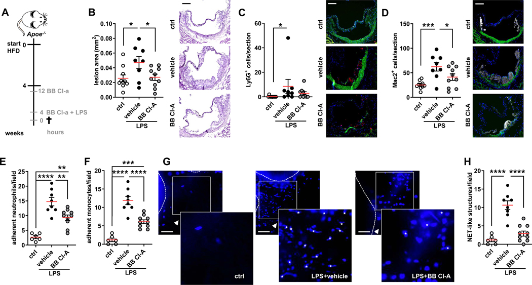
Background: Acute infection is a well-established risk factor of cardiovascular inflammation increasing the risk for a cardiovascular complication within the first weeks after infection. However, the nature of the processes underlying such aggravation remains unclear. Lipopolysaccharide derived from Gram-negative bacteria is a potent activator of circulating immune cells including neutrophils, which foster inflammation through discharge of neutrophil extracellular traps (NETs). Here, we use a model of endotoxinemia to link acute infection and subsequent neutrophil activation with acceleration of vascular inflammation Methods: Acute infection was mimicked by injection of a single dose of lipopolysaccharide into hypercholesterolemic mice. Atherosclerosis burden was studied by histomorphometric analysis of the aortic root. Arterial myeloid cell adhesion was quantified by intravital microscopy.

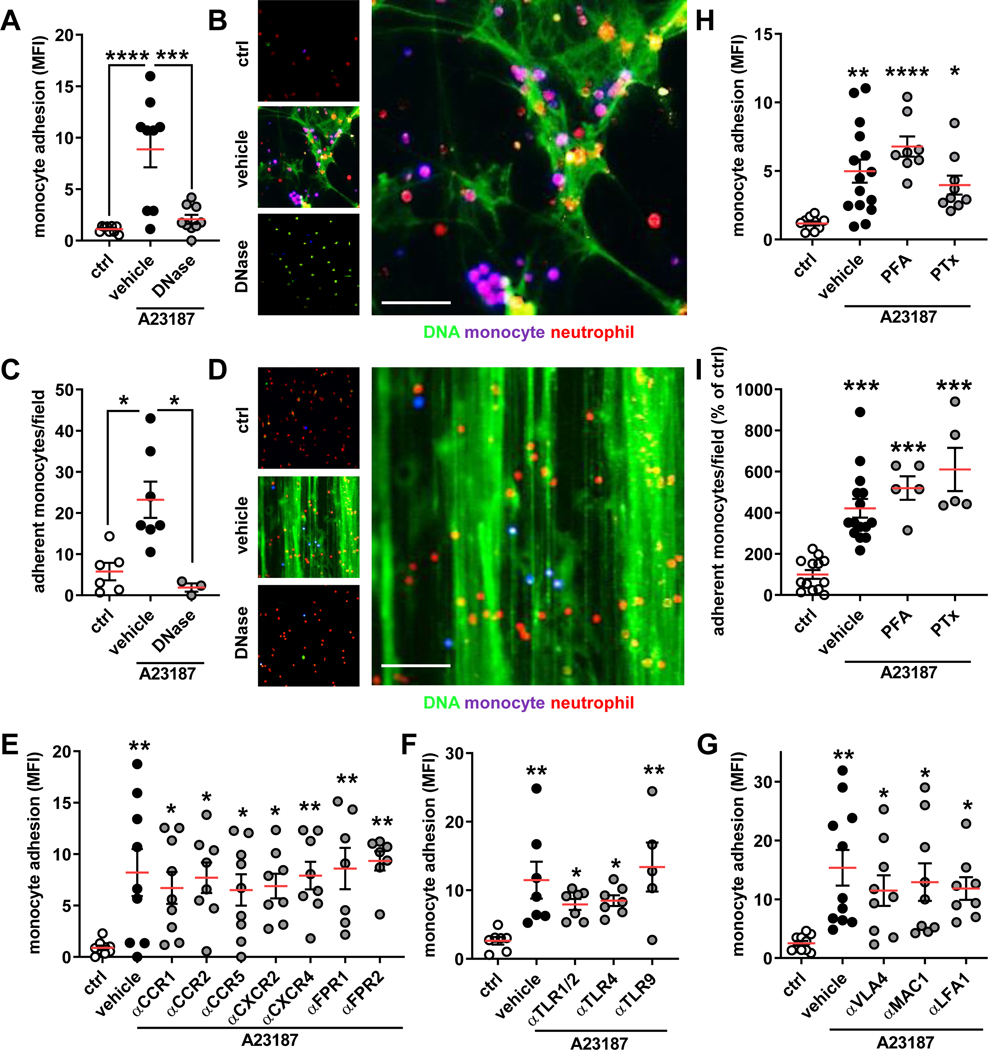
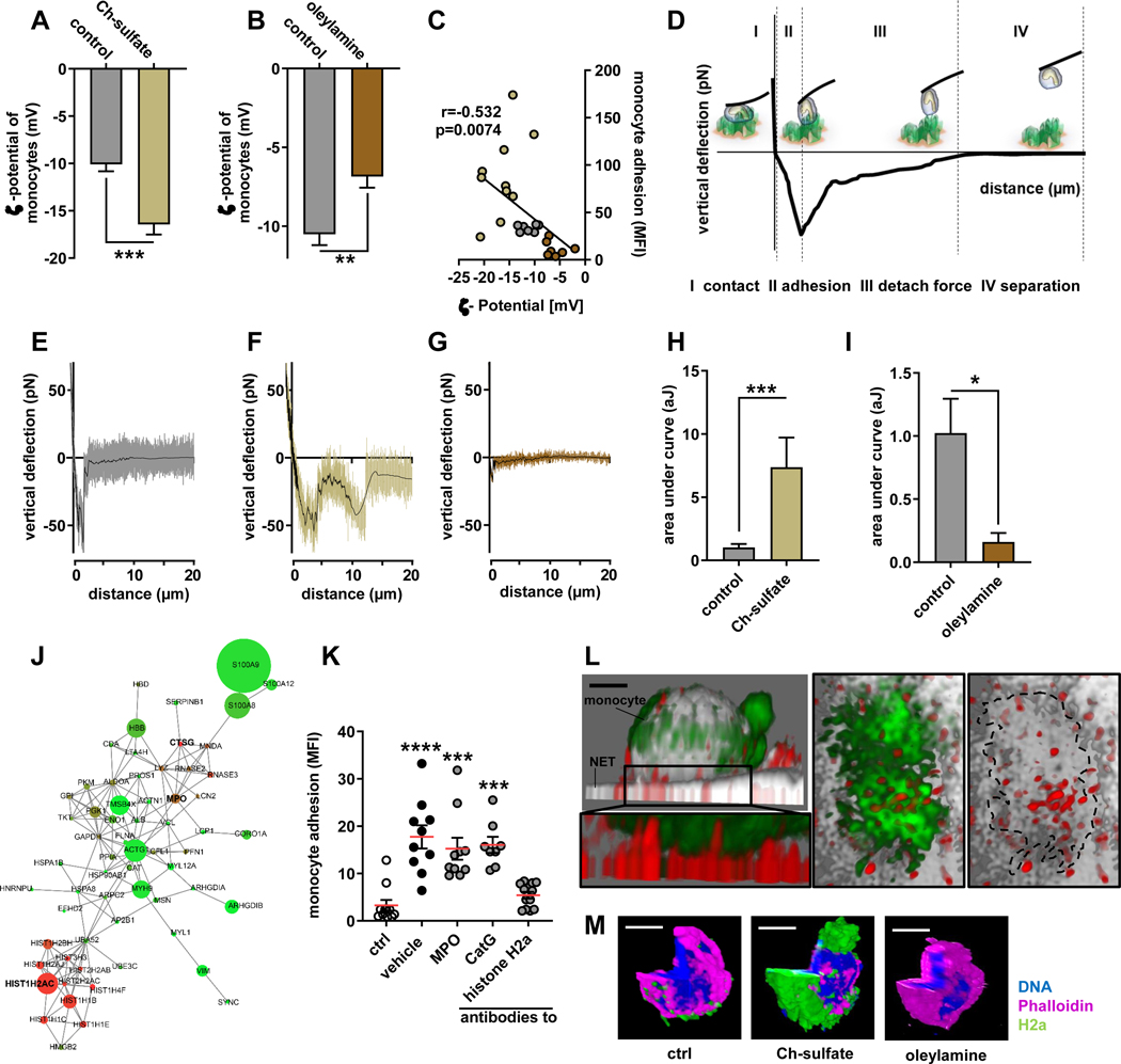
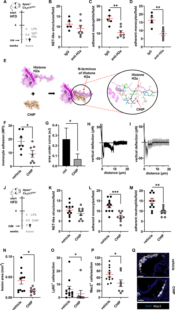
Results: Lipopolysaccharide treatment rapidly enhanced atherosclerotic lesion size by expansion of the lesional myeloid cell accumulation. Lipopolysaccharide treatment led to the deposition of NETs along the arterial lumen, and inhibition of NET release annulled lesion expansion during endotoxinemia, thus suggesting that NETs regulate myeloid cell recruitment. To study the mechanism of monocyte adhesion to NETs, we used in vitro adhesion assays and biophysical approaches. In these experiments, NET-resident histone H2a attracted monocytes in a receptor-independent, surface charge-dependent fashion. Therapeutic neutralization of histone H2a by antibodies or by in silico designed cyclic peptides enables us to reduce luminal monocyte adhesion and lesion expansion during endotoxinemia.

Conclusions: Our study shows that NET-associated histone H2a mediates charge-dependent monocyte adhesion to NETs and accelerates atherosclerosis during endotoxinemia.

Keywords: atherosclerosis; extracellular trap; histones; inflammation; neutrophils; sepsis.


Figures

Figure 1:
Figure 1:. Endotoxinemia accelerates atherosclerosis.
(A) Experimental setup. Apoe−/− or Apoe−/−Cx3cr1GFP mice were fed a HFD for 4 weeks and treated with either PBS (ctrl) or with LPS (1mg/kg BW). Another LPS-treated group received a BB Cl-amidine (BB Cl-A, 1mg/kg BW) 12 h before and along with LPS injection. (B) Aortic root lesion size analyzed in HE-stained sections. Representative images, scale bar 50 μm. (C/D) Lesional neutrophils (Ly6G+ cells) (C) and macrophages (Mac2+ cells) (D) quantified in root sections. Representative immunofluorescence images showing lesional neutrophil (red), Mac2+ positive cells (grey) and nuclei (DAPI, blue), scale bar 50 μm. (E/F) Quantification of luminally adherent neutrophils (E) and monocytes (F). (G) Representative intravital microscopy (IVM) images of the carotid artery of Apoe−/−Cx3cr1GFP mice. DNA is stained with DAPI, scale bar 50 μm. NET-like structures as identified by automized image analyzes are marked up with Asterisks. (H) Count of luminal NET-like structures in left common carotid artery. Data are analyzed by one-way ANOVA with Tukey’s multiple comparisons test (B, D, E, F, H) or Kruskal-Wallis test with Dunn’s multiple comparisons test (C); *p≤0.05, **p≤0.01, ***p≤0.001, ****p≤0.0001. All data are presented as mean±SEM.
Figure 2:
Figure 2:. Monocyte adhesion to NETs is receptor independent.
In vitro monocyte adhesion to expelled NETs under static or flow conditions. (A/B) Monocytes were added to neutrophils (ctrl) or NETting neutrophils (induced by A23187) and adhesion was quantified by a fluorescence plate reader. NETs were also degraded by DNase I. Representative microscopic image (B) of monocytes (violet) adherent to NETs (green; neutrophils red), scale bar 50 μm. (C/D) Monocytes were perfused at 0.5 dyne/cm2 over neutrophils, NETs, or degraded NETs and their adhesion was quantified manually. Representative microscopic image (D) of monocytes (violet) adherent to NETs (green; neutrophils red), scale bar 100 μm. (E-G) Monocytes where pre-treated with antagonists or antibodies to chemokine receptors (E), Toll-like receptors (F), or integrins (G) prior to incubation with NETs. (H/I) Monocytes fixed with PFA or treated with pertussis toxin (PTx) were used in static (H) or flow (I) adhesion assays. Data are analyzed by one-way ANOVA with Tukey’s multiple comparisons test (A, H, G) or Kruskal-Wallis test with Dunn’s multiple comparisons test (C, E, F, I); *p≤0.05, **p≤0.01, ***p≤0.001, **** p≤0.0001. All data are presented as mean±SEM.
Figure 3:
Figure 3:. Monocyte adhesion to NETs in regulated by cationic histone H2a.
(A/B) ζ-potential analysis of isolated monocytes treated with Ch-sulfate (A) or oleylamine (B). (C) Pearson correlation of monocyte adhesion to NETs versus monocyte ζ-potential. (D) Scheme of single cell atomic force microscopy (AFM) force spectroscopy. Monocytes were probed on expelled NETs at 200 pN contact force. (E-G) Representative force curves of native monocytes (E) or monocytes treated with Ch-sulfate (F) or oleylamine (G) probed on NETs. (H/I) Quantification of the area under the curve reflecting the energy required to rupture the monocyte-NET interaction. (J) Proteome analysis of NET resident proteins. Circle size reflects protein abundance, while color codes for charge (red cationic, green anionic). (K) Monocyte adhesion to NETs pre-incubated with antibodies to indicated NET-associated proteins. (L) Representative immunofluorescence confocal microscopy image showing the tight interaction between NET resident histone H2a and a NET-bound monocyte, scale bar 3 μm. Displayed is a xy projection (DNA gray, monocyte green, histone H2a red) and a zoom-in underneath (monocyte green, histone H2a red). The right two panels represent a top view (xz) of the zoom-in area thus visualizing the interface between the monocyte (green) and NET-resident histone H2a (red). In the right panel the monocyte is outlined (dashed line). (M) Representative confocal microscopy images of histone H2a binding monocyte in a charge dependent-manner, DNA blue, monocyte purple, histone H2a green, scale bar 5 μm. Data are analyzed by unpaired t-test (A, B, H, I) or Kruskal-Wallis test with Dunn’s multiple comparisons test (K); *p≤0.05, **p≤0.01, ***p≤0.001. All data are presented as mean±SEM. HNP, human neutrophil peptides; MPO, myeloperoxidase; PR3, proteinase 3; NE, neutrophil elastase; CatG, cathepsin G.
Figure 4:
Figure 4:. Neutralization of histone H2a inhibits endotoxin-induced arterial myeloid cell recruitment and atheroprogression.
(A) Experimental outline. Apoe−/−Cx3cr1GFP were fed a HFD for 4 weeks, treated with LPS (1 mg/kg, 4h) and injected with isotype respective control (IgG), or a histone H2a-targeting antibody (anti-H2a). (B-D) Intravital microscopy was used to quantify luminal NET-like structures (B) in the left carotid artery as well as adherent neutrophils (C) and monocytes (D).(E) The structure of histone H2a (magenta), CHIP (orange) and the histone H2a-CHIP complex which was derived from protein-protein docking and molecular dynamic simulation. CHIP bound and interacted with the N-terminal part of histone H2a. The electrostatic interactions between Arg or Lys of Histone H2a (green sticks) with Glu or Asp of CHIP (cyan sticks) as well as hydrogen bonds (displayed as dash lines) help to stabilize the complex formation between histone H2a and CHIP. (F-I) Pharmacological interruption of histone H2a monocyte-binding was validated in static adhesion assays (F) as well as by single cell force spectroscopy (G-I). (J-Q) In vivo validation of therapeutic effect of CHIP in endotoxin-accelerated atherosclerosis. (J) Experimental outline. Apoe−/−Cx3cr1GFP were fed a HFD for 4 weeks, treated with LPS (1 mg/kg, 4h) and injected with vehicle or CHIP (5 mg/kg). (K-M) Intravital microscopy was used to quantify luminal NET-like structures (K) in the left carotid artery as well as adherent neutrophils (L) and monocytes (M). (N) Aortic root lesion size analyzed after HE staining. Quantification of Lesional neutrophils (Ly6G+ cells) (O), and macrophages (Mac2+ cells) (P). (Q) Representative immunofluorescence images showing lesional Mac2+ cells (grey) and nuclei (DAPI, blue), scale bar 50 μm. Data are analyzed by Mann-Whitney test (B, C, D, F, G, O) or unpaired t-test (K-M, N, P); *p≤0.05, **p≤0.01, ***p≤0.001. All data are presented as mean ±SEM.

References

    1. Fayad ZA, Swirski FK, Calcagno C, Robbins CS, Mulder W, Kovacic JC. Monocyte and Macrophage Dynamics in the Cardiovascular System: JACC Macrophage in CVD Series (Part 3). J Am Coll Cardiol. 2018;72:2198–2212. doi: 10.1016/j.jacc.2018.08.2150. - DOI - PMC - PubMed
    1. Park J, Wysocki RW, Amoozgar Z, Maiorino L, Fein MR, Jorns J, Schott AF, Kinugasa-Katayama Y, Lee Y, Won NH, et al. Cancer cells induce metastasis-supporting neutrophil extracellular DNA traps. Sci Transl Med. 2016;8:361ra138. doi: 10.1126/scitranslmed.aag1711. - DOI - PMC - PubMed
    1. Winter C, Silvestre-Roig C, Ortega-Gomez A, Lemnitzer P, Poelman H, Schumski A, Winter J, Drechsler M, de Jong R, Immler R, et al. Chrono-pharmacological Targeting of the CCL2-CCR2 Axis Ameliorates Atherosclerosis. Cell Metab. 2018;28:175–182.e5. doi: 10.1016/j.cmet.2018.05.002. - DOI - PubMed
    1. Ortega-Gomez A, Salvermoser M, Rossaint J, Pick R, Brauner J, Lemnitzer P, Tilgner J, de Jong RJ, Megens RT, Jamasbi J, et al. Cathepsin G Controls Arterial But Not Venular Myeloid Cell Recruitment. Circulation. 2016;134:1176–1188. doi: 10.1161/CIRCULATIONAHA.116.024790. - DOI - PMC - PubMed
    1. Alard JE, Ortega-Gomez A, Wichapong K, Bongiovanni D, Horckmans M, Megens RT, Leoni G, Ferraro B, Rossaint J, Paulin N, et al. Recruitment of classical monocytes can be inhibited by disturbing heteromers of neutrophil HNP1 and platelet CCL5. Sci Transl Med. 2015;7:317ra196. doi: 10.1126/scitranslmed.aad5330. - DOI - PubMed

Publication types

Substances