. 2020 Nov 10;19(1):206.
doi: 10.1186/s12934-020-01469-z.

An automated oxystat fermentation regime for microoxic cultivation of Magnetospirillum gryphiswaldense

Affiliations

An automated oxystat fermentation regime for microoxic cultivation of Magnetospirillum gryphiswaldense

Cornelius N Riese et al. Microb Cell Fact. .

Abstract

Background: Magnetosomes produced by magnetotactic bacteria represent magnetic nanoparticles with unprecedented characteristics. However, their use in many biotechnological applications has so far been hampered by their challenging bioproduction at larger scales.

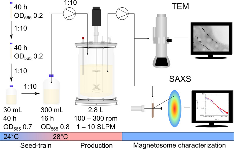
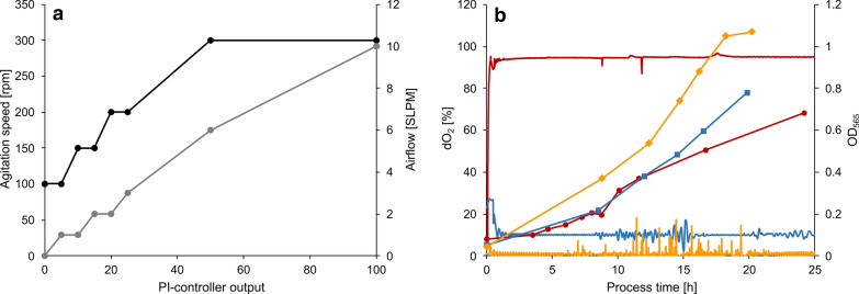
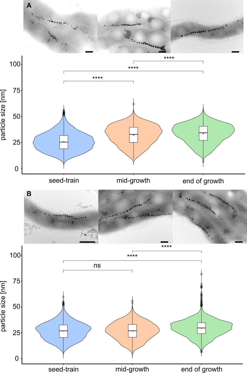
Results: Here, we developed an oxystat batch fermentation regime for microoxic cultivation of Magnetospirillum gryphiswaldense in a 3 L bioreactor. An automated cascade regulation enabled highly reproducible growth over a wide range of precisely controlled oxygen concentrations (1-95% of air saturation). In addition, consumption of lactate as the carbon source and nitrate as alternative electron acceptor were monitored during cultivation. While nitrate became growth limiting during anaerobic growth, lactate was the growth limiting factor during microoxic cultivation. Analysis of microoxic magnetosome biomineralization by cellular iron content, magnetic response, transmission electron microscopy and small-angle X-ray scattering revealed magnetosomal magnetite crystals were highly uniform in size and shape.

Conclusion: The fermentation regime established in this study facilitates stable oxygen control during culturing of Magnetospirillum gryphiswaldense. Further scale-up seems feasible by combining the stable oxygen control with feeding strategies employed in previous studies. Results of this study will facilitate the highly reproducible laboratory-scale bioproduction of magnetosomes for a diverse range of future applications in the fields of biotechnology and biomedicine.

Keywords: Magnetosome biomineralization; Magnetosomes; Magnetospirillum gryphiswaldense; Oxystat fermentation.


Conflict of interest statement

The authors declare that they have no competing interests.

Figures

Fig. 1
Fig. 1
Overview over the oxystat fermentation approach including seed-train, production with different stirrer speeds (rounds per minute = rpm) as well as airflow rates (standard liter per minute = SLPM) and magnetosome characterization by transmission electron microscopy (TEM) and small-angle X-ray scattering (SAXS). See text for more details
Fig. 2
Fig. 2
a Programmed cascade for automated dO2 control by dynamic adjustment of agitation (black) and aeration (grey) with compressed air (21% dO2). b Representative fermentations at 95% (red circles), 10% (blue squares) and 1% (orange diamonds) dO2. OD565 is indicated by filled symbols. dO2 is indicated by solid lines
Fig. 3
Fig. 3
TEM micrographs of cells grown under a oxic (95% dO2, scalebar 1 µm), b microoxic (1% dO2, scalebar, 500 nm) and b anoxic (0% dO2, scalebar 1 µm) conditions. Growth and substrate consumption triplicates at dO2 tensions of 95% (red), 1% (orange) and 0% (blue). d Growth by OD565. e Lactate concentration in mM. f Nitrate concentration in mM. g Magnetic response Cmag. h Iron content of the cell pellet in µg mgdw−1
Fig. 4
Fig. 4
a Transmission electron micrographs of representative cells (scale bar 200 nm) and magnetosome particle sizes under anoxic (0% dO2) conditions at different timepoints of the process. Seed-train = 0 h (n = 2925), mid-growth = 15 h (n = 3069) and end of growth = 20 h (n = 3058). b Transmission electron micrographs (scale bar 1 µm (left micrograph) and 200 nm) and magnetosome particle sizes under microoxic (1% dO2) conditions at different timepoints of the process. Seed-train = 0 h (n = 3421), mid-growth = 14 h (n = 2267) and end of growth = 18 h (n = 3180). In box plots, the box indicates the interquartile range, the bar indicates the median, and the red dot represents the mean. Grey dots represent data points above or below the 95th and 5th percentile. The violin plots show the magnetosome particle size distribution of measured particle sizes. For statistical comparison of particle sizes, Wilcoxon rank sum test was performed (****, p < 0.0001; ns, not significant)
Fig. 5
Fig. 5
Transmission electron micrographs and magnetosome particle sizes at process end among seed-train (n = 2925), microoxic (1% dO2) (n = 3180) and anoxic (0% dO2) conditions (n = 3025) with representative TEM micrographs of cells under the respective conditions (scale bar 200 nm). In box plots, the box indicates the interquartile range, the bar indicates the median, and the red dot represents the mean. Grey dots represent data points above or below the 95th and 5th percentile. The violin plots show the magnetosome particle size distribution of measured particle sizes. For statistical comparison of particle sizes, Wilcoxon rank sum test was performed (****, p < 0.0001)

Similar articles

Cited by

References

    1. Lefèvre CT, Bazylinski DA. Ecology, diversity, and evolution of magnetotactic bacteria. Microbiol Mol Biol Rev. 2013;77:497–526. doi: 10.1128/MMBR.00021-13. - DOI - PMC - PubMed
    1. Uebe R, Schüler D. Magnetosome biogenesis in magnetotactic bacteria. Nat Rev Microbiol. 2016;14:621–637. doi: 10.1038/nrmicro.2016.99. - DOI - PubMed
    1. Barber-Zucker S, Keren-Khadmy N, Zarivach R. From invagination to navigation: the story of magnetosome-associated proteins in magnetotactic bacteria. Protein Sci. 2015;25:338–351. doi: 10.1002/pro.2827. - DOI - PMC - PubMed
    1. Lohße A, Ullrich S, Katzmann E, Borg S, Wanner G, Richter M, Voigt B, Schweder T, Schüler D. Functional analysis of the magnetosome island in Magnetospirillum gryphiswaldense: the mamAB operon is sufficient for magnetite biomineralization. PLoS ONE. 2011;6:e25561. doi: 10.1371/journal.pone.0025561. - DOI - PMC - PubMed
    1. Lohße A, Borg S, Raschdorf O, Kolinko I, Tompa É, Pósfai M, Faivre D, Baumgartner J, Schüler D. Genetic dissection of the mamAB and mms6 operons reveals a gene set essential for magnetosome biogenesis in Magnetospirillum gryphiswaldense. J Bacteriol. 2014;196:2658–2669. doi: 10.1128/JB.01716-14. - DOI - PMC - PubMed

Supplementary concepts

LinkOut - more resources