Review
. 2020 Nov 9;8(1):189.
doi: 10.1186/s40478-020-01062-w.

PINK1/PARKIN signalling in neurodegeneration and neuroinflammation

Affiliations
Review

PINK1/PARKIN signalling in neurodegeneration and neuroinflammation

Peter M J Quinn et al. Acta Neuropathol Commun. .

Abstract

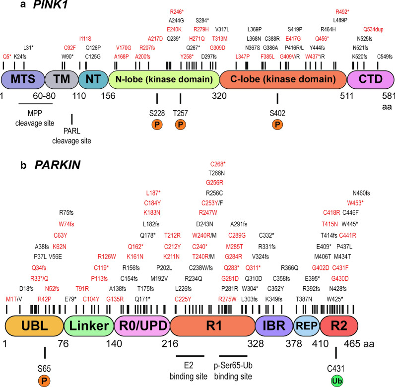
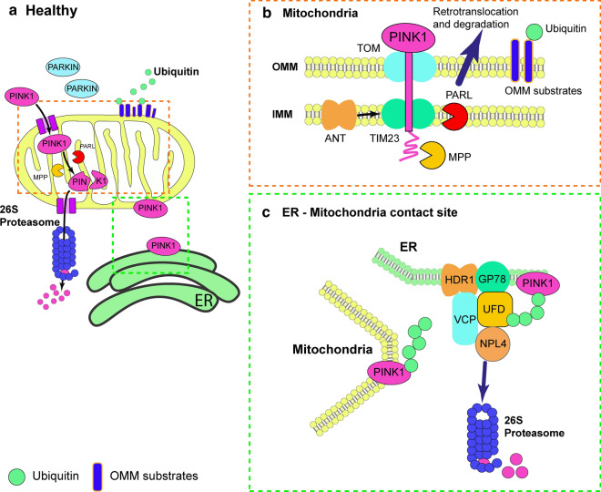
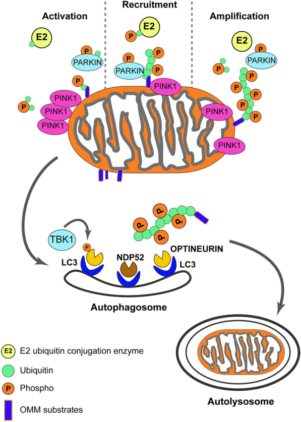
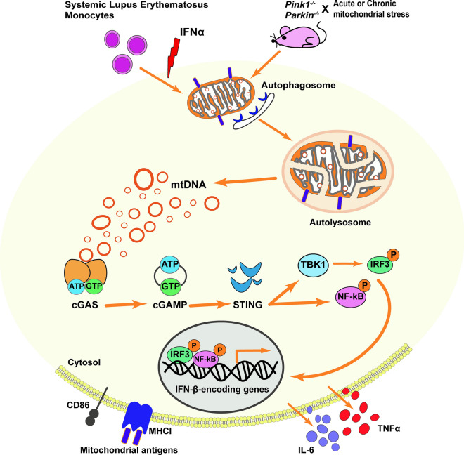
Mutations in the PTEN-induced kinase 1 (PINK1) and Parkin RBR E3 ubiquitin-protein ligase (PARKIN) genes are associated with familial forms of Parkinson's disease (PD). PINK1, a protein kinase, and PARKIN, an E3 ubiquitin ligase, control the specific elimination of dysfunctional or superfluous mitochondria, thus fine-tuning mitochondrial network and preserving energy metabolism. PINK1 regulates PARKIN translocation in impaired mitochondria and drives their removal via selective autophagy, a process known as mitophagy. As knowledge obtained using different PINK1 and PARKIN transgenic animal models is being gathered, growing evidence supports the contribution of mitophagy impairment to several human pathologies, including PD and Alzheimer's diseases (AD). Therefore, therapeutic interventions aiming to modulate PINK1/PARKIN signalling might have the potential to treat these diseases. In this review, we will start by discussing how the interplay of PINK1 and PARKIN signalling helps mediate mitochondrial physiology. We will continue by debating the role of mitochondrial dysfunction in disorders such as amyotrophic lateral sclerosis, Alzheimer's, Huntington's and Parkinson's diseases, as well as eye diseases such as age-related macular degeneration and glaucoma, and the causative factors leading to PINK1/PARKIN-mediated neurodegeneration and neuroinflammation. Finally, we will discuss PINK1/PARKIN gene augmentation possibilities with a particular focus on AD, PD and glaucoma.

Keywords: Alzheimer’s disease; Mitophagy; Neurodegeneration; PARKIN; PINK1; Parkinson’s disease.


Conflict of interest statement

The authors declare that they have no conflict of interest.

Figures

Fig. 1
Fig. 1
Schematic representations of PINK1 and PARKIN domains and disease-related mutations. a PINK1 is composed by 581 amino acids, encompassing the mitochondrial targeting sequence (MTS), transmembrane region (TM), N-terminal regulatory region (NT), N-lobe of the kinase domain, C-lobe of the kinase domain and the C-terminal domain (CTD). Mitochondrial processing peptidase (MPP) and presenilin-associated rhomboid-like (PARL) cleavage sites and PINK1 auto-phosphorylation sites are depicted in the figure (S228, T257, S402). b PARKIN is formed by 465 amino acids with a ubiquitin-like domain (UBL), linker, really-interesting-new-gene (RING)/unique Parkin domain (R0/UPD), RING1 (R1), in-between-RING (IBR), repressor element of Parkin (REP), and a RING2 (R2) domain. E2 co-enzyme and p-Ser65-Ub binding sites, as well as Ser65 phosphorylation and Cys431 catalytic sites, are displayed. Disease-associated mutations collected from the movement disorder society genetic mutation (www.mdsgene.org/) and ClinVAR (www.ncbi.nlm.nih.gov/clinvar/) databases are displayed on top of schematic representation. In red are depicted the mutations considered pathogenic
Fig. 2
Fig. 2
The canonical PINK1/PARKIN pathway. a and b In healthy mitochondria, PINK1 is constitutively imported via translocase of the outer membrane (TOM)/translocase of the inner membrane (TIM)23 complexes to the inner mitochondrial membrane (IMM), cleaved by two proteases (mitochondrial processing peptidase (MPP) and presenilin-associated rhomboid-like (PARL)) and retro-translocated to the cytosol. Cleaved PINK1 is then degraded by the ubiquitin/proteasome system. While Parkin remains inactive in the cytosol. (a and c) PINK1 is also present at the mitochondria-endoplasmic reticulum (ER) interface, where it interacts with the endoplasmic-reticulum-associated protein degradation (ERAD) machinery. At the ER, PINK1 degradation by the proteasome is controlled by the ERAD E3 ubiquitin ligases HRD1 and gp78 and by the ERAD-associated proteins VCP, UFD1, andUFD2A
Fig. 3
Fig. 3
PINK1/PARKIN-directed quality control in damaged mitochondria. After damage, PINK1 is no longer imported into the inner mitochondrial membrane (IMM) and accumulates on the outer mitochondrial membrane (OMM). Here, a supercomplex composed by TOM complex subunits and PINK1 homodimers is formed, facilitating PINK1 autophosphorylation and activation. Once activated, PINK1 phosphorylates ubiquitinated substrates on the OMM and PARKIN enable its E3 ubiquitin ligase functions in concert with E2 ubiquitin-conjugating enzymes. PINK1-mediated phosphorylation of ubiquitin phospho-Ser65- ubiquitin on OMM substrates acts as the PARKIN receptor for its recruitment from the cytosol. PINK1 and PARKIN initiate a positive feedback loop, resulting in the coating of damaged mitochondria with phospho-ubiquitin chains. Individual OMM proteins decorated with poly-ubiquitin can be extracted from the membrane and degraded by the 26 S proteasome. Phospho-ubiquitin chains are bound by two mitophagy adaptors, nuclear domain 10 protein 52 (NDP52) and optineurin. Phosphorylation of optineurin by TANK Binding Kinase 1 (TBK1) enhances its binding to ubiquitin chains and promotes selective autophagy of damaged mitochondria. The two adaptors recruit autophagosomes via microtubule-associated protein 1A/1B-light chain 3 (LC3) binding, allowing the engulfment of dysfunctional mitochondria resulting in their direct degradation in lysosome
Fig. 4
Fig. 4
PINK1/PARKIN-signalling and inflammation. Mice lacking Parkin or Pink1 upon acute (exhaustive exercise-induced) or chronic (mitochondrial DNA (mtDNA) mutation-induced) mitochondrial stress present inflammation due to the activation of the stimulator of interferon genes (STING) as result from the accumulation of mtDNA mutations and release of mtDNA into the cytosol. While, in systemic lupus erythematosus excessive IFNα damages mitochondrial respiration, leading to oxidative stress that impairs lysosomal degradation and obstructs autophagic clearance. Undegraded mtDNA from mitochondria, interact with the cytosolic DNA sensor cGAS in a sequence-independent way, promoting a conformational change of cGAS to catalyse the formation of 2,3-cyclic GMP-AMP (cGAMP). The cGAS activation, as well as cGAMP synthase, activate STING, recruiting binding kinase 1 (TBK1) as well as interferon regulatory factor 3 (IRF3). The IRF3 then displaces to the nucleus and induces immune-stimulated genes and type I IFN expression. The nuclear factor kappa-light-chain-enhancer of activated B cells (NF-κB) signalling can also be activated by STING. In the absence of PARKIN and PINK1, high levels of mitochondrial antigens are presented to major histocompatibility complex (MHC) class I molecules in macrophages and dendritic cells triggering an adaptive immune response

References

    1. Benda C. Ueber dier Spermatogenese de Verbebraten und höherer Evertebraten, II. Theil: die Histiogenese der Spermien. Arch Anat Physiol Arch Anat Physiol. 1898;73:393–398.
    1. Bernard G, Bellance N, James D, Parrone P, Fernandez H, Letellier T, et al. Mitochondrial bioenergetics and structural network organization. J Cell Sci. 2007;120(5):838–848. doi: 10.1242/jcs.03381. - DOI - PubMed
    1. Sjostrand FS. Electron microscopy of mitochondria and cytoplasmic double membranes. Nature. 1953;171(4340):30–32. doi: 10.1038/171030a0. - DOI - PubMed
    1. Palade GE. An electron microscope study of the mitochondrial structure. J Histochem Cytochem. 1953;1(4):188–211. doi: 10.1177/1.4.188. - DOI - PubMed
    1. Cogliati S, Enriquez JA, Scorrano L. Mitochondrial cristae: where beauty meets functionality. Trends Biochem Sci. 2016;41(3):261–273. doi: 10.1016/j.tibs.2016.01.001. - DOI - PubMed

Publication types

MeSH terms